
Tubular prostheses

a technology of tubular prosthesis and prosthesis, which is applied in the field of artificial replacements, can solve the problems of many complications, no replacements currently available, and no available replacement of esophageal tissu

Active Publication Date: 2014-11-06
YALE UNIV +1
View PDF4 Cites 7 Cited by
  • Summary
  • Abstract
  • Description
  • Claims
  • Application Information

AI Technical Summary

Benefits of technology

This patent describes a new way to make artificial airways, esophagus, and urinary conduits that can be used to replace damaged or diseased tissue. These artificial airways and conduits are made by implanting a tubular stent and extracellular matrix into a recipient. The stent can be made from a natural or synthetic material, and the extracellular matrix can be produced and secreted by non-autologous smooth muscle cells. The extracellular matrix can be formed as a tube or a tube with a stent inside it. These new surgical tools provide better options for repairing damaged anatomical conduits.

Problems solved by technology

There are no replacements currently available fir trachea in humans.
Currently, there is no available replacement for esophageal tissue.
Both of these procedures have many complications and a viable esophageal replacement is certainly medically needed.
Though widely used, ileal conduits pose many problems that can lead to short-term and long-term complications [Konety, Allareddy, V., Influence of post-cystectomy complications on cost and subsequent outcome.
In the short term, patients may suffer from complications at the bowel harvest site, including anastomotic leaks and peritonitis.
In addition, ileal urinary conduits may suffer from ischemia and necrosis, which can lead to perforation, anastomotic breakdown, and leakage of urine from the conduit.

Method used


Image

  • Tubular prostheses
  • Tubular prostheses
  • Tubular prostheses

Examples


example , 1

Example, 1

Tracheal PGA Scaffold Preparation

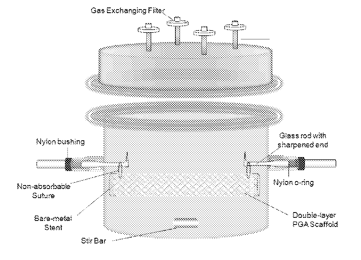
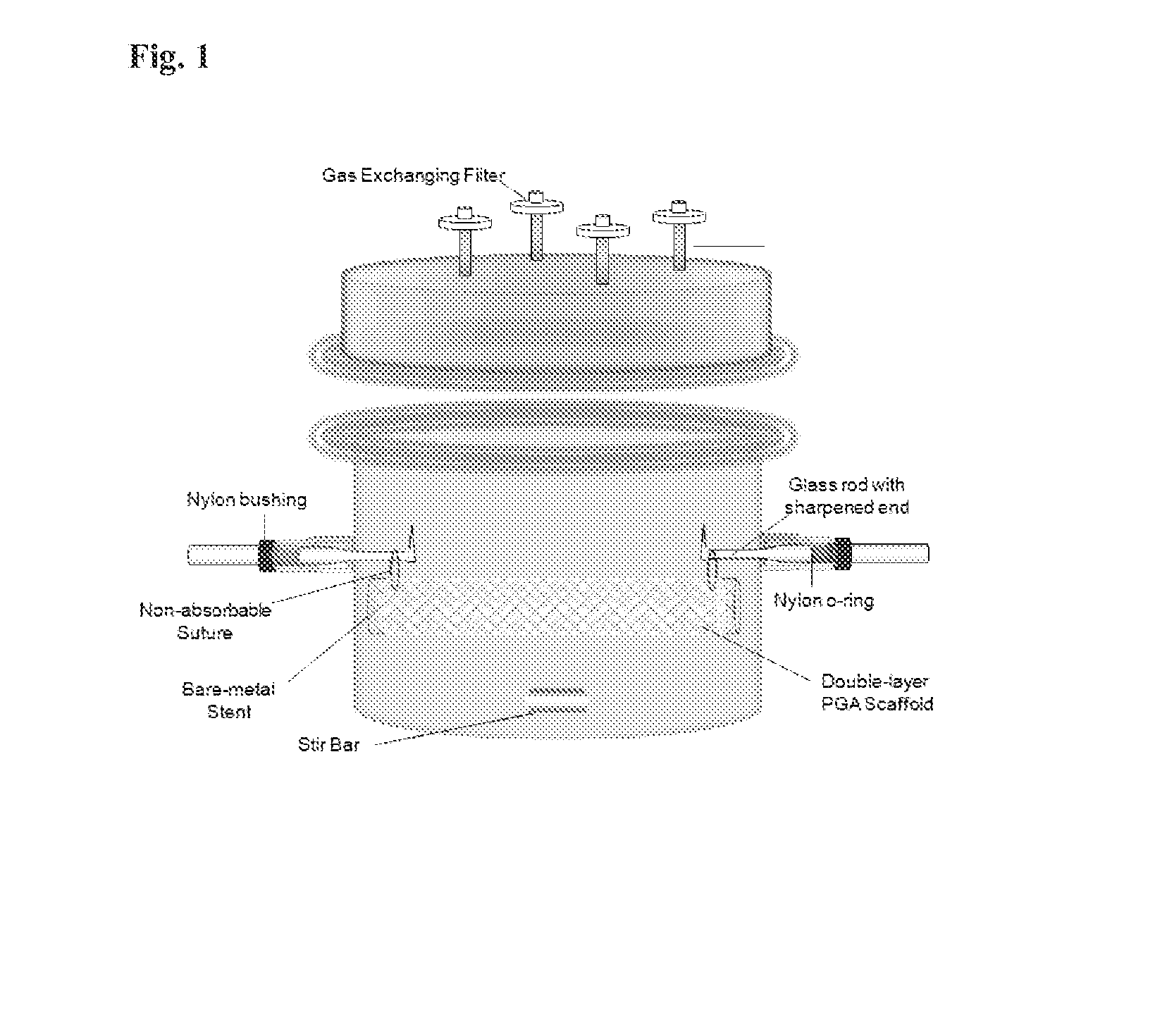
[0055]A poly(glycolic) acid (PGA) sheet is cut into 5.4 cm×8.5 cm and 5.2 cm×8.5 cm pieces. 5.2 cm×8.5 cm PGA mesh is rolled into a tube and inserted inside a bare-metal stent (1.7×8.5 cm). 5.4 cm×8.5 cm PGA mesh is then sewn around the bare-metal stent using the absorbable PGA suture, sandwiching the stent between the two layers of PGA mesh. A crochet needle is applied carefully throughout the PGA / stent construct to interlace the two PGA layers together. A non-absorbable suture is sewn through both ends of PGA / stent construct to suspend the construct inside a specially designed bioreactor for trachea reconstruction. The construct is then dipped into 1M NaOH solution for 2 minutes to treat the surface of the PGA mesh followed by three rinsing in distilled water. The PGA / stent construct is then assembled in the bioreactor as shown in

example 2

Smooth Muscle Cells (SMC) Seeding and Tracheal Culture Maintenance

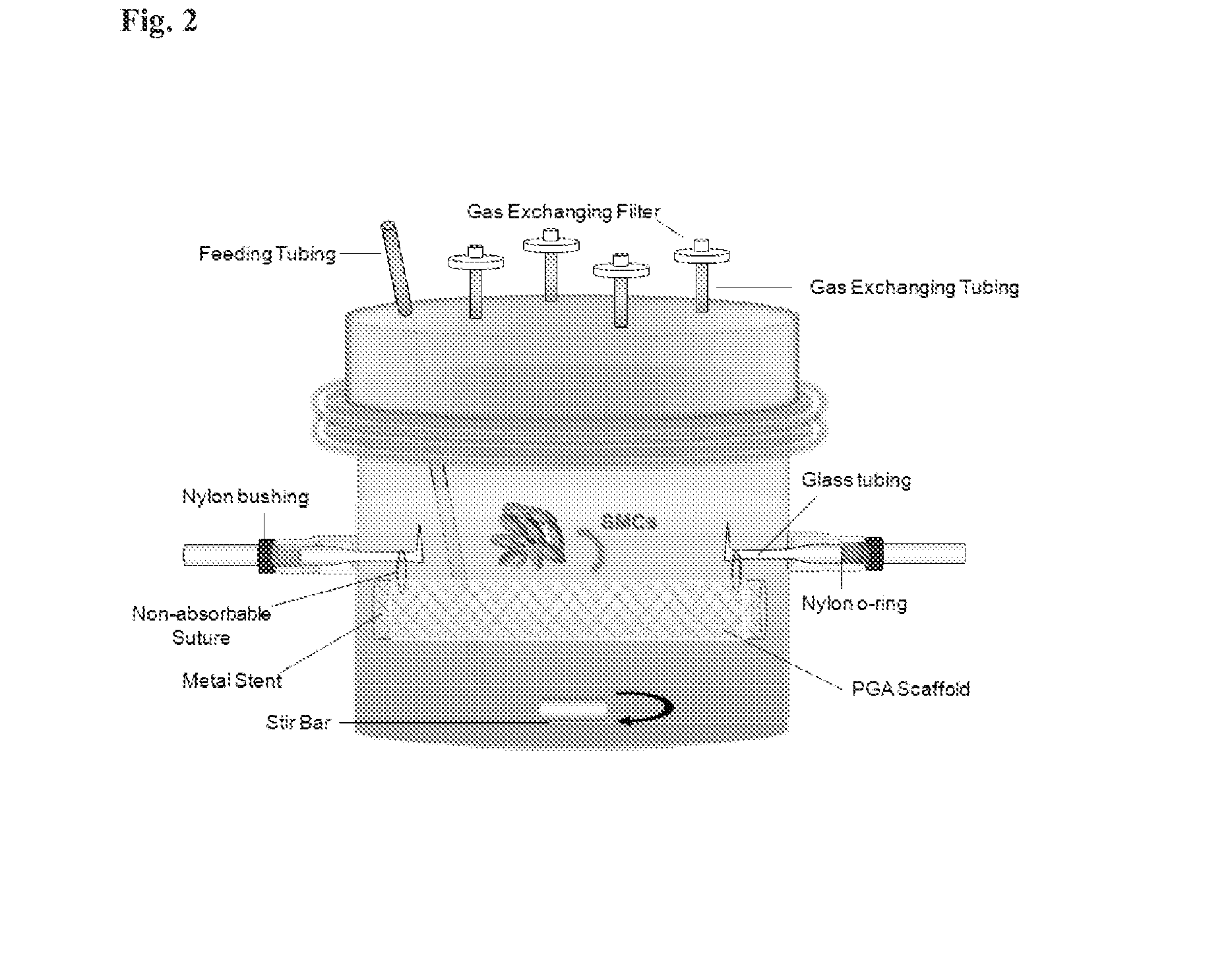
[0056]Primary SMCs were isolated from dog aortas and expanded in T-75s in 20% fetal bovine serum (FBS) low glucose Dulbecco's Modified Eagle's Medium. 120 million SMCs of P2 and P3 were re-suspended in 7 ml of medium and seeded onto the PGA / Stent construct inside the bioreactor, as shown in FIG. 2. The construct was cultured inside the bioreactor statically for 12 weeks in 1.3 L of low glucose Dulbecco's Modified Eagle's Medium with 20% FBS, basic fibroblast growth factor (10 ng / ml), platelet derived growth factor (10 ng ml), L-ascorbic acid, copper sulfate, HEPES, L-proline, L-alanine, L-glycine, and Penicillin G (FIG. 2), Medium was changed 1.5 times per week and ascorbic acid was supplemented three times per week.

example 3

Decellularization of Engineered Trachea

[0057]Engineered trachea (6 cm in length) was first incubated in 250 mL CHAPS buffer (8 mM CHAPS, 1M NaCl, and 25 mM EDTA in PBS) for 45 minutes at 37 C.° under high-speed agitation, followed by thorough sterile PBS rinsing. The engineered trachea was further treated with 250 mL sodium dodecyl sulfate (SDS) buffer (1.8 mM SDS, 1M NaCl, and 25 mM EDTA in PBS) for 45 minutes at 37 C.° with high-speed agitation. The engineered trachea then underwent 2 days of washing in PBS to completely remove the residual detergent. All decellularization steps were performed under sterile conditions. The decellularized engineered trachea was stored in sterile PBS containing penicillin 100 U / mL and streptomycin 100 mg / mL at 4 C.°.


PUM


Abstract

Tubular prostheses are provided for use in airways, upper digestive, and urinary tracts. Each of these uses has its own specific sets of biological specifications, based on what it must contain and exclude and the physical and chemical pressures and stresses to which it is subjected. The prostheses may be made from allogeneic cells. Thus they can be manufactured and stored prior to an individual's personal need arising.

Description

TECHNICAL FIELD OF THE INVENTION[0001]This invention is related to the area of artificial replacements for diseased or damaged anatomical conduits. In particular, it relates to conduits of fluids, solids, and gasses.BACKGROUND OF THE INVENTION[0002]Currently, there are about 2,000 patients per year in the U.S. who need replacement tracheal tissue. Causes for this include tracheal cancer, invasive infections of the trachea or bronchi, and trauma. There are no replacements currently available fir trachea in humans. At best, when a segment of trachea is resected, the only surgical option is to “pull together” the two ends of the trachea and sew them together, hoping that the anastomosis does not “pull apart” thereafter.[0003]Currently in the U.S., approximately 4,000 patients per year need an esophageal replacement. This is due primarily to esophageal cancer, though trauma and infection are causes a small number of cases of esophageal replacement. Currently, there is no available repla...

Claims


Application Information

Patent Timeline
Patent Type & Authority Applications(United States)
IPC IPC(8): A61F2/20
CPCA61F2/20A61F2002/046A61L27/3633A61L27/3679A61L27/3687A61L27/3826A61L27/3882A61L27/48A61L2430/40A61L2430/22
Inventor NIKLASON, LAURAHUANG, ANGELAZHAO, LIPINGDAHL, SHANNON
Owner YALE UNIV
Learn More