Methods for medical imaging

a medical imaging and method technology, applied in the field of medical imaging, can solve the problems of limited success in humans, lack of response ability, and use of fluorescein, and achieve the effect of improving the depth of penetration and imaging quality

Inactive Publication Date: 2015-01-29
THE TRUSTEES OF THE UNIV OF PENNSYLVANIA
View PDF4 Cites 22 Cited by
  • Summary
  • Abstract
  • Description
  • Claims
  • Application Information

AI Technical Summary

Benefits of technology

[0016]The invention further provides a method further comprising imaging devices to convert the near-infrared signal to a visible signal. The invention further provides a method wherein the imaging device is selected from the group consisting of devices which can be mounted over the patient, hand-held devices, devices which are attached to a long lens system, minimally invasive cameras, telescopes, endoscopes, esophagoscopes, colonoscopes, laparoscopes, thoracoscope long lens, capsule endoscopes, and combinations thereof. The invention further provides a method wherein the imaging device is ingested or implanted in the subject. The invention fur

Problems solved by technology

(Hoffman 2005) These approaches are highly useful to study the biology of tumors, but, they have had limited success in humans due to the lack of tumor access, toxicity, dearth of clinical approved probes and paucity of large scale imaging devices.
(Schaafsma 2011, Schulz 2010, Choi 2010) A recent approach in humans utilized a folate-fluorescein fluorophore preparation specific to ovarian tumors but was limited by false negatives in folate-receptor negative tumors and the use of fluorescein which can be difficult to differentiate from autofluorescence.
Neovasculature in cancer tissues and inflammatory lesions have higher pressure concentrations and lack the ability to respond to vasoactive mediato

Method used


Image

  • Methods for medical imaging
  • Methods for medical imaging
  • Methods for medical imaging

Examples


example 1

NIR Labeling of Small Animals In Vivo

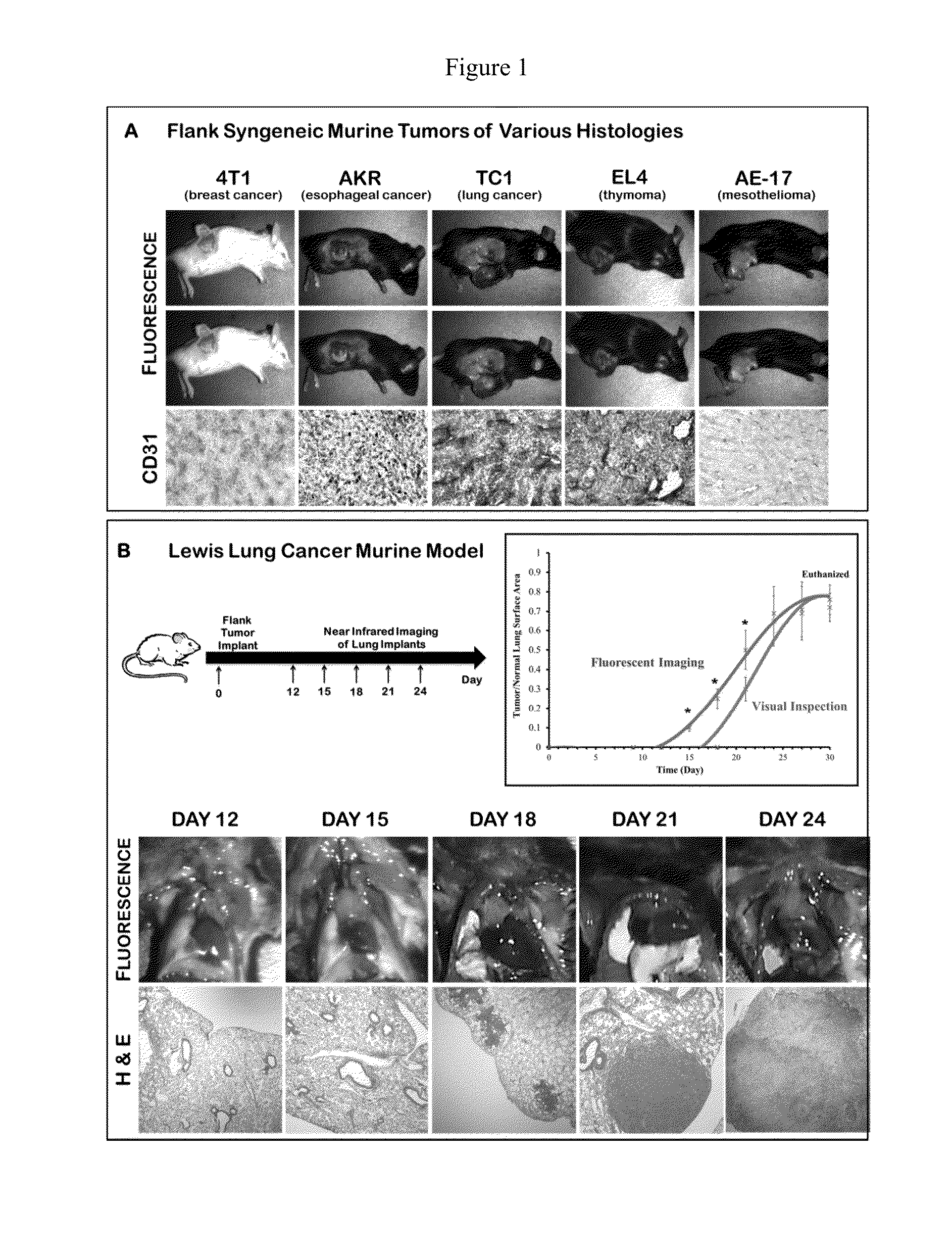
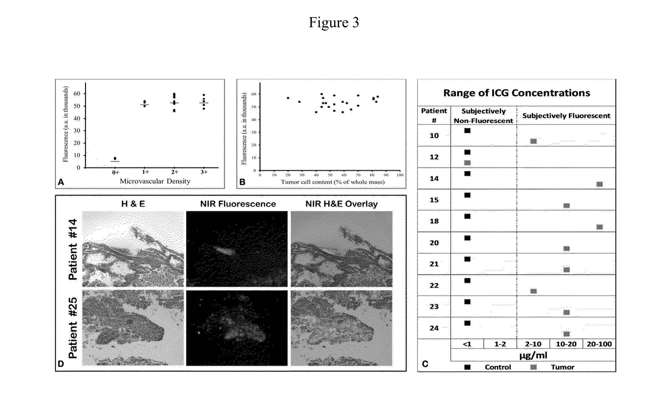
[0128]Initially, in order to determine if the EPR effect could deliver a MR contrast agent to solid tumors, we conducted a proof-of-concept study in multiple animal models of malignant disease. Fifty female C57bl / 6 mice were injected with five different syngeneic cancer cell lines (4T1 breast cancer, TC1 lung cancer, EL4 thymoma, AE17 mesothelioma, AKR esophageal cancer) into their flanks. After the tumors were well established (˜200 mm3), 7.5 mg / kg of ICG was administered via the tail vein. The next day, a tissue spectrometer was used to semi-quantitate fluorescence from the tumor, surrounding tissue and 12 organs. (Mohs 2010) The mean fluorescence from the tumor was 54,238 arbitrary units (au) (range 46,283-60,000). The mean fluorescence from surrounding normal tissues and organs averaged 4863±1254 au. Tumors were harvested, sectioned and assayed for microvessel density to determine if there was a correlation between tumor vascularity and fluor...

example 2

Tumor Fluorescence During Surgical Resection

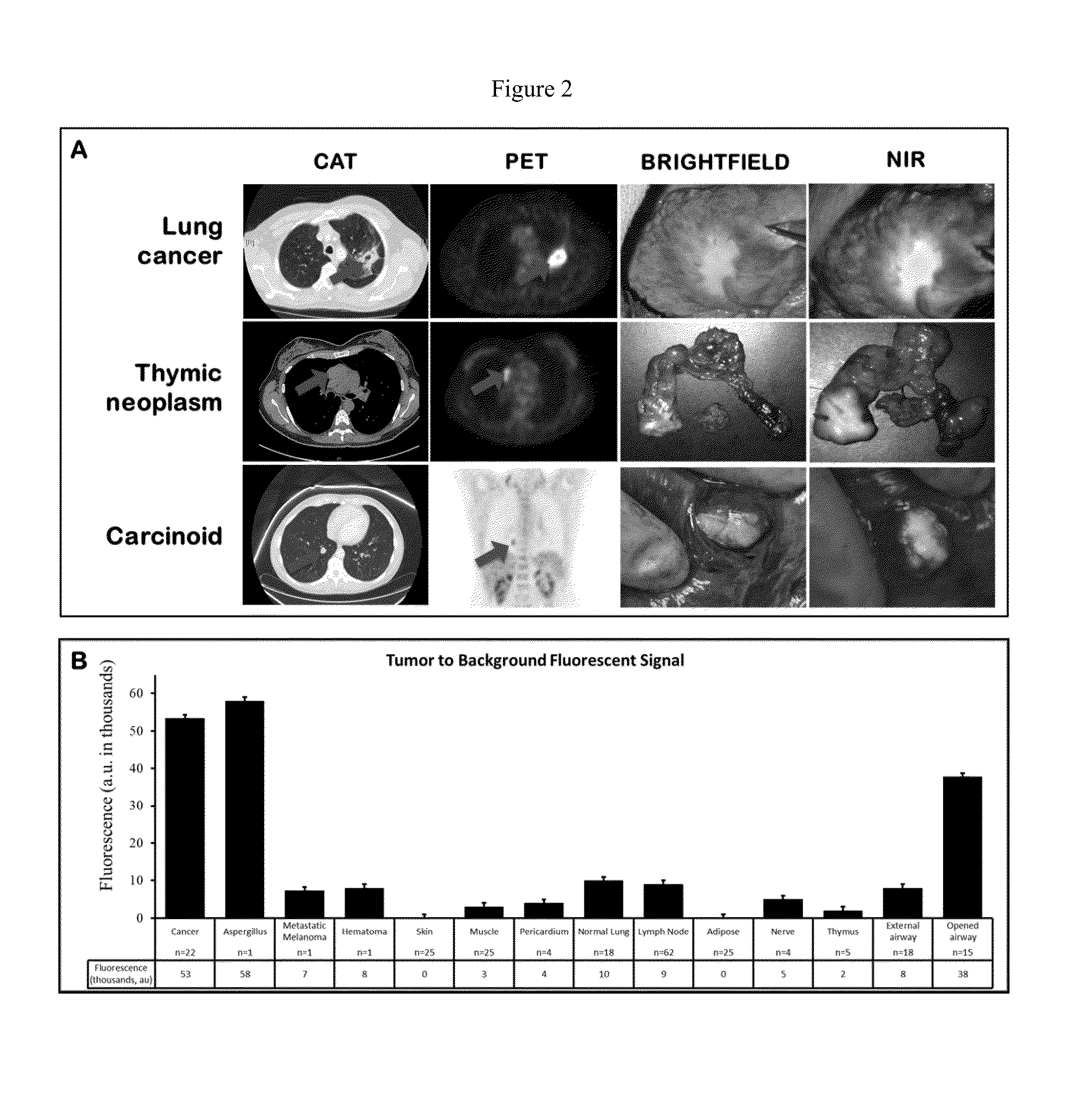
[0131]To determine if indocyanine green would be delivered to human tumors, patients undergoing cancer surgery were first imaged in vivo at the onset of the operation. At the time of surgery, the chest was opened and inspected by standard visualization and manual palpation. In all cases, the surgeon could immediately see or feel the tumor. A dual camera head with a brightfield and a NIR output was then used to visualize tumor fluorescence (FIG. 7). In 16 out of 27 (59%) cases, the dual camera head could detect tumor fluorescence at various depths of penetration into the tissue. In the remaining 11 cases, the tumor was located too deep in the organs to image by NIR. The deepest tumor that could be detected was ˜1 cm from the surface of the organ. Attempts to quantitate tumor fluorescence in vivo were not feasible for several reasons including variations in operating room conditions, lack of miniaturized tissue spectrometer with safe laser l...

example 3

Ex Vivo Analysis of Tumor Fluorescence

[0132]Following in vivo imaging, the patients then underwent a standard-of-care surgical resection of the tumor. Once removed from the patient, the specimen was examined, opened, biopsied and analyzed ex vivo (FIG. 2a). Every case was photo-documented both by brightfield imaging and NIR imaging. Qualitatively, NIR imaging revealed strong fluorescence in 25 out of 27 (92%) masses. Then, the hand held spectrometer was used to semi-quantitate tissue fluorescence. Each tumor had 4 measurements at four perpendicular locations and the center of the tumor (total of 20 measurements / tumor). Mean fluorescence in the human tumors was 53,304±4193 au in 25 out of 27 masses (93%) (FIG. 2b). We conjectured that the center of the tumor might be less fluorescent than the periphery due to necrosis or, conversely, that the center of the tumor might be more fluorescent than the periphery due to increased ICG retention. We found neither to be true. The fluorescence ...


PUM


Abstract

The invention relates to methods of identifying, detecting, and locating a tissue(s), nodule(s) or mass(es) and its draining lymph nodes that is/are suspected to be abnormal, typically a neoplasm (i.e., cancer, malignancy, premalignancy) in an individual undergoing an invasive procedure (i.e., surgery or endoscopy) or a non-invasive procedure (ie. radiology). The method involves the use of, for example, indocyanine green (ICG). The uptake of this dye is different by diseased tissues and lymph nodes compared to non-diseased tissues when administered at the appropriate combination of dose and time and monitored with an appropriate device that can excite and capture the signal.

Description

BACKGROUND OF THE INVENTION[0001]1. Field of Invention[0002]The invention relates to methods of identifying, detecting, and locating a tissue(s), nodule(s) or mass(es) and its draining lymph nodes that is / are suspected to be abnormal, typically a neoplasm (i.e., cancer, malignancy, premalignancy) in an individual undergoing an invasive procedure (i.e., surgery or endoscopy) or a non-invasive procedure (ie. radiology). The method involves the use of, for example, indocyanine green (ICG). The uptake of this dye is different by diseased tissues and lymph nodes compared to non-diseased tissues when administered at the appropriate combination of dose and time and monitored with an appropriate device that can excite and capture the signal.[0003]2. Description of Related Art[0004]The invention relates to methods of identifying, detecting, and locating a tissue, nodule or mass that is suspected to be abnormal, typically a neoplasm (i.e., cancer, malignancy, premalignancy) in an individual u...

Claims


Application Information

Patent Timeline
IPC IPC(8): A61K49/00A61N5/10A61B5/00
CPCA61N5/1077A61B5/0071A61K49/0034
Inventor SINGHAL, SUNIL
Owner THE TRUSTEES OF THE UNIV OF PENNSYLVANIA
Why Eureka
Try Eureka