Methods of Treating Serosal Cancer

a serosal cancer and cancer technology, applied in the field of serosal cancer treatment methods, can solve the problems of limited csc assays in vitro, mesenchymal, endothelial, etc., and achieve the effects of improving patient quality of life, increasing patient survival time, and increasing in vivo half li

Inactive Publication Date: 2015-01-29
SLOAN KETTERING INST FOR CANCER RES
View PDF0 Cites 7 Cited by
  • Summary
  • Abstract
  • Description
  • Claims
  • Application Information

AI Technical Summary

Benefits of technology

[0013]As described in WO2011 / 057034 (to Moore et al.), the identification and isolation of clonally pure serosal cancer stem cells lead to the new insights for treating serosal cancer. In this application, the present invention is directed to methods of treating serosal cancer in a patient undergoing chemotherapy by administering a hyaluronan synthase inhibitor, a hyaluronidase, a collagenase, or a combination thereof, for a time and in an amount to augment the chemotherapy, to improve patient quality of life, to increase patient survival time and / or to cause remission of symptoms, wherein the chemotherapy comprises administering to the patient a therapeutically-effective amount of a compound selected from the group consisting LBH-589 (Panobinostat), NVP-AUY922, LAQ824 (NVP-LAQ824, Dacinostat), colchicine, brefeldin A and diphenyleneiodonium chloride, or a combination thereof. The hyaluronan synthase inhibitor, hyaluronidase and collagenase target degradation or inhibition of the glycocalyx and can be PEGylated or otherwise modified to increase their in vivo half life. In certain embodiments, the serosal cancer is ovarian cancer or a metastasis of ovarian cancer.
[0023]The extensive characterization of the catenae lead to the discovery of multiple ways to identify catenae and to follow their fate, including by identification of specific surface antigens, catena gene signatures, surfaceome-related catena gene signatures, surfaceome-related catena protein signatures, miRNA-related catena signatures, catena cluster-defining gene signatures, exosomal catena protein signatures, secretome catena protein signatures, glycocalyx signatures, activated phosphoprotein expression, and identification of a low molecular weight complex of hyaluronan and collagen that binds to an anti-COL1A2 antibody. These properties have lead to a variety of methods to identify and / or monitor for the presence of serosal cancer stem cells in a patient sample and provides the ability for personalized medicine approaches to serosal cancer therapy, including the ability to alter a therapeutic regimen of the invention in response to the presence (or appearance) of serosal cancer stem cells.

Problems solved by technology

Unfortunately, unless CSCs can be eradicated, they may proliferate again and generate the cancer, leading to relapse.
CSCs are thought to be particularly resistant to chemotherapy and radiation, making them particularly difficult to eliminate even with treatment that can efficiently destroy the bulk of the tumor and produce remission.
However, in vitro CSC assays are limited by the problem of an unknown and probably variable “plating efficiency” dependent on provision of, e.g., the appropriate combination and concentration of growth factors, morphogens and / or interactive niche components.
Furthermore, any in vivo assay has a “seeding efficiency” depending how efficient the cells are in localizing to their correct “niche.” If CSCs are injected into non-orthotopic sites (e.g., subcutaneously) lacking the appropriate “niche” or microenvironment (mesenchymal, endothelial), their numbers may be underestimated due to death or terminal differentiation.
Unfortunately, current screening methods for the detection of early stage ovarian cancer are inadequate.
The presence of cancer cells in effusions within the serosal (peritoneal, pleural, and pericardial) cavities is a clinical manifestation of advanced stage cancer and is associated with poor survival.
Unlike the majority of solid tumors, particularly at the primary site, cancer cells in effusions are not amenable to surgical removal and failure in their eradication is one of the main causes of treatment failure [Davidson, 2007].
While there have been some recent reports of isolation of subpopulations of cells from ovarian cancer that appeared to be enriched for cells capable of initiating tumors when transplanted into immunodeficient mice [Szotek, 2006; Zhang, 2008; Bapat, 2005], there have been no reports of clonally pure cells that can be maintained in their stem cell state in a tissue culture system.
The lack of an in vitro system to maintain and expand clonally pure cells without differentiation has hindered the gene expression profiling and proteomics analysis of serosal cancer stem cells.
Furthermore, lack of an in vitro culture system for CSC expansion has slowed down the development of high throughput drug screenings with potential to identify novel compounds that specifically target CSCs.

Method used


Image

  • Methods of Treating Serosal Cancer
  • Methods of Treating Serosal Cancer
  • Methods of Treating Serosal Cancer

Examples


example 1

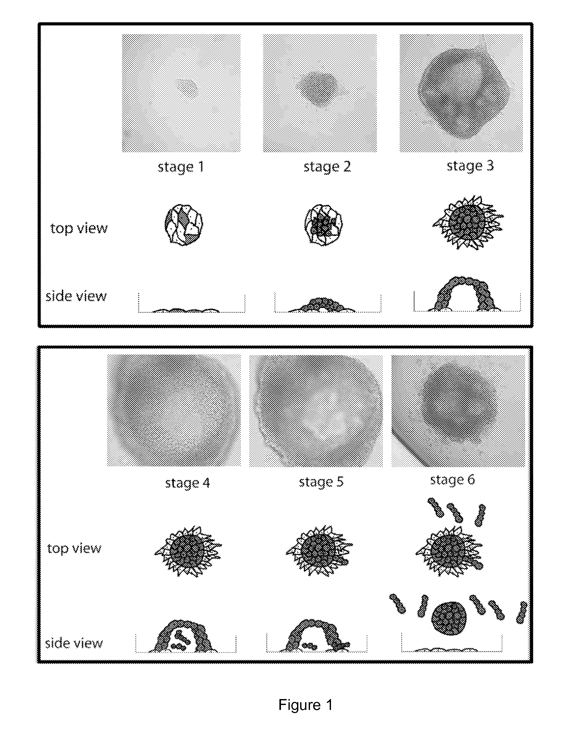
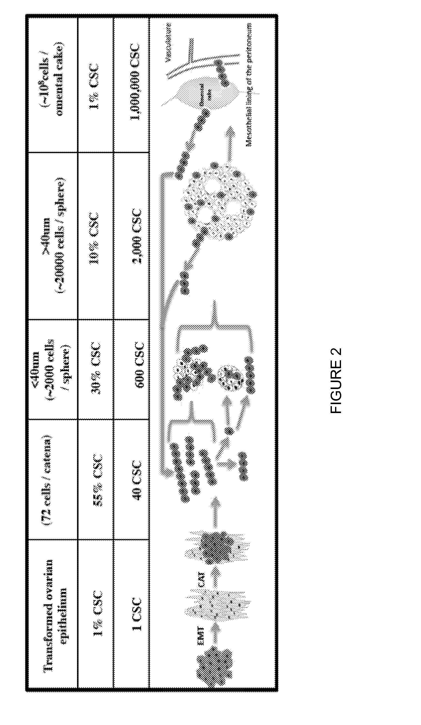
Catena and Spheroid Formation from Cancer Patient Ascites

1. Catena Formation

[0137]Serosal cancer samples from pleural, pericardial or ascites fluids containing tumor cells were obtained from cancer patients with metastatic cancer. Tumor cells were harvested by centrifugation at 1200 rpm for 10 min. The serosal fluid was removed and stored at −20° C. The harvested tumor cells were put into tissue culture flasks with serosal fluid from the same patient mixed 1:1 with serum-containing media. Free-floating chains of tumor cells were immediately observable under the microscope. The chains remained in suspension for many weeks. The tumor cells were cultured at 37° C. for several weeks and each week, the free-floating chains of cells in suspension were separated from the attached cells and replated into a new flask with the same combination of serosal fluid and serum-containing media. In these studies, as few as 100 of these free-floating cells from primary serosal tumor samples were able ...

example 2

Screening Catenae for Drug Sensitivity

1. Methods

[0139]Ovcar3-GTL-derived catenae were tested for their ability to self-propagate in flat bottom 384-well microtiter plates (Corning). Cultures of Ovcar3-GTL catenae were mechanically or enzymatically dissociated to single cells. For mechanical dissociation, catena cultures were pipetted vigorously, an equal volume of M5-FCS media was added to decrease the viscosity, and the cells were pelleted. For enzymatic dissociation, catena cultures were incubated at 5 mg / ml collagenase IV (Invitrogen) for 10 min at 37° C. followed by centrifugation to pellet the cells. Cells were resuspended in M5-FCS to produce homogenous cultures of single cells which were seeded in 50 μL aliquots per well at the indicated cell densities and grown for the indicated times before addition of test compounds or other reagents.

[0140]To assess cell growth, cells were observed under the microscope and manually counted using a hemocytometer or were treated with alamarB...

example 3

Glycocalyx Analysis

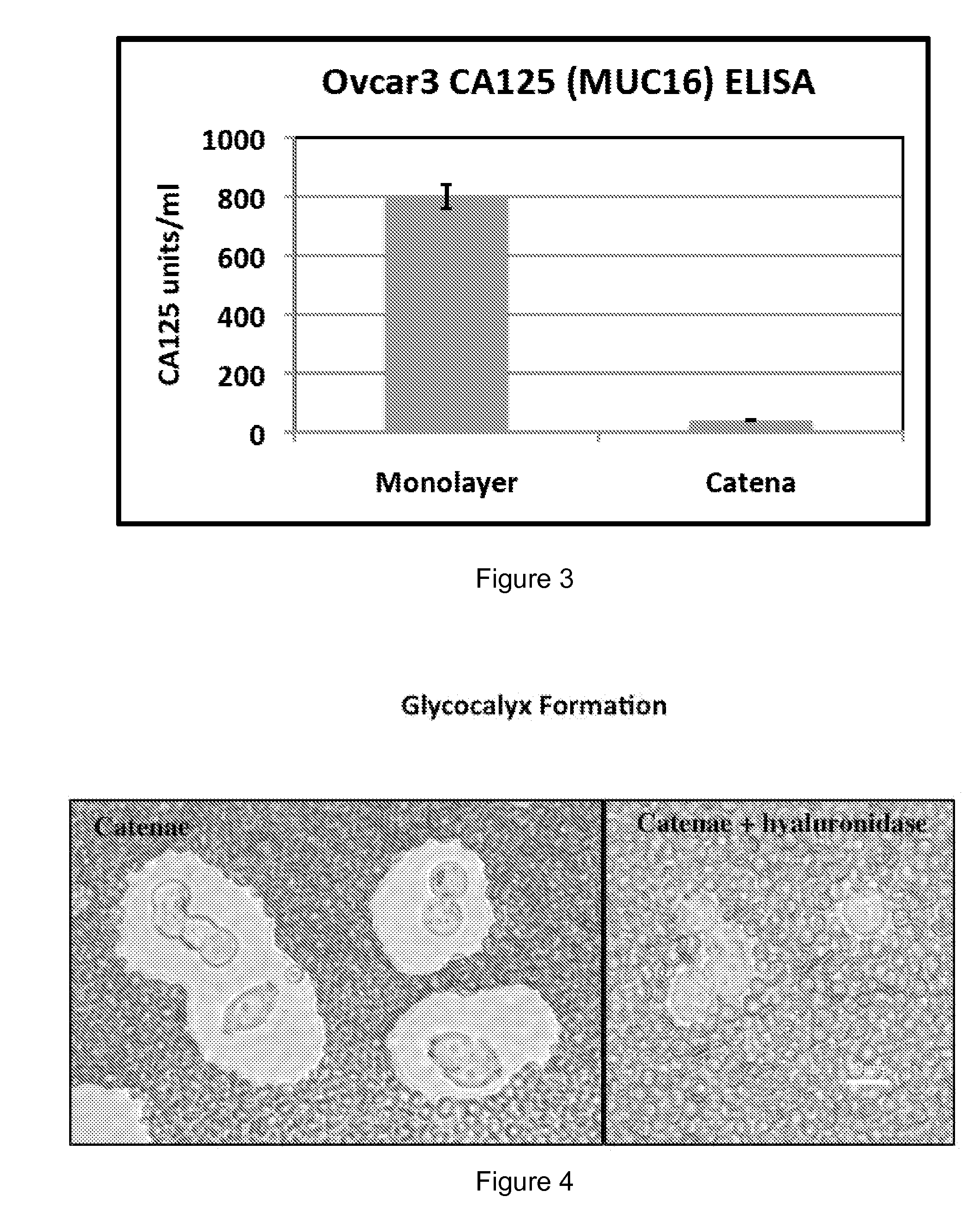
[0157]The catena and spheroid cultures became increasingly viscous at high cell density. Without passage, the catena cultures became so viscous that harvesting the suspension cells was difficult even after a long incubation with collagenase-IV and / or strenuous mechanical dissociation, suggesting that the presence of a glycocalyx coat around the catenae and spheroids was generating the viscous (or mucinous) media. The cells and culture media were examined for the presence of mucins and hyaluronan.

[0158]Initial FACS analysis for the mucin CA125 (the protein product of the MUC16 gene), a biomarker for different types of cancer, indicated that CA125 was not expressed on the surface of catenae. Likewise, ELISA experiments showed that CA125 was not secreted by catenae (FIG. 3). In contrast, Ovcar3-GTL epithelial cells were 98% positive for CA125 by FACS and secreted 800 units / ml of CA125 into culture media. For the ELISA, cell supernatants were collected by spinning the...


PUM

PropertyMeasurementUnit
timeaaaaaaaaaa
timeaaaaaaaaaa
sizesaaaaaaaaaa

Abstract

The discovery of clonally pure populations of serosal cancer stem cells (CSCs) as well as methods of producing CSCs, culturing the CSCs and using them in screening assays, has lead to the development of methods of treating serosal and ovarian cancers by targeting removal or inhibition of the glycocalyx coat surrounding such cells, and includes combination therapies using particular chemotherapeutics in conjunction with glycocalyx inhibitors, as well as the same new chemotherapy treatments without targeting the glycocalyx, where the chemotherapeutic agent is any one of LBH-589 (Panobinostat), NVP-AUY922, LAQ824 (NVP-LAQ824, Dacinostat), colchicine, brefeldin A, diphenyleneiodonium chloride, any combination thereof or another agent identified herein. These treatment methods of the invention can also be used in combination with radiation treatment or other conventional cancer therapy.

Description

[0001]This application claims priority under 35 U.S.C. §119(e) to U.S. Ser. No. 61 / 470,958, filed Apr. 1, 2011, which is incorporated herein by reference in its entirety.FIELD OF THE INVENTION[0002]The discovery of clonally pure populations of serosal cancer stem cells (CSCs) as well as methods of producing CSCs, culturing the CSCs and using them in screening assays, has lead to the development of methods of treating serosal and ovarian cancers by targeting removal or inhibition of the glycocalyx coat surrounding such cells, and includes combination therapies using particular chemotherapeutics in conjunction with glycocalyx inhibitors, as well as the same new chemotherapy treatments without targeting the glycocalyx, where the chemotherapeutic agent is any one of LBH-589 (Panobinostat), NVP-AUY922, LAQ824 (NVP-LAQ824, Dacinostat), colchicine, brefeldin A, diphenyleneiodonium chloride, any combination thereof or another agent identified herein. These treatment methods of the invention...

Claims


Application Information

Patent Timeline
Patent Type & Authority Applications(United States)
IPC IPC(8): A61K38/48A61K47/48A61K31/337A61K31/4045A61N5/10A61K31/165A61K31/365A61K31/03G01N33/50A61K38/47A61K31/5377
CPCA61K38/4886G01N2800/7028A61K47/48215A61K31/337A61K31/4045A61K31/5377A61K31/165A61K31/365A61K31/03G01N33/5011A61N5/1001C12Y302/01035C12Y304/24007G01N2800/52A61K38/47A61K31/16A61K31/23A61K45/06A61K47/60A61P35/00A61K2300/00
Inventor MOORE, MALCOLM A.S.ERTEM, SERVER A.
Owner SLOAN KETTERING INST FOR CANCER RES
Why Eureka
Try Eureka