
Mesh Pouches for Implantable Medical Devices

a medical device and mesh pouch technology, applied in the field of mesh pouches for implantable medical devices, can solve the problems of little that can be done, antibiotics are effective at preventing implant-related infections, and scar tissue formation is excessive, so as to reduce the swelling and inflammation associated with implant-or-operation complications, reduce the effect of scar tissue formation, and attenuation of pain

Active Publication Date: 2015-03-26
TYRX
View PDF2 Cites 14 Cited by
  • Summary
  • Abstract
  • Description
  • Claims
  • Application Information

AI Technical Summary

Benefits of technology

The invention provides coated surgical meshes that can be used to secure, provide pain relief, and prevent complications associated with implantation of pacemakers, defibrillators, and other medical devices. The coated meshes release drugs such as anesthetics, anti-inflammatory agents, and antimicrobial agents that can reduce pain, inflammation, and prevent bacterial growth. The biodegradable polymer coatings help in promoting tissue healing and recovery.

Problems solved by technology

However, such devices are often associated with post-surgical complications including post-implant infection, pain, excessive scar tissue formation and shrinkage of the prosthesis or mesh.
Patients will occasionally be given systemic antibiotics prophylactically; however, literature review of clinical trials does not indicate that systemic antibiotics are effective at preventing implant-related infections.
Post-operative infection is tied to three elements: lack of host defense mechanisms, surgical site and bacteria present at the time of device implantation.5 The general health of the patient (i.e., the host factor) is always important; however, since many patients requiring surgery are compromised in some way—and there is little that can be done to mitigate that factor—controlling the other two factors becomes important.

Method used


Image

  • Mesh Pouches for Implantable Medical Devices
  • Mesh Pouches for Implantable Medical Devices
  • Mesh Pouches for Implantable Medical Devices

Examples


example 1

Antibiotic Release from DTE-DT Succinate Coated Mesh

A. Preparation of Mesh by Spray-Coating

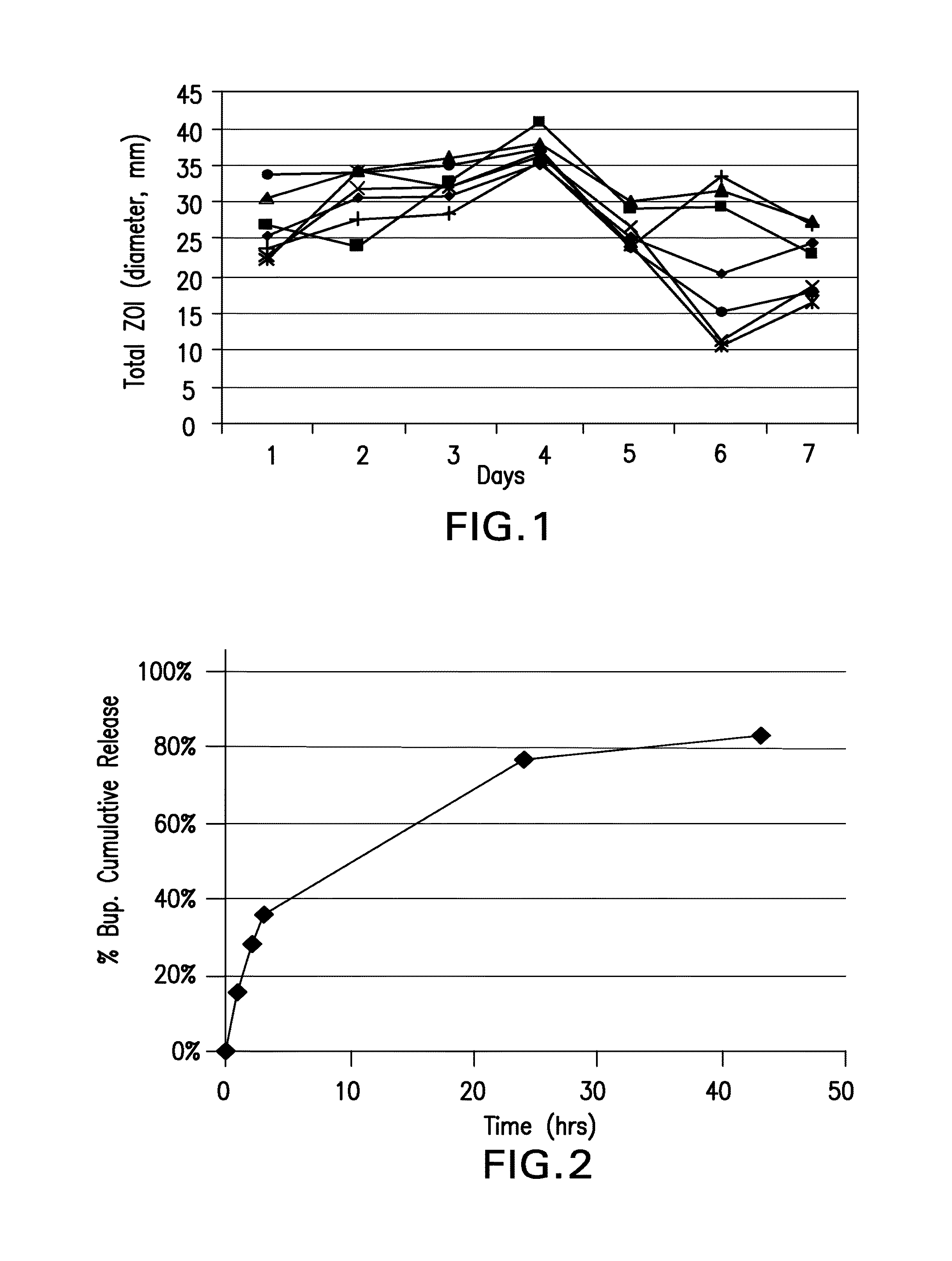
[0110]A 1% solution containing a ratio of 1:1:8 rifampin:minocycline:polymer in 9:1 tetrahydrofuran / methanol was spray-coated onto a surgical mesh by repeatedly passing the spray nozzle over each side of the mesh until each side was coated with at least 10 mg of antimicrobial-embedded polymer. Samples were dried for at least 72 hours in a vacuum oven before use.

[0111]The polymers are the polyarylates P22-xx having xx being the % DT indicated in Table 3. In Table 3, Rxx or Mxx indicates the percentage by weight of rifampin (R) or minocycline (M) in the coating, i.e., R10M10 means 10% rifampin and 10% minocycline hydrochloride with 80% of the indicated polymer. Table 3 provides a list of these polyarylates with their % DT content, exact sample sizes, final coating weights and drug coating weights.

TABLE 3Polyarylate Coated Meshes with Rifampin and Minocycline HClAvg. CoatingCoatingSampleCoating P...

example 2

Bupivacaine Release from DTE-DT Succinate Coated Mesh

A. Preparation of Mesh

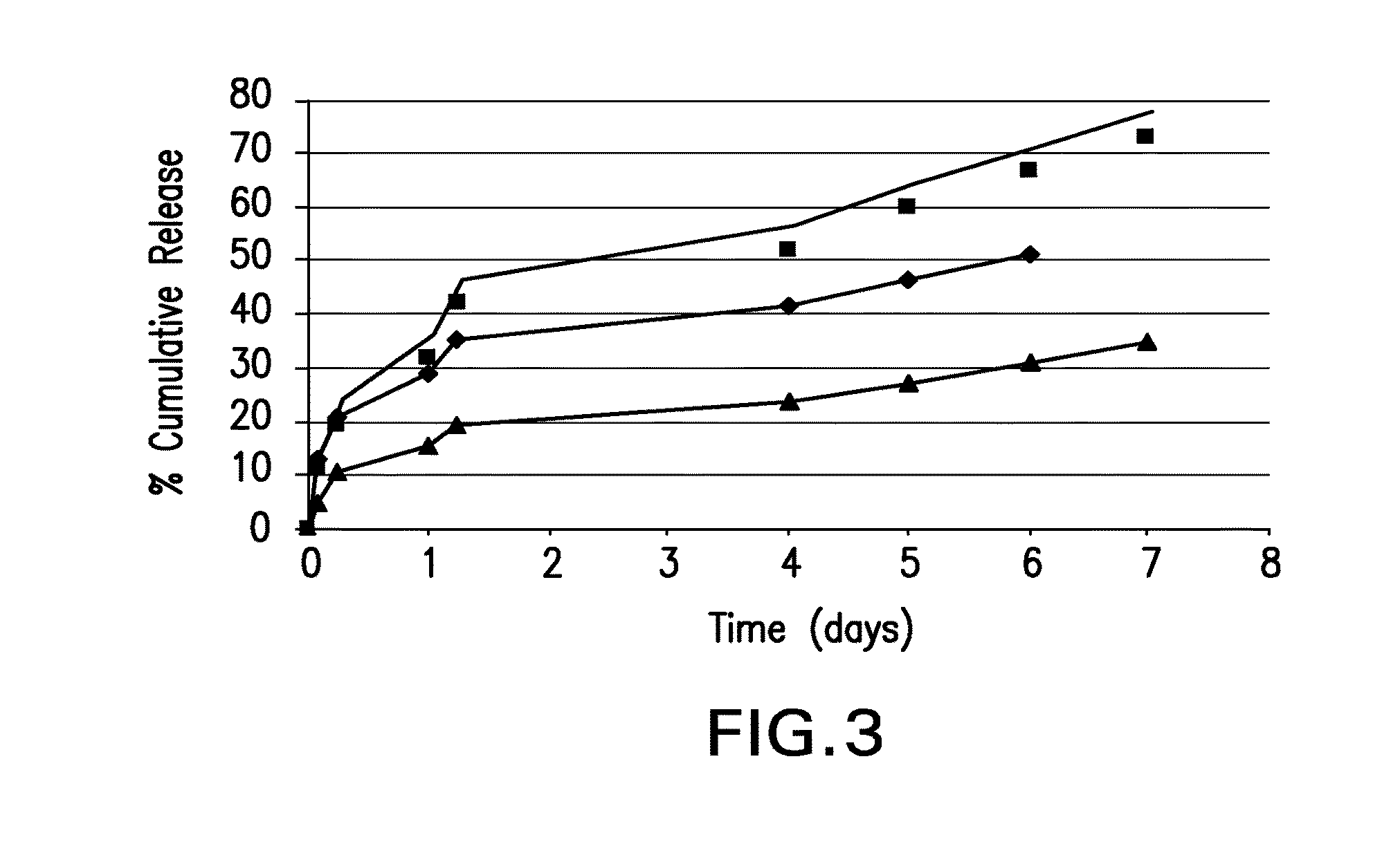
[0116]For the experiment shown in FIG. 2, a first depot coating containing 540 mg of bupivacaine HCl as a 4% solution with 1% P22-27.5 polyarylate in a mixture of THF Methanol was spray coated onto a mesh. A second layer consisting of 425 mg of the same polyarylate alone was deposited on top of the first layer.

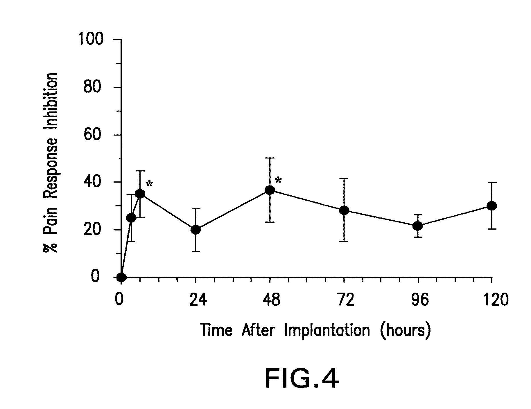
[0117]For the experiment shown in FIG. 3, a solution of approximately 4% bupivacaine in DTE-DT succinate polymer having 27.5% DT was sprayed onto a mesh using the indicted number of passes followed by the indicated number of dips into a solution of the same polyarylate in THF:Methanol (9:1)

B. Anesthetic Release

[0118]Pre-weighed pieces of mesh were placed in PBS at 37° C. and a sample withdrawn periodically for determination of bupivacaine by HPLC. FIG. 2 shows the cumulative release of bupivacaine into PBS from the multilayer polyarylate coating as a function of time. Nearly 80% of the bupivacaine had b...

example 3

In Vivo Bupivacaine Release from DTE-DT Succinate Coated Meshes

A. Overview

[0120]Rats with jugular cannulas for pharmacokinetic studies were surgically implanted with a 1×2 cm P22-27.5 polyarylate-coated mesh containing 7.5 mg of bupivacaine / cm2. Before surgery, baseline pin-prick responses to nociception were measured at the planned surgical incision site, and baseline blood samples were obtained. A hernia was created by incision into the peritoneal cavity during via subcostal laparotomy, and a Lichtenstein non-tension repair was performed using the bupivacaine-impregnated polyarylate-coated mesh. Blood samples were drawn at 3, 6, 24, 48, 72, 96, and 120 hours after implantation. Prior to drawing blood, the rats were subjected to a pin prick test to assess dermal anesthesia from bupivacaine release. The behavioral results indicate that moderate levels of dermal anesthesia appeared from 3 to 120 hours, with the amount at 6 and 48 hours significantly above baseline (p<0.05). Pharmacok...


PUM

PropertyMeasurementUnit
timeaaaaaaaaaa
flow rateaaaaaaaaaa
surgical timeaaaaaaaaaa

Abstract

Biodegradable polymer-coated surgical meshes formed into pouches are described for use with cardiac rhythm management devices (CRMs) and other implantable medical devices. Such meshes are formed into a receptacle, e.g., a pouch or other covering, capable of encasing, surrounding and / or holding the cardiac rhythm management device or other implantable medical device for the purpose of securing it in position, inhibiting or reducing bacterial growth, providing pain relief and / or inhibiting scarring or fibrosis on or around the CRM or other implantable medical device. Preferred embodiments include surgical mesh pouches coated with one or more biodegradable polymers that can act as a stiffening agent by coating the filaments or fibers of the mesh to temporarily immobilize the contact points of those filaments or fibers and / or by increasing the stiffness of the mesh by at least 1.1 times its original stiffness. The pouches of the invention can also provide relief from various post-operative complications associated with their implantation, insertion or surgical use, and, optionally, include one or more drugs in the polymer matrix of the coating to provide prophylactic effects and / or alleviate side effects or complications associated with the surgery or implantation of the CRM or other implantable medical device.

Description

[0001]This application is a continuation-in-part of U.S. Ser. No. 11 / 672,929, filed Feb. 8, 2007, which claims priority under 35 U.S.C. §119(e)(5) of U.S. Provisional Patent Application No. 60 / 864,597, filed Nov. 6, 2006 and U.S. Provisional Patent Application No. 60 / 772,827, filed Feb. 8, 2006; this application also claims priority under 35 U.S.C. §119(e)(5) to U.S. Provisional Patent Application No. 60 / 864,597, filed Nov. 6, 2006, each of which are incorporated herein by reference.FIELD OF THE INVENTION[0002]Biodegradable polymer-coated surgical meshes formed into pouches are described for use with cardiac rhythm management devices (CRMs) and other implantable medical devices IMDs). Such meshes are formed into a receptacle, e.g., a pouch or other covering, capable of encasing, surrounding and / or holding the CRM or other IMD for the purpose of securing it in position, inhibiting or reducing bacterial growth, providing pain relief and / or inhibiting scarring or fibrosis on or around ...

Claims


Application Information

Patent Timeline
Patent Type & Authority Applications(United States)
IPC IPC(8): A61L31/16A61N1/375A61L27/34A61F2/00A61L31/10A61L31/14
CPCA61L31/16A61L31/10A61L31/146A61L27/34A61L31/148A61L2300/404A61N1/375A61B2019/4878A61L2300/41A61L2300/406A61L2300/402A61F2/0063A61B2090/0815A61F2210/0004A61L31/048A61L31/14A61L2420/08A61N1/37512C08L23/12C08L67/04C08L69/00C08L71/02
Inventor BUEVICH, FATIMADO, FRANKMCJAMES, WILLIAMPULAPURA, SATISHEDELMAN, WILLIAMMOSES, ARIKHADIAMOND, MASONTIMOTHY, SHARI
Owner TYRX
Learn More