Quantitative analysis of contact-dependent cell-to-cell transfer and disease transmission

a cell-to-cell transfer and quantitative analysis technology, applied in the field of quantitative analysis of cell-to-cell transfer transport processes, can solve the problems of difficult, if not impossible, difficult to create homogeneous conditions with equal spacing between cells, and fragile structures of tnts between mammalian cells, and achieve high accuracy

Inactive Publication Date: 2015-06-04
BERGEN TEKNOLOGIOVERFORIN AS
View PDF1 Cites 6 Cited by
  • Summary
  • Abstract
  • Description
  • Claims
  • Application Information

AI Technical Summary

Benefits of technology

[0020]A main advantage of the disclosed method over flow cytometric analysis lies therein that it allows a clear differentiation between transfers wherein the traceable substance is located within the volumes defined by the plasma membranes of the cells, and transfers wherein the traceable substance adhere to the external plasma membrane. Another advantage is that the cell-to-cell substance can be quantified with high accuracy. An accurate quantification of the cell-to-cell substa...

Problems solved by technology

The quantitative analysis of contact-dependent cell-to-cell transfer however has proven very difficult, if not impossible, as de novo formed TNTs between mammalian cells are very delicate structures and easily disrupted.
Furthermore it is difficult to create homogeneous conditions with equal spacing between cells in order to visualize and count TNTs.
Above all, flow cytometric analysis demands a disruption of the spatial organization of the cultured cells so that a number of biologically interesting parameters are lost.
The flow cytometric methods can further not reliably differentiate between contact-dependent cell-to-cell s...

Method used


Image

  • Quantitative analysis of contact-dependent cell-to-cell transfer and disease transmission
  • Quantitative analysis of contact-dependent cell-to-cell transfer and disease transmission
  • Quantitative analysis of contact-dependent cell-to-cell transfer and disease transmission

Examples


example 1

Determination of Contact-Dependent Cell-to-Cell Organelle Transfer

a) Cell Culture, Staining and Co-Culture

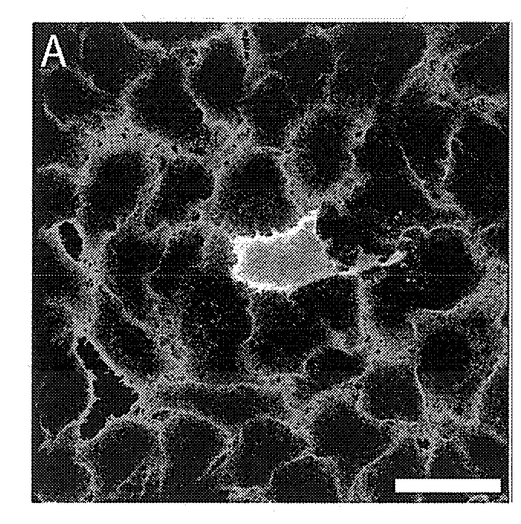
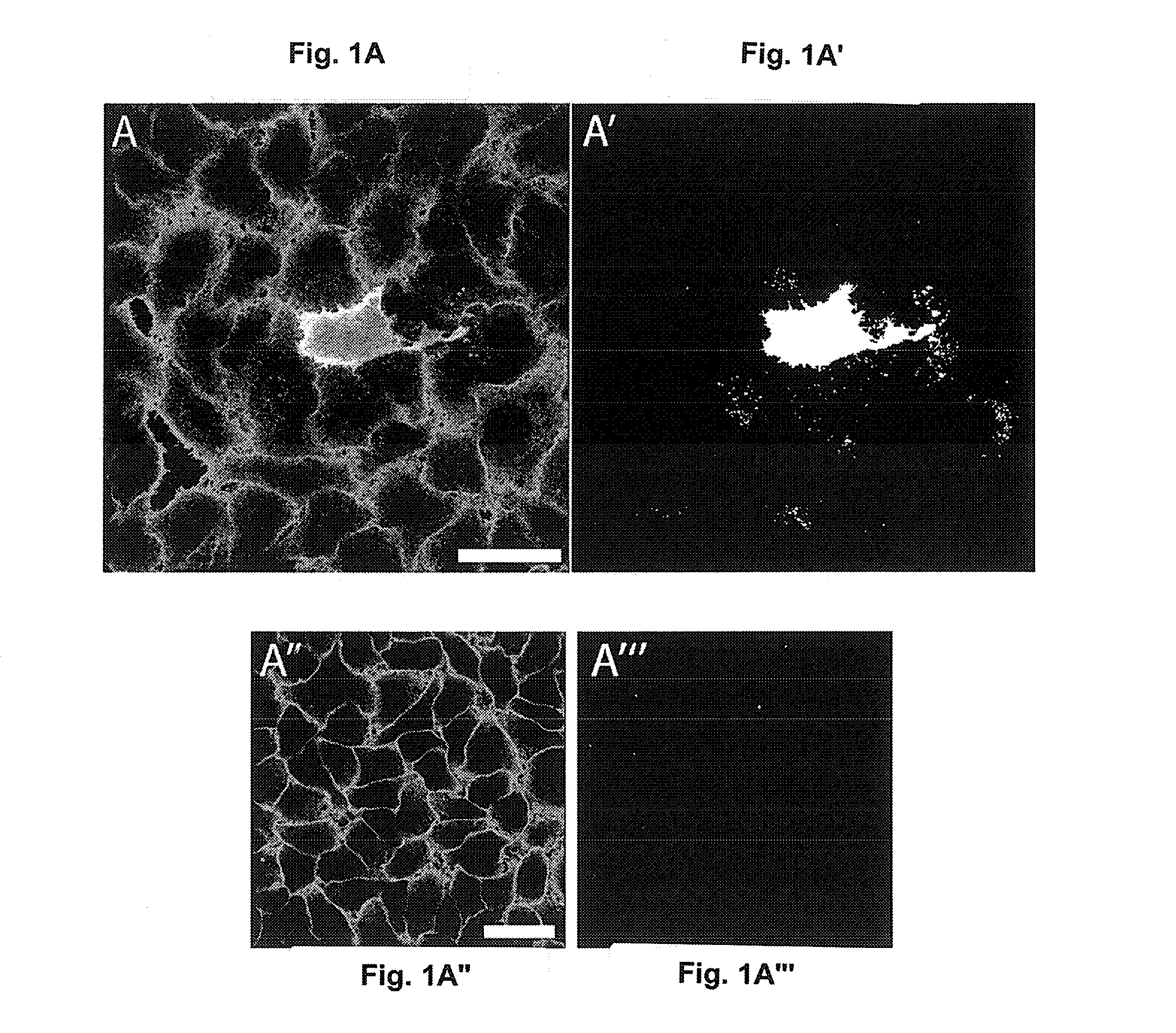
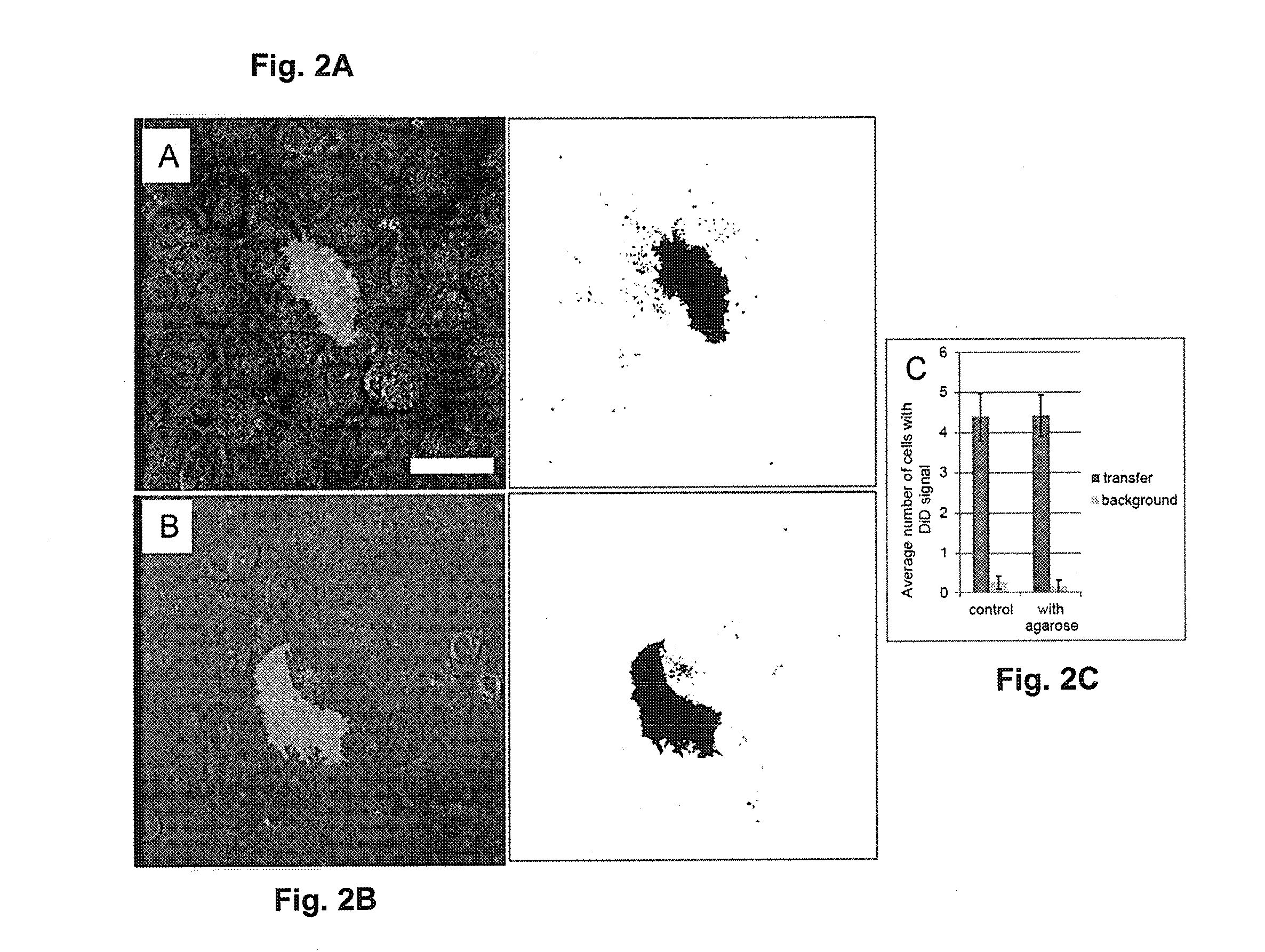
[0098]HeLa-Kyoto cells were cultured in DMEM:F12 supplemented with 10% fetal calf serum and 1% penicillin / streptomycin (Invitrogen Detection Technologies, Carlsbad, Calif., USA) on 24-well microscopy plates (Greiner Bio-One GmbH). Cells were maintained under humidified air supplemented with 5% CO2 at 37° C. and plated with a density of approximately 35 000 cells / cm2. After 24 hours, 200 ng (5 μl) / ml lipid analog Vybrant® DiD Cell-Labelling solution (Invitrogen Detection Technologies, Carlsbad, Calif., USA) was added to the plated cells in culture medium, incubated at 37° C. for 20 min wherein tilted 5 times. After the incubation period, the culture medium was exchanged 4 times within 2 hours. Donor DiD-stained cells were washed twice with phosphate buffered saline PBS (Sigma-Aldrich, St. Louis, Mo. 63103, USA) and harvested by trypsinization (0.25% trypsin / EDTA) (Sigma-Aldrich, ...

example 2

Contact-Dependent Transfer of Transferrin Receptor

[0122]Several studies have shown the presence of organelles stained with fluorescent endocytosed lipid analogues and their subsequent transfer to connected cells, suggesting that endosomal sub-compartments may play a role in TNT-mediated organelles transfer (Gurke et al., Exp Cell Res. 2008 10; 314(20):3669-83). We addressed this question by testing whether the transfer of the Transferrin receptor (Tf-R), a well-known marker of recycling endosomes, occurs between cell in low donor / acceptor ratio co-cultures at confluence. For this purpose, we transiently transfected the transferrin receptor fused to mCherry (Tf-RmCherry) in HeLa-Kyoto cells, which has been widely used to study intracellular trafficking of the transferrin receptor, and subsequently imaged.

a) Plasmid mCherry-Tagged Transferrin Receptor and Transfection

[0123]HeLa-Kyoto cells, provided by Dr. R. Pepperkok (EMBL, Heidelberg, Germany), were cultured in DMEM (GIBCO®, Invitr...

example 3

Manipulation of Cell-to-Cell Transfer by siRNA Knockdown

[0142]To investigate the transfer of DiD-stained organelles under gene knockdown, 24-well normal culture plates and 24-well plates with microscopy suitable bottom were coated with gelatine containing siRNAs and lipofectamine by the following procedure. 0.2% w / v Gelatine was dissolved in water and filtered (0.45 μm pore size). 1.37 g Sucrose was dissolved in OptiMEM. 6 μl Sucrose / OptiMEM solution was mixed with 1.75 μl Lipofectamine™ 2000, 1.75 μl water and 10 μl silence-select siRNA (3 μM), and this mix incubated for 20 minutes at room temperature together with 14.5 μl gelatine solution. A 2-step 1:50 dilution in water was carried out, resulting in 1.6 ml volume. 300 μL solution / well were dried in a vacuum centrifuged at 60° C., followed by a cooling step through application of vacuum at 37° C. and stored in sealed boxes containing silica gel drying pearls as desiccant for up to several months. For experimental setup, HeLa-Kyot...


PUM


Abstract

A method for a microscope-based quantitative analysis of contact-dependent cell-to-cell transfer between cells. Single donor cells containing a traceable marker are cultured in a lawn of acceptor cells to allow cell-to-cell transfers. The donor and acceptor cells are then imaged and/or stained for microscopy to determine the location, shape and volumes of donor cells and of surrounding acceptor cells within selected regions of interest. The spatial distribution of the traceable marker in each volumetric picture element of the region of interest is determined and analyzed to determine quantitatively and qualitatively the substance transfer. The method allows a reliable quantitative evaluation of the effect of an active agent on cell-to-cell molecular communication and disease transmission.

Description

FIELD OF THE INVENTION[0001]The invention relates to a method for quantitative analysis of transport processes between cells.BACKGROUND OF THE INVENTION[0002]Molecular communication and substance transfer between mammalian cells can take place through a variety of pathways. The paracrine and endocrine signalling pathways for example require exocytosis into extracellular fluids or blood circulation followed by an uptake of the released substance by other cells expressing the respective receptors. The intercellular transport of materials by extracellular fluids does not require cells to be physical contact to each other. For communication and material transfer through direct contact, the mammalian organisms provide gap- and tight- and adhesive junctions.[0003]Tunnelling nanotubes (hereinafter TNTs) have been described as a transient form of contact-dependent cell-to-cell communication and biological material transfer (Rustom et al., Science 2004, 303(5660):1007-10). TNTs have been sho...

Claims


Application Information

Patent Timeline
IPC IPC(8): G01N33/50G01N15/08
CPCG01N33/5032G01N2015/086G01N2015/0846G01N15/08G01N33/5005G01N21/6458G01N2021/6439
Inventor KOGEL, TANJAHODNELAND, ERLENDGERDES, HANS-HERMANNFREI, DOMINIK MICHAELBURTEY, ANNE
Owner BERGEN TEKNOLOGIOVERFORIN AS
Why Eureka
Try Eureka