Detection of Brain Injury

a brain injury and brain injury technology, applied in the field of brain injury detection, can solve the problems of tbi-caused neuronal damage or cte progression, no non-invasive diagnostic methods or tools, synaptic dysfunction and loss, neurite retraction and axonal degeneration, etc., to improve the long-term prognosis and good or poor long-term survival prospects

Active Publication Date: 2015-12-03
UNIVERSITY OF MONTANA
View PDF3 Cites 12 Cited by
  • Summary
  • Abstract
  • Description
  • Claims
  • Application Information

AI Technical Summary

Benefits of technology

[0012]An “expression profile” or “hybridization profile” of a particular sample is essentially a fingerprint of the state of the sample; while two states may have any particular gene similarly expressed, the evaluation of a number of genes simultaneously allows the generation of a gene expression profile that is unique to the state of the cell. That is, normal tissue may be distinguished from abnormal (e.g., diseased or injured) tissue, and within abnormal tissue, different prognosis states (for example, good or poor long term survival prospects) may be determined. By comparing expression profiles of tissue (e.g., blood, tissue biopsy or necropsy sample, or cerebral spinal fluid) in different states, information regarding which genes are important (including both up- and down-regulation of genes) in each of these states is obtained. The identification of sequences that are differentially expressed in tissue, as well as differential expression resulting in different prognostic outcomes, allows the use of this information in a number of ways. For example, a particular treatment regime may be evaluated (e.g., to determine whether a therapeutic drug acts to improve the long-term prognosis in a particular patient). Similarly, diagnosis may be done or confirmed by comparing patient samples with known expression profiles. Furthermore, these gene expression profiles (or individual genes) allow screening of drug candidates that alter or normalize tissue expression profiles to impart a clinical benefit.

Problems solved by technology

Unfortunately, no non-invasive diagnostic methods or tools currently exist to evaluate TBI-caused neuronal damage or CTE progression.
However profound loss of neurons and brain atrophy are late-occurring events in the pathogenesis of the disease and are preceded by metabolic changes such as hyperphosphorylation of tau and deposition of neurofibrillary tangles presumably leading to synaptic dysfunction and loss, neurite retraction and axonal degeneration.

Method used


Image

  • Detection of Brain Injury
  • Detection of Brain Injury
  • Detection of Brain Injury

Examples


example 1

Gene Array Analysis Before and after TBI in Rodent Model

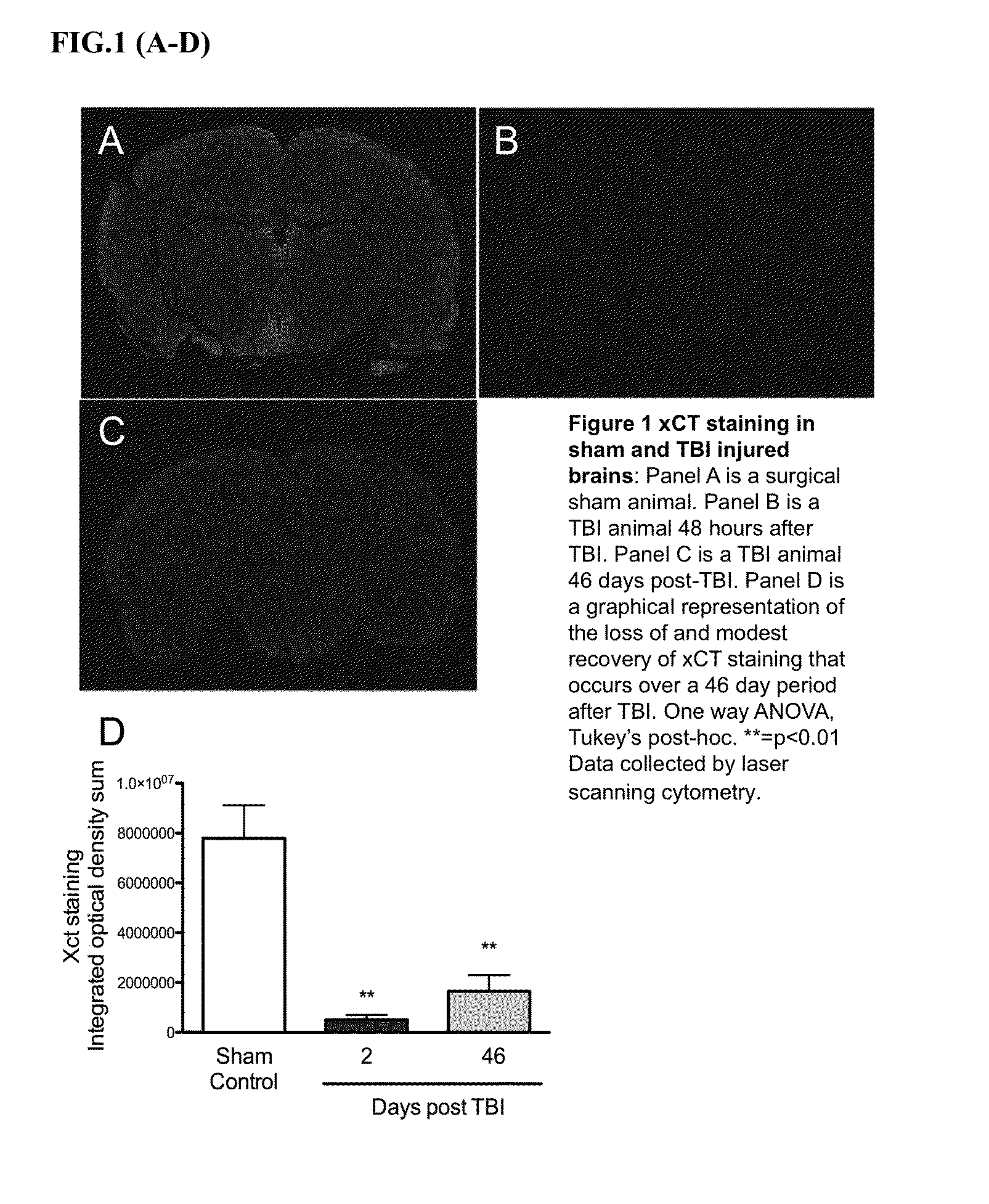
[0087]System xc− is a cystine / glutamate antiporter comprised of two distinct subunits xCT and 4F2hc (SLC3A2) and a member of the heteromeric amino acid transporter (HAT) family. Under physiological conditions, System xc− mediates the exchange of extracellular L-cystine and intracellular L-glutamate across the plasma membrane. In the CNS, the influx of L-cystine represents the critical rate limiting step in the biosynthesis of glutathione (GSH) while the concurrent efflux of L-glutamate serve as a non-vesicular route of excitatory neurotransmitter release to initiate excitatory amino acid (EAA) signalling. GSH serves as the key cellular antioxidant responsible for scavenging reactive oxygen species (ROS) that develop as a result of physiological cellular metabolism. Thus a global loss of System xc− activity would result in decreased intracellular glutathione levels, leaving the CNS vunerable to oxidative stress due to an increas...

example 2

miRNA Expression Profiles of Oxidative Stress and Antioxidant Defense Genes

[0089]To follow up this study, we performed a gene array analysis to determine if oxidative stress genes up-regulate to compensate for the loss of GSH (FIG. 4). From this study we found exactly the opposite occurred; TBI resulted in a down-regulation of key anti-oxidant defense genes leaving neurons critically susceptible to ROS damage. In an effort to elucidate the mechanism of how the xCT subunit and anti-oxidant defense genes were down-regulated by TBI we performed a microRNA (miRNA) expression profile study on rat (R. norvegus) cortical brain tissue following neuronal injury in a lateral fluid percussion TBI model (FIG. 5). In light of our prior findings, we chose miRNAs that specifically interact with anti-oxidant defense genes and xCT. A specific cluster of miRNAs that were significantly (2-13 fold) up or down regulated as a result of TBI was discovered (Table 3). Further studies indicate all of these h...

example 3

miRNA Expression Profiles of Human Peripheral Blood Plasma

[0090]Approximately 3 mL of blood was collected from each subject and processed as described below. Total RNA was isolated and prepared from 200 μL plasma according to the miRNeasy Serum / Plasma Kit (50) protocol according to the manufacturer's instructions (Qiagen). cDNA was prepared using the isolated total RNA. Real-time PCR was performed using the Qiagen miScript SYBR Green PCR kit and custom miScript miRNA PCR Array on a Bio-Rasd iQ5 cycler. Data analysis was performed using the Qiagen Data Analysis Center. The following plasma samples were obtained:

Sample SourceDescriptionn =ControlNo TBI, non-athlete14Football PlayersNo TBI with 3 months49Soccer PlayersNo TBI with 3 months19Acute TBI24-72 post-TBI4Chronic TBI3 months post-TBI6

All plasma samples were obtained by voluntary donation according to the Institutional Review Board protocols of The University of Montana. Samples were obtained at random (control group), from stud...


PUM

PropertyMeasurementUnit
timeaaaaaaaaaa
Northern blotaaaaaaaaaa
sizeaaaaaaaaaa

Abstract

The present invention provides minimally invasive methods of detecting, diagnosing, and assessing neuronal damage associated with traumatic brain injury (TBI) or chronic traumatic encephalopathy (CTE). Specific species of microRNAs (miRNA), small, noncoding RNA molecules that play gene regulatory functions, are correlated with cellular damage and oxidative stress following TBI or CTE, allowing for rapid, minimally-invasive diagnosis and assessment of brain injury. The early identification and longitudinal assessment of neuronal damage in subjects suffering from or at risk of suffering from a TBI (e.g., football players, boxers, military personnel, fall victims) will improve clinical outcomes by guiding critical medical and behavioral decision making.

Description

CROSS-REFERENCE TO RELATED APPLICATIONS[0001]This application claims the benefit of the filing date of U.S. Provisional Patent Application Ser. No. 61 / 970,613, filed Mar. 26, 2014, the disclosure of which is incorporated by reference in its entirety.REFERENCE TO SEQUENCE LISTING[0002]This application includes as part of the originally filed subject matter a Sequence Listing electronically submitted via EFS-Web as a single text file named “UM014002SL.txt”. The Sequence Listing text file was created on Mar. 25, 2015 and is 122 kb in size. The contents of the Sequence Listing are hereby incorporated by reference.BACKGROUND OF THE INVENTION[0003]The acute and chronic molecular effects of mild TBI (mTBI) have not been well studied or characterized. Over the past several decades it has become increasingly clear that repetitive mTBI is capable of altering the biochemical activity of the brain in ways that cannot be detected by current methodologies. Highlighting this issue is the definitiv...

Claims


Application Information

Patent Timeline
Patent Type & Authority Applications(United States)
IPC IPC(8): C12Q1/68
CPCC12Q2600/178C12Q1/6883C12Q2600/112C12Q2600/118C12Q2600/158A61P25/28A61P39/06
Inventor PATEL, SARJUBHAIRAU, THOMAS
Owner UNIVERSITY OF MONTANA
Why Eureka
Try Eureka