Angiotensins in muscular dystrophy
- Summary
- Abstract
- Description
- Claims
- Application Information
AI Technical Summary
Benefits of technology
Problems solved by technology
Method used
Image
Examples
example 1
Administration of Angiotensin (1-7), PanCyte, or Linear PanCyte in the Mdx Mouse Model of Duchenne Muscular Dystrophy
[0238]In this Example, mdx mice, a known and accepted model of Duchenne Muscular Dystrophy (DMD) are used to assess the effects of several angiotensin (1-7) peptides and an angiotensin (1-7) receptor agonist, AVE0991, on the muscle degeneration typically observed in DMD patients. See, Dangain and Vrbova, Muscle development in mdx mutant mice, 1984, Muscle Nerve 7: 700-704; see also Tanabe et al., Skeletal muscle pathology in X-chromosome-linked muscular dystrophy (mdx) mouse, 1986, Acta Neuropathol, 69:91-95; Kobayashi et al., Endpoint measures in the mdx mouse relevant for muscular dystrophy pre-clinical studies, 2012, Acta Materialia, 22: 34-42. Specifically, the mdx mouse has a point mutation within its dystrophin gene, which leads to disruption of dystrophin production and results in almost no functional dystrophin being present in the mouse. The lack of functiona...
example 2
Administration of Ang (1-7) in the Sgcd− / − Mouse Model of Muscular Dystrophy
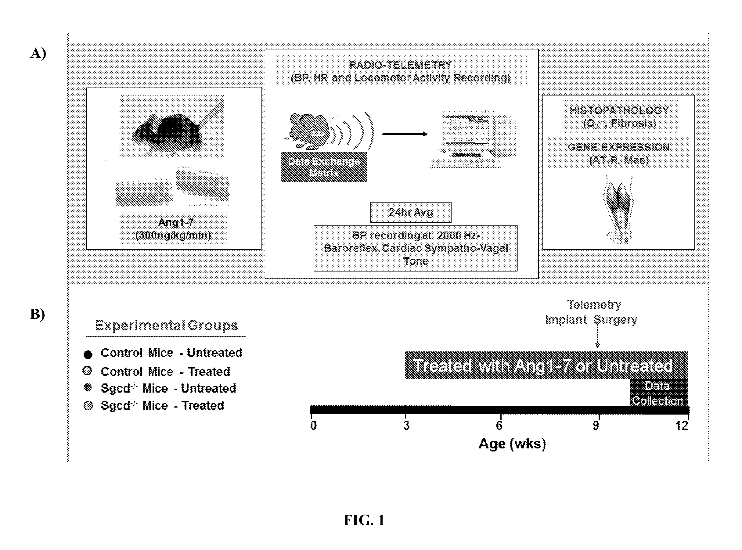
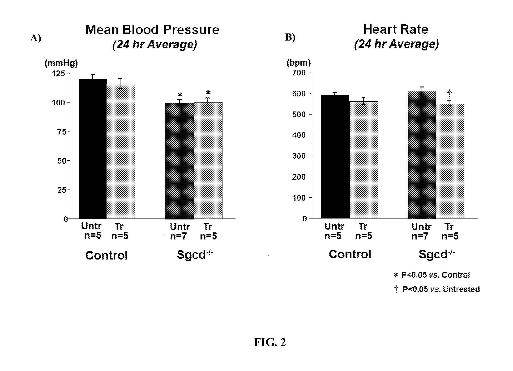
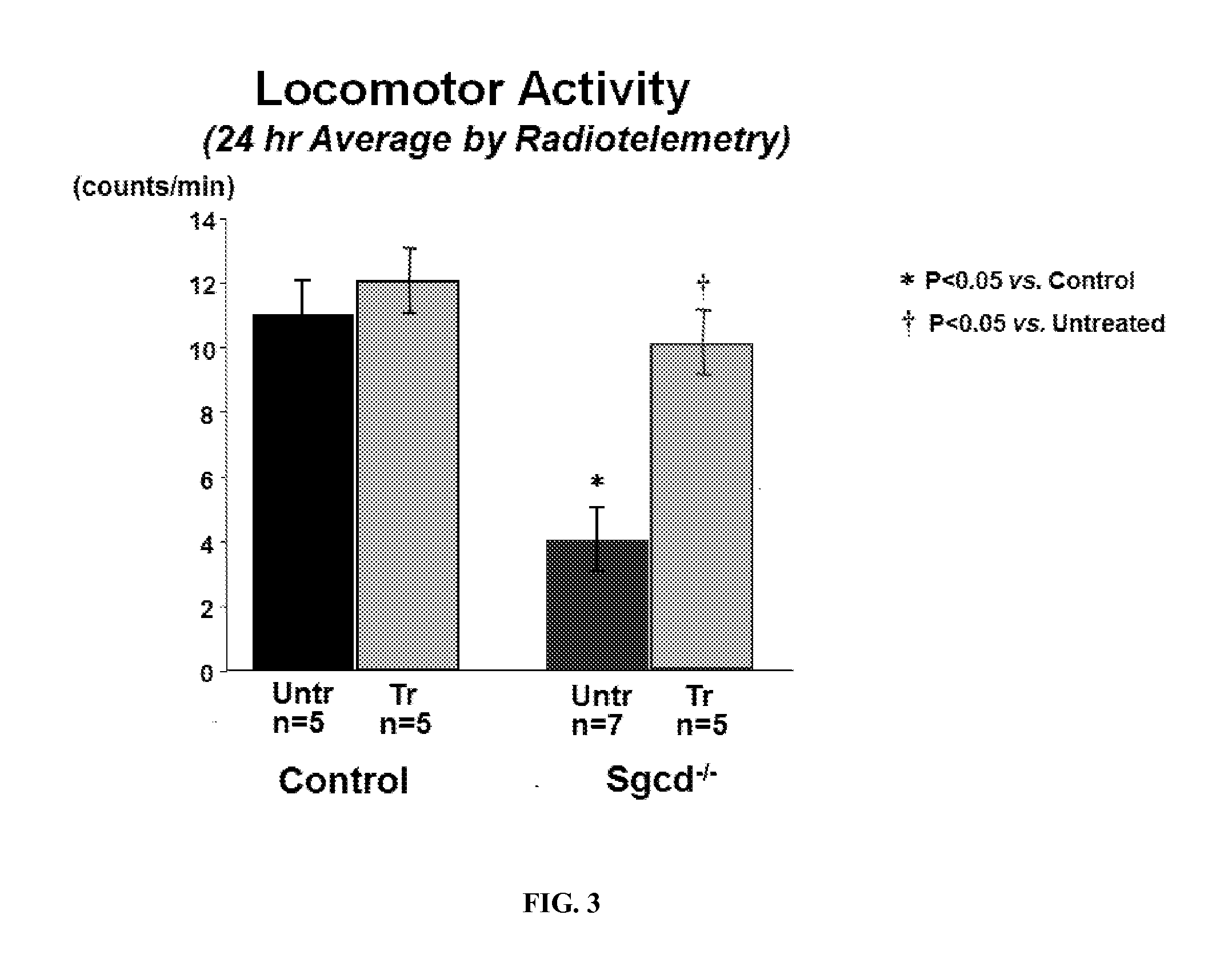
[0244]In this Example, Sgcd − / − mice, a known model of limb-girdle muscular dystrophy, were used to assess the effects of angiotensin (1-7) peptides on skeletal muscle fibrosis, blood pressure, heart rate, cardiac activity, baroreflex sensitivity, oxidative stress, AT1R receptor expression, and resting cardiac and vagal sympathetic tone. Sgcd − / − mice are missing a key component of the sarcoglycan complex, specifically, they are missing a link between the F-actin cytoskeleton and the extracellular matrix. This defect causes muscle wasting and weakness due to a reduction in muscle fiber integrity and dysregulation in cell signaling in muscle cells. It has been previously shown that this defect results in a reduction of locomotor activity and autonomic dysfunction at a young age. The autonomic dysfunction is thought to lead to cardiac dysfunction later in life.
[0245]To examine the effects of angiotensin (1-7) ...
example 3
Oral Administration of PanCyte and Linear PanCyte Compositions to Mdx Mice
[0259]In this example, specific angiotensin (1-7) peptides, namely PanCyte (SEQ ID NO: 22) and Linear PanCyte (also referred to as “TXA301”; SEQ ID NO: 2) are used in exemplary oral formulations to assess the effect of oral administration on the condition of mdx mice. In this Example, oral gavage is used to administer agent according to known methods, and separate aqueous formulations of PanCyte and Linear PanCyte are each made with citric acid, laurolyl carnitine, and hydroxypropyl methylcellulose phthalate according to methods described in U.S. Provisional Patent Application 61 / 701,972, filed Sep. 17, 2012.
[0260]A total of 80 mdx mice separated into eight groups, including a PBS control and a naked Angiotensin (1-7) peptide control, are used in this example according the design shown in Table 2:
TABLE 2Study DesignRoute ofDosingGroupAgentDoseNAdmin.Frequency1VehiclePBS10OralDailyControl(PBS)2Angiotensin (1-7)...
PUM
| Property | Measurement | Unit |
|---|---|---|
| Frequency | aaaaa | aaaaa |
| Frequency | aaaaa | aaaaa |
| Frequency | aaaaa | aaaaa |
Abstract
Description
Claims
Application Information
 - R&D
- Intellectual Property
- Life Sciences
- Materials
- Tech Scout
- Unparalleled Data Quality
- Higher Quality Content
- 60% Fewer Hallucinations
Browse by: Latest US Patents, China's latest patents, Technical Efficacy Thesaurus, Application Domain, Technology Topic, Popular Technical Reports.
© 2025 PatSnap. All rights reserved.Legal|Privacy policy|Modern Slavery Act Transparency Statement|Sitemap|About US| Contact US: help@patsnap.com


