Compressive plenoptic microscopy

a plenoptic microscopy and compression technology, applied in the field of medical imaging, can solve the problems of reducing the accuracy of image enhancement, requiring fast controlled mechanical scanning, and significantly limiting temporal resolution, so as to achieve fast quantification of activity and maximize information

Active Publication Date: 2017-01-05
RGT UNIV OF CALIFORNIA
View PDF18 Cites 12 Cited by
  • Summary
  • Abstract
  • Description
  • Claims
  • Application Information

AI Technical Summary

Benefits of technology

[0016]An aspect of the technology of this disclosure is a computational imaging method that takes advantage of plenoptic acquisition to maximize information, but does not attempt to reconstruct a volume image. The effects of optical scattering are preserved throughout the entire data processing sequence. The technology operates in two primary steps. A first initialization step identifies individual neurons by their optical signature and provides a reliable estimate of their position in space. In the second step, a fast quantitative algorithm uses calibration data to quickly quantify activity in each identified neuron at video frame-rate. The technology can be implemented on a light field microscope, as an add-on device by inserting a micro-lens array in the imaging path of a fluorescence microscope, or with a on-a-chip, lens-free imaging apparatus that incorporates the plenoptic element directly onto or within a focal distance away from the optical sensor: a board-level low-noise camera or optical detector array. It is also possible to substitute the micro-lens array for other types of plenoptic encoding phase masks such as engineered diffusers and random surface patterns with proper calibration.
[0017]The advantages of the technology of this disclosure include: (a) high spatial resolution at the scale of a single neuron; (b) high temporal resolution, determined by the frame-rate of the video camera (100Hz demonstrated in our prototype); (c) simultaneous monitoring of multiple neurons (500 neurons successfully detected with our prototype) in large volumes (a 200 micrometer cube); (d) simple optical design (as a standalone device, or as an add-on for fluorescence microscope), with no fine alignment required; (e) efficient processing that enables real time quantification of neural activity during experiments, which allows for possible brain machine interfaces; and (f) good performance, even in heavy optical scattering conditions.
[0018]In a preferred embodiment, the imaging method of the present description incorporates a learning step with training data. The technology allows the user to either accept to process data after acquisition is completed, or to initiate a fully automated startup calibration procedure prior to stimulating the subject tissue.

Problems solved by technology

However, this method observes blood flow instead of directly targeting neurons, leading to poor spatial and temporal resolution.
Two-photon microscopy provides high resolution images in 3D, but requires fast controlled mechanical scanning which considerably limits temporal resolution.
It is significantly faster than two-photon methods, but still limited to observing one focal depth level at a time.
Alternative options with a single microscope objective are possible, at the expense of an additional loss in resolution.
Volume image reconstruction has been implemented for functional brain imaging but existing techniques are unable to correct for optical scattering.
Consequently, volume image reconstruction through thick brain tissue leads to blurred reconstructions and losses of information, and the shape of individual neurons can not be reconstructed accurately.

Method used


Image

  • Compressive plenoptic microscopy
  • Compressive plenoptic microscopy
  • Compressive plenoptic microscopy

Examples


Embodiment Construction

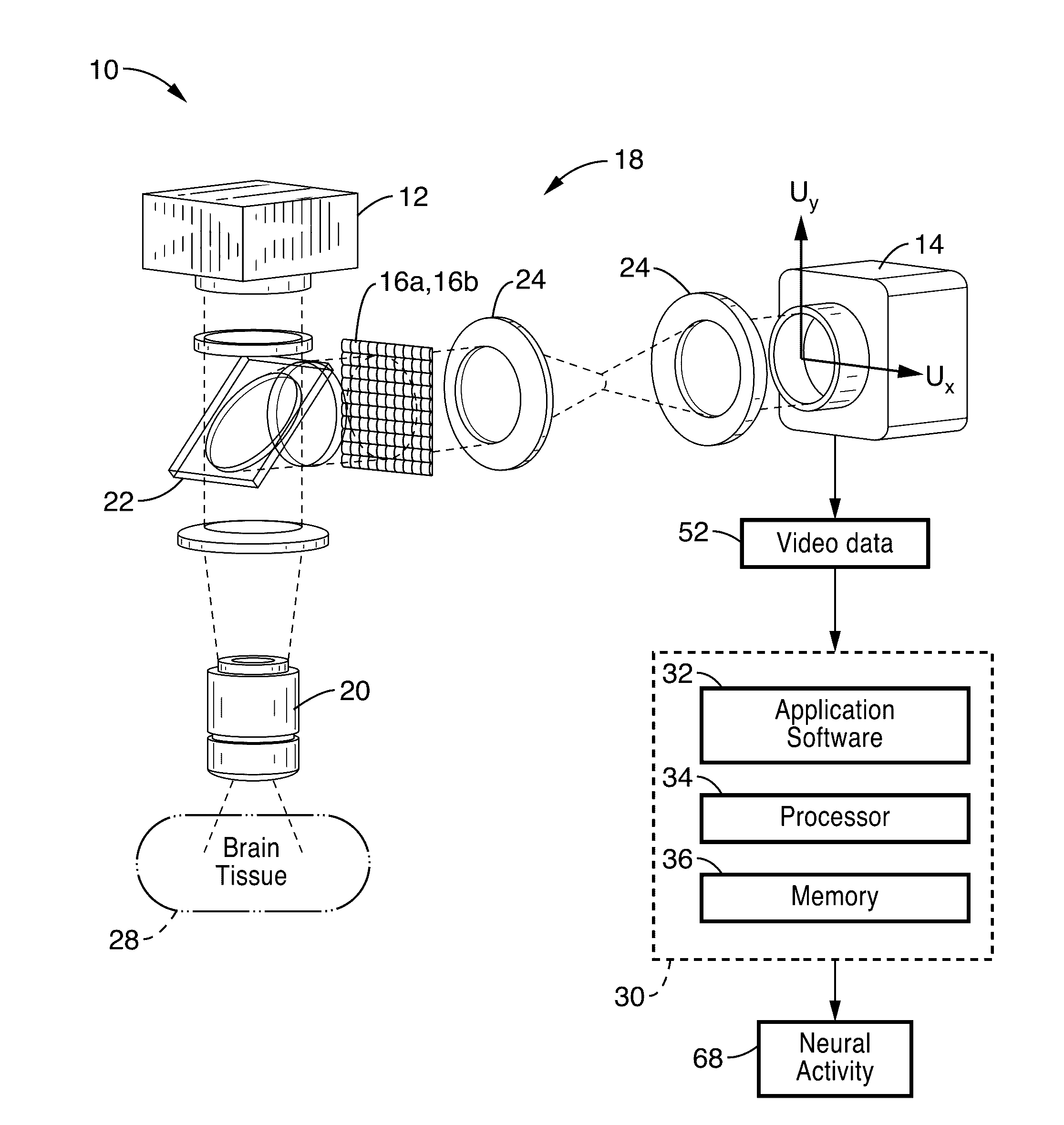
[0031]FIG. 1 shows a schematic view of an embodiment of a plenoptic imaging system 10 in accordance with the present description. In the embodiment shown in FIG. 1, the system 10 is shown implemented with a fluorescence microscope 18, which may comprise a device specific for use with light field microscopy, or may alternatively comprise a modified fluorescence microscope with an engineered phase mask placed in the imaging plane. It is also appreciated that other devices, such as lens-free fluorescence imager 100 shown in FIG. 10, may also be used in place of microscope 18.

[0032]As seen in FIG. 1, an excitation light source 12 (e.g. LED at 488 nm) is directed at brain tissue 28 via intervening optics (e.g. fluorescence filter cube 22 (which may comprise dichroic mirrors / filters) and objective 20. It is to be noted that the brain tissue mass 28 is shown in FIG. 1 in block form; however, the non-invasive methods of the present description are configured to direct light from source 12 i...


PUM


Abstract

A system and method for quantitative functional neuroimaging through thick brain tissue in live animals. A computational imaging method is disclosed that uses plenoptic image acquisition including a first initialization step that identifies individual neurons by their optical signature and provides a reliable estimate of their position in space and a second stimulation-based image processing step that used acquired calibration data to quickly quantify activity in each identified neuron at video frame-rate.

Description

CROSS-REFERENCE TO RELATED APPLICATIONS[0001]This application claims priority to, and the benefit of, U.S. provisional patent application Ser. No. 62 / 188,626 filed on Jul. 4, 2015, incorporated herein by reference in its entirety.STATEMENT REGARDING FEDERALLY SPONSORED RESEARCH OR DEVELOPMENT[0002]Not ApplicableINCORPORATION-BY-REFERENCE OF COMPUTER PROGRAM APPENDIX[0003]Not ApplicableNOTICE OF MATERIAL SUBJECT TO COPYRIGHT PROTECTION[0004]A portion of the material in this patent document is subject to copyright protection under the copyright laws of the United States and of other countries. The owner of the copyright rights has no objection to the facsimile reproduction by anyone of the patent document or the patent disclosure, as it appears in the United States Patent and Trademark Office publicly available file or records, but otherwise reserves all copyright rights whatsoever. The copyright owner does not hereby waive any of its rights to have this patent document maintained in ...

Claims


Application Information

Patent Timeline
Patent Type & Authority Applications(United States)
IPC IPC(8): G02B21/36A61B5/00G02B21/16G06T7/00G06T15/08
CPCG02B21/367A61B5/0042A61B5/0071A61B5/4064A61B5/7246A61B5/743A61B2560/0223G02B3/0056G02B21/16G02B21/361G02B27/0075G06T7/0016G06T2207/10016G06T2207/10052G06T2207/10056G06T2207/30016
Inventor WALLER, LAURAADESNIK, HILLELPEGARD, NICOLAS
Owner RGT UNIV OF CALIFORNIA
Why Eureka
Try Eureka