Eureka AIR delivers breakthrough ideas for toughest innovation challenges, trusted by R&D personnel around the world.

Method of producing in vitro testicular constructs and uses thereof

a testicular and in vitro technology, applied in the field of in vitro testicular construct production, can solve the problems of loss of function and cell-type-specific gene expression, inability to maintain highly complex signaling interactions of existing in vitro models, and inability to support human in vitro systems

Active Publication Date: 2017-04-20
WAKE FOREST UNIV HEALTH SCI INC
View PDF3 Cites 14 Cited by
  • Summary
  • Abstract
  • Description
  • Claims
  • Application Information

AI Technical Summary

Benefits of technology

The patent describes a new composition of cells that can be used in artificial testicular constructs. These constructs can be used to screen compounds for pharmacological or toxicological activity. The composition includes spermatogonial stem cells, Sertoli cells, and Leydig cells, which are all immortalized cells. These cells are combined in a specific ratio to mimic the natural composition of cells in a testis. The composition can also include peritubular cells and can have a diameter of 10-30 microns. The total number of cells in the construct is between 10,000 and 100,000. The artificial testicular constructs can produce testosterone, express markers of spermatogonial cells and Leydig cells, and can screen compounds for their effects on spermatogonial stem cell fate. The compositions can be stored in a hydrogel and can be deposited in a chamber for further use. The device can also be packaged and protected for storage.

Problems solved by technology

However, in these systems, the enzymatic digestion and mechanical disruption of native tissue required to isolate, characterize, and expand the various cell types often resulted in a loss of functionality and cell-type-specific gene expression.
However, existing in vitro models are predominately inadequate for maintaining the highly complex signaling interactions of the mammalian testis.
Currently, no human in vitro system exists that is able to support the paired functionality of both spermatogenesis and steroidogenesis within the same model system.

Method used


Image

  • Method of producing in vitro testicular constructs and uses thereof
  • Method of producing in vitro testicular constructs and uses thereof
  • Method of producing in vitro testicular constructs and uses thereof

Examples


example 1

Human Testis Material

[0091]Human testis tissue was obtained from The National Disease Research Interchange (NDRI). Testicular tissue was used for human testis extracellular matrix (ECM) extraction or cut into small segments to be used for cryopreservation and immunohistochemical testing. For cryopreservation, tissue fragments (˜2-5 mm) were frozen in 1× minimum essential medium containing 8% DMSO, 20% fetal bovine serum (FBS) slowly overnight using a Mr. Frosty container inside a −80° C. freezer. Cryotubes were moved to liquid nitrogen (−196° C.) for long-term storage. For all of cell isolations, cryopreserved tissue was used. Tissue pieces used for immunohistochemical staining were fixed in 4% paraformaldehyde and paraffin-embedded. Morphology of testes, stained by Hematoxylin and Eosin (H&E), showed normal spermatogenesis in all patient samples used in this study. All human materials in this study were used under regulation and approval of Institutional Review Board (IRB) of the W...

example 2

Testicular Cell Isolation, Culture and Cryopreservation of Spermatogonial Stem Cell (SSC) Lines

[0092]Preparation and Culturing of SSC.

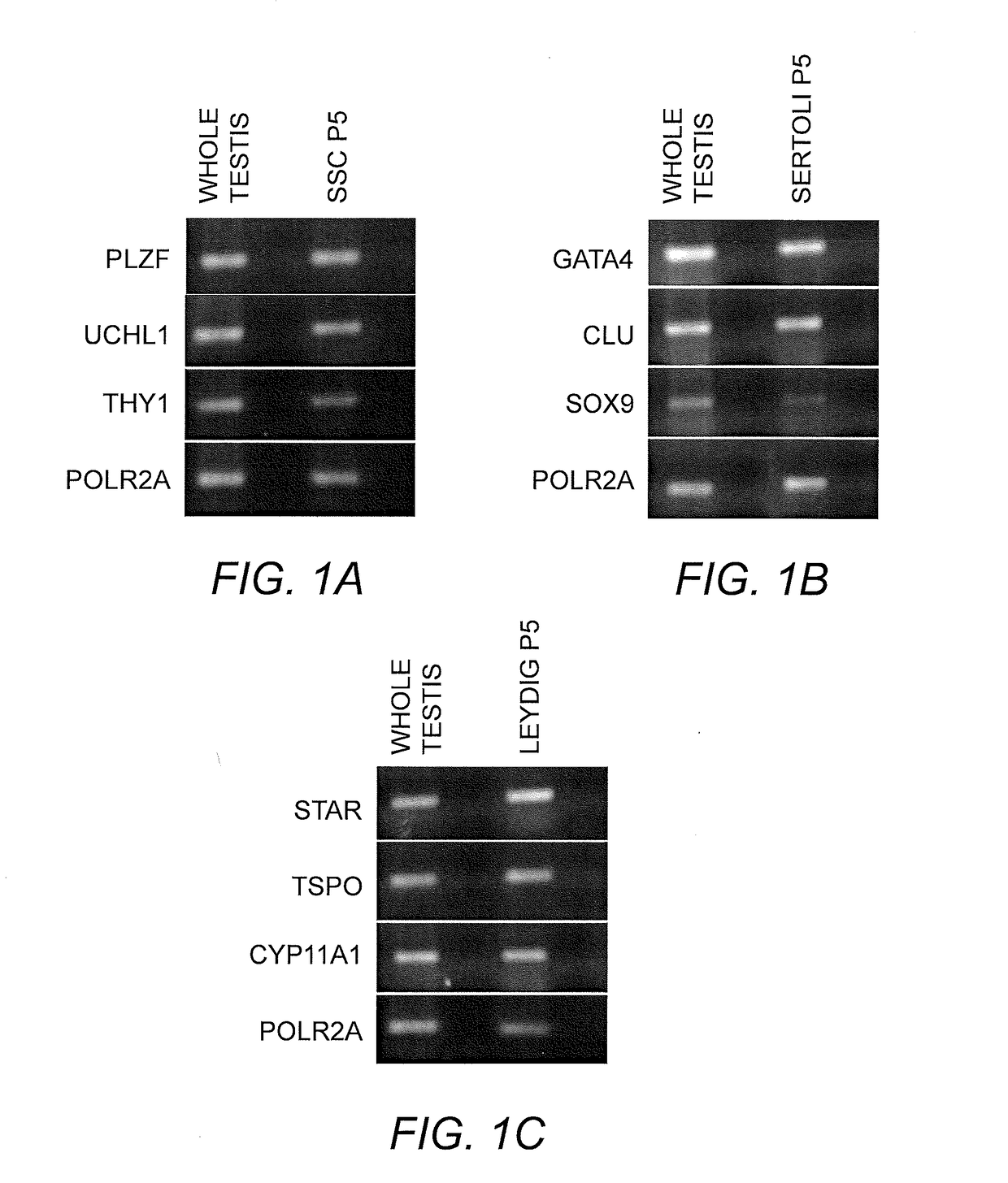
[0093]Spermatogonial cell lines were isolated and cultured as previously described using a modified SSC propagation media that included growth factors necessary for maintenance of spermatogonia in their undifferentiated state (Sadri-Ardekani, H., et al., Propagation of human spermatogonial stem cells in vitro. JAMA: The Journal of the American Medical Association, 302(19):2127-34 (2009); Sadri-Ardekani, H., et al., In vitro propagation of human prepubertal spermatogonial stem cells. JAMA: The Journal of the American Medical Association, 305(23):2416-8 (2011)) Briefly, previously cryopreserved testicular tissue segments of about 100 mg to 200 mg were thawed and enzymatically digested using a combination of collagenase, hyaloronidase, and trypsin. A two-step enzymatic digestion was performed and isolated testicular cells were cultured at 37° C., 5% CO2 ...

example 3

Testicular Cell Isolation, Culture, Immortalization and Cryopreservation of Primary Human Sertoli Cell Lines

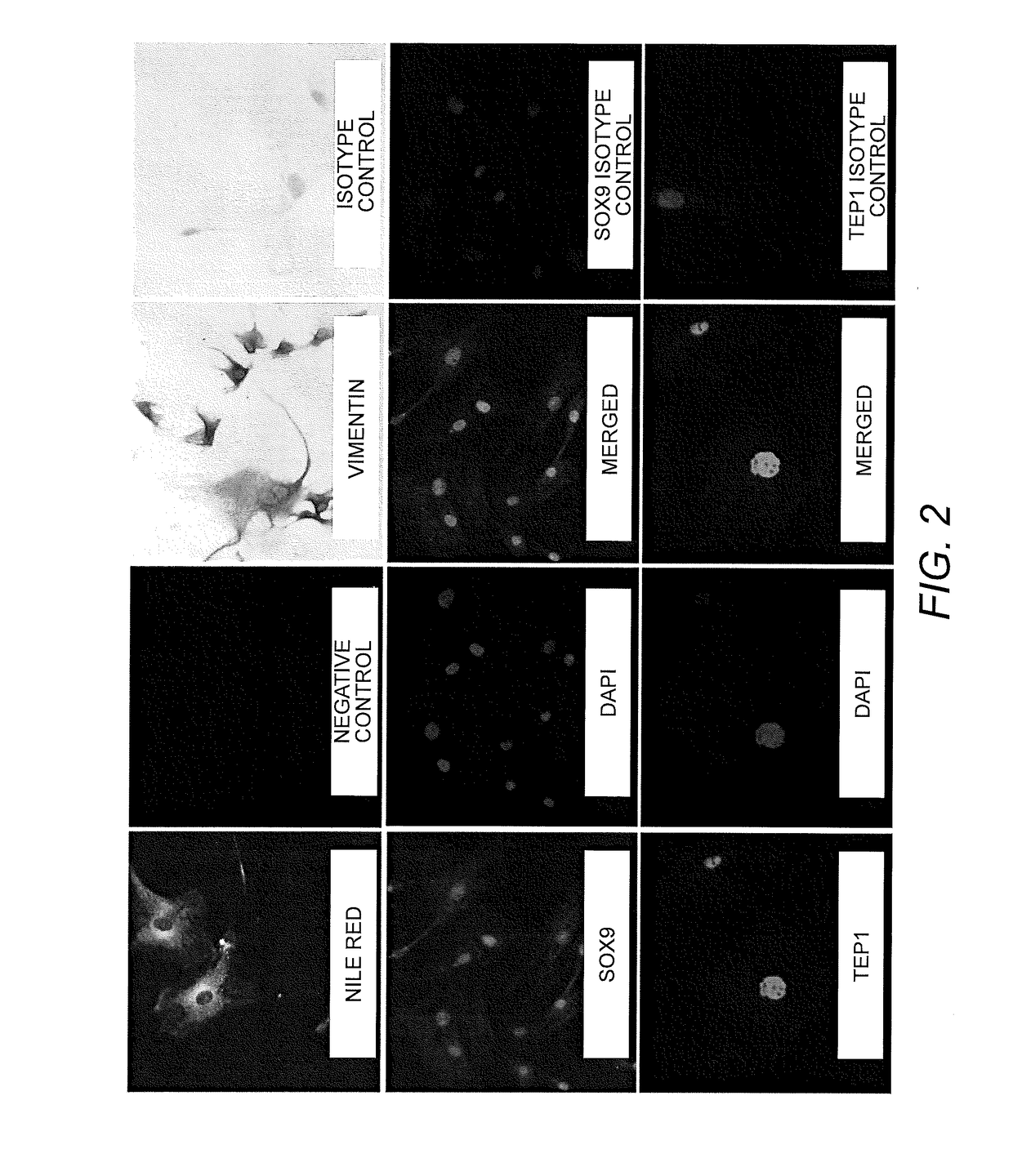
[0096]Preparation and Culturing of Sertoli Cell Lines. Human Sertoli cells were isolated from human tissue as previously described by Chui et al. (Chui, K., et al., Characterization and functionality of proliferative human Sertoli cells. Cell Transplant 20(5):619-35(2011)). Briefly, testicular samples were obtained within 24 hours of death. These tissues were washed first with ice-cold calcium / magnesium-free Hank's balanced salt solution (HBSS) containing 100 U / mL penicillin and 100 μg / mL streptomycin. The tunica albuginea was removed and separated brown tubule tissue was transferred to an Erlenmeyer flask in HBSS. These tissues were shaken at 275-325 rpm for 15 minutes to separate the interstitial cells from the tubules. The supernatant was removed following removal from shaker and 0.25% trypsin and 0.1% collagenase type IV (Sigma) were added with 300 rpm shaking for 20 minut...


PUM


Abstract

A cell composition composed of spermatogonial stem cells, Sertoli cells, Leydig cells and optionally peritubular cells, is provided, as is a culture composition, artificial testicular construct, hydrogel composition, and device containing the same. A method for using the device as a physiologically relevant in vitro model of human testicular function to screen compounds for pharmacological or toxicological activity is also provided.

Description

RELATED APPLICATIONS[0001]This application claims the benefit of priority from U.S. Provisional Patent Application Ser. Nos. 62 / 242,046, filed Oct. 15, 2015, and 62 / 275,031, filed Jan. 5, 2016, the contents of which are incorporated herein by reference in their entireties.GOVERNMENT SUPPORT[0002]This invention was made with Government support under Grant No. W81XWH-13-2-0052 awarded by the Armed Forces Institute for Regenerative Medicine. The US Government has certain rights to this invention.BACKGROUND OF THE INVENTION[0003]The mammalian testis maintains an elaborate communication network between several cell types and subpopulations that cooperatively drive differentiation of male germ cells to haploid gametes, and the production of androgens via the process of steroidogenesis. This dual functionality reflects activities of two distinct testicular regions, the seminiferous tubules and interstitial tissue of the testis. Primitive spermatogonia confined to the basement membrane of t...

Claims


Application Information

Patent Timeline
IPC IPC(8): C12N5/076A01N1/02
CPCC12N5/061A01N1/021C12N2513/00C12N2510/04C12N2501/998A01N1/0221C12N5/0697C12N2533/90C12N2506/246G01N2500/10G01N2510/00G01N2520/00
Inventor PENDERGRAFT, SAMUELSADRI-ARDEKANI, HOOMANATALA, ANTHONYBISHOP, COLIN
Owner WAKE FOREST UNIV HEALTH SCI INC
Why Eureka
Eureka Blog
Learn More