Composition for inducing direct transdifferentiation of somatic cell into vascular progenitor cell, and use thereof
Active Publication Date: 2017-05-11
UNIST ULSAN NAT INST OF SCI & TECH
View PDF0 Cites 0 Cited by
Summary
Abstract
Description
Claims
Application Information
AI Technical Summary
This helps you quickly interpret patents by identifying the three key elements:
Problems solved by technology
Method used
Benefits of technology
Benefits of technology
The invention describes a way to make vascular cells from other types of cells in the body. This process is faster and safer than previous methods, which can result in fewer side effects when using stem cells in therapy.
Problems solved by technology
In this regard, treatment through the transplantation of functional vascular cells is problematic because effective methods of differentiating vascular cells are not readily available and because it is difficult to obtain large amounts of cells.
Also, methods of inducing the differentiation of embryonic stem cells (ESCs) and induced pluripotent stem cells into vascular cells have been proposed, but are disadvantageous because the efficiency of induction into cells of interest is low and there is the risk of activating tumor genes from the embryonic stem cells or pluripotent stem cells upon differentiation into specific cells.
Furthermore, although induced pluripotent stem cells are able to avoid ethical issues associated with embryonic destruction and are resistant to immune system rejection when transplanted, they are undesirably liable to form teratoma, as in the embryonic stem cells.
Also, methods of preparing vascular progenitor cells from somatic cells through direct transdifferentiation have not yet been reported.
Method used
Image
Click on the blue label to locate the original text in one second.
Reading with bidirectional positioning of images and text.
Examples
example 1
Preparation of Induced Vascular Progenitor Cells (iVPCs)
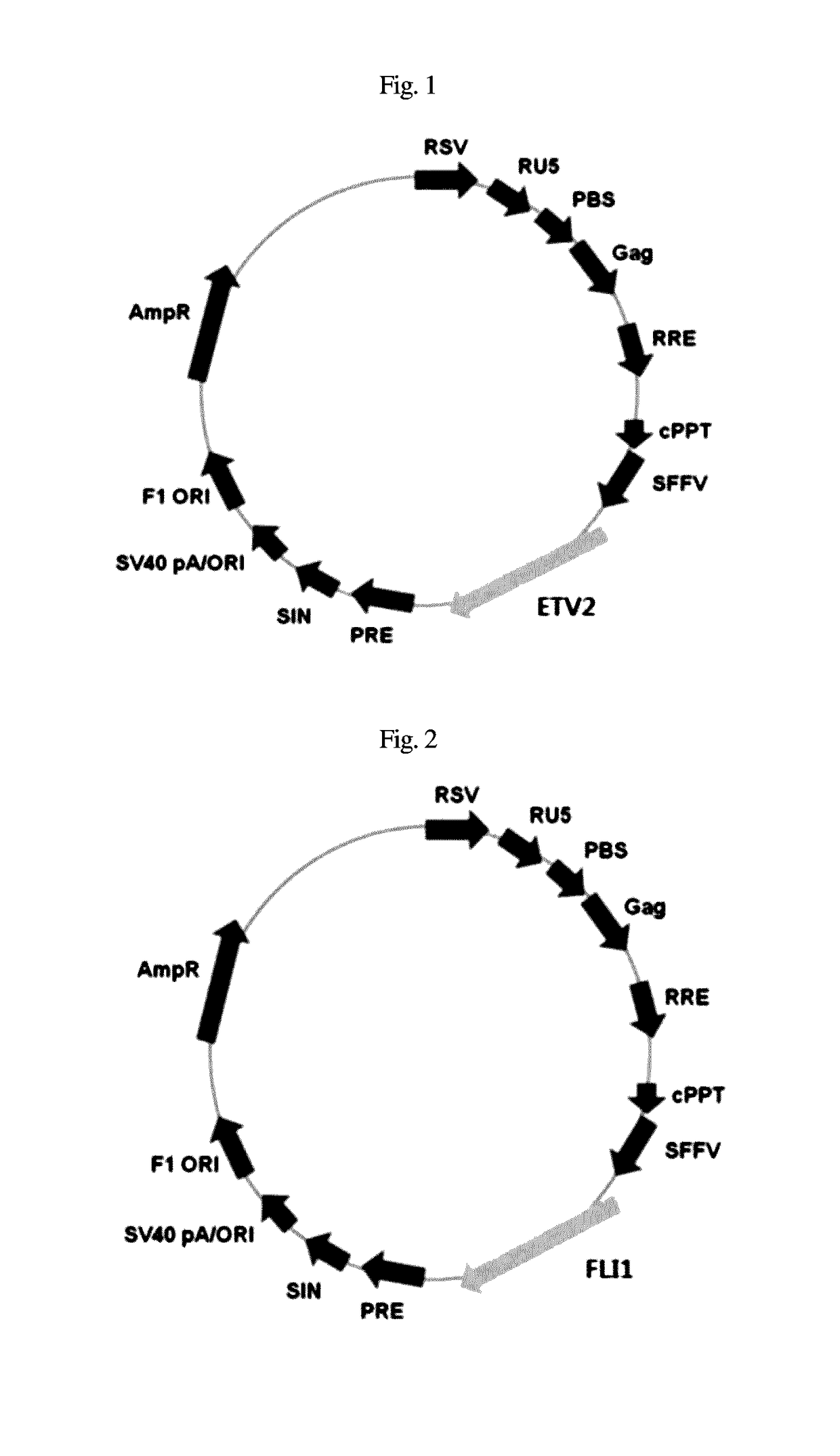
[0050]293 cells were infected with an SFFV promoter, that is, an SF-based lentiviral vector encoding complementary DNA of ETV2 and FLI1, and with a packaging-defective helper plasmid using a Fugene 6 transfectionreagent (Roche). After 48 hr, a viral supernatant was obtained according to the method disclosed in Zaehres, H. & Daley, G. Q., (2006), Methods Enzymol 420, 49-64.
[0051]The dermal fibroblasts were aliquoted at a density of 1×10⁴ cells into a 0.1% gelatin-coated 6-well plate and cultured for 24 hr, together with the viral supernatant that contained ETV2 and FLI1 (1:1) and was supplemented with 6 μg / ml protaminesulfate (Sigma). The transduction efficiency was calculated using SF-GFP control virus.
[0052]Two days after injectio...
example 2
Expression of ETV2 and FLI1 using RT-PCR and Phase-Contrast Microscopy
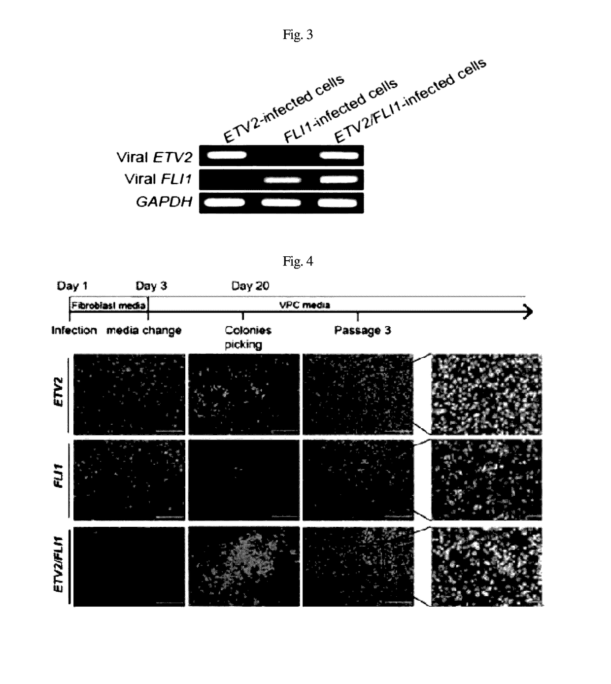
[0053]Five days after infection, the expression of ETV2 and FLI1 was observed using RT-PCR.
[0054]Specifically, total RNA was extracted from each cell using an RNeasy kit (Qiagen), five days after the infection, and cDNA was synthesized using Omniscript RT (Qiagen). PCT was performed using a TaQ DNA polymerase recombinant (Invitrogen).
[0055]After RT-PCR, expression was confirmed through loading on agarose gel, using GAPDH as a control (FIG. 3).
[0056]As illustrated in FIG. 4, using phase-contrast microscopy, colonies began to appear in the cell populations infected with ETV2 and ETV2 / FLI1 within 10 to 11 days after the infection, and the number of colonies increased over time.
[0057]The present inventors observed colonies in the cell populations infected with FLI1 within 30 days after the infection. In order to proliferate the colonies, the cell populations were physically separated and cultured in a gelatin-coated d...
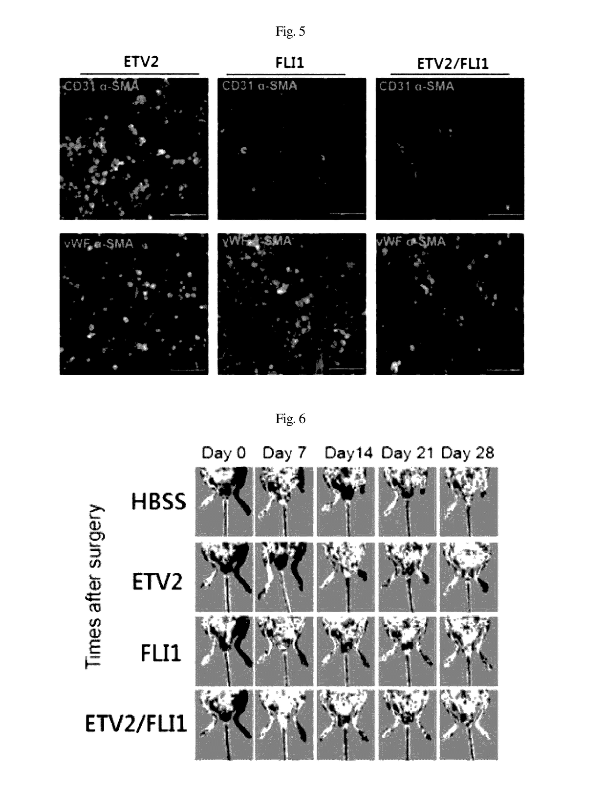
[0058]In order to perform immunocytochemistry, the cells were immobilized for 10 min in 4% para-formaldehyde and treated with 0.1% Triton X-100 for 10 min so as to be permeable. The cells were cultured for 30 min in a 4% FBS / PBS blocking solution and then reacted for 1 hr with a primary antibody diluted with the blocking solution at room temperature. The primary antibody used was as follows: vWF (1:400, Abcam), CD31 (1:200, Chemicon) and α-SMA (1:200, Abcam).
[0059]After reaction with the primary antibody, the cells were washed three times with 0.05% PBST (tween20 / PBS). Thereafter, a secondary fluorescent antibody was diluted with PBS and then reacted with the cells for 1 hr (Alexa Fluor 488 and 568; 1:1000, Molecular Probes). The cells were washed three times with 0.05% PBST, and the nuclei were counterstained for 15 sec using Hoechst 33342 (Thermo Scientific). After the staining, the cells were observed using a...
PUM
Abstract
The present invention relates to a composition for inducing direct transdifferentiation of a somatic cell into a vascular progenitorcell and a use thereof and, more specifically, to a composition for inducing direct transdifferentiation of a somatic cell into a vascular progenitorcell, a pharmaceutical composition for the prevention or treatment of ischemic vascular diseases, a cell therapeutic agent for the prevention or treatment of ischemic vascular diseases, a composition for screening a therapeutic drug for ischemic vascular diseases, a 3D printing biological material composition for the production of an artificial tissue for the treatment of ischemic vascular diseases, and a method for direct transdifferentiation of a somatic cell into a vascular progenitor cell. By producing a vascular progenitor cell by direct transdifferentiation of a somatic cell according to the present invention, it is possible to reduce the production period of the vascular progenitor cell and to avoid the formation of teratoma, which is a side effect of an induced pluripotent stem cell, thereby minimizing the side effects of a stem cell therapeutic agent
Description
TECHNICAL FIELD[0001]The present invention relates to a composition for inducing the direct transdifferentiation of somatic cells into vascular progenitor cells, including at least one selected from the group consisting of a protein of each of direct transdifferentiation factors ETV2 and FLI1, a nucleic acid molecule encoding the protein, and a vector for expressing the protein by introducing the nucleic acid molecule encoding the protein, and to a method for the direct transdifferentiation of somatic cells into vascular progenitor cells and vascular cells using the above composition. In addition, the present invention relates to a pharmaceutical composition, a cell therapy agent, a drug screening composition, or a 3D printing biomaterial composition for the production of artificial tissue, each of which includes vascular progenitor cells and vascular cells induced by the above method for the direct transdifferentiation of somatic cells, thereby being used to prevent or treat ischem...
Claims
Application Information
Patent Timeline
Application Date:The date an application was filed.
Publication Date:The date a patent or application was officially published.
First Publication Date:The earliest publication date of a patent with the same application number.
Issue Date:Publication date of the patent grant document.
PCT Entry Date:The Entry date of PCT National Phase.
Estimated Expiry Date:The statutory expiry date of a patent right according to the Patent Law, and it is the longest term of protection that the patent right can achieve without the termination of the patent right due to other reasons(Term extension factor has been taken into account ).
Invalid Date:Actual expiry date is based on effective date or publication date of legal transaction data of invalid patent.

