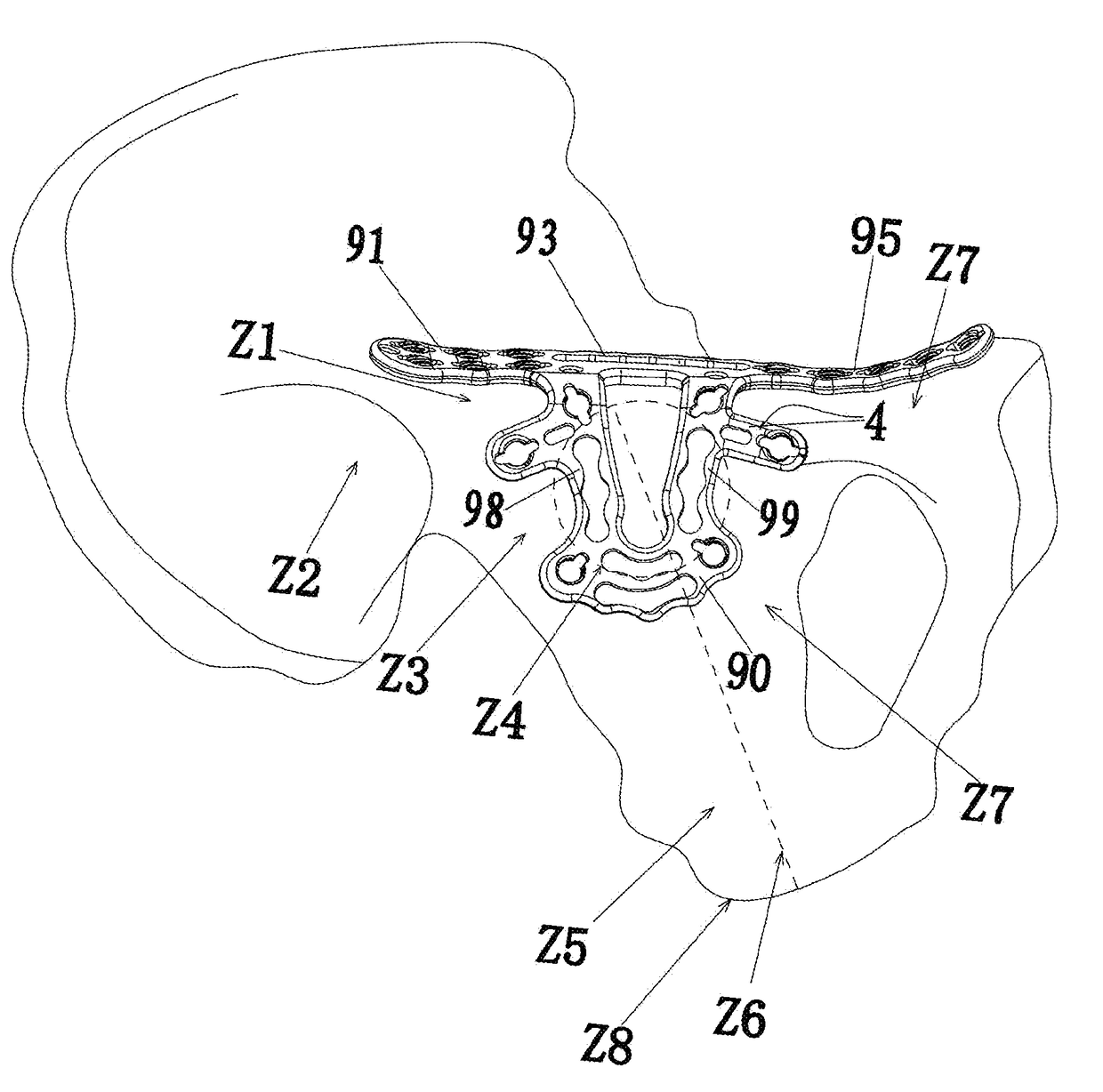
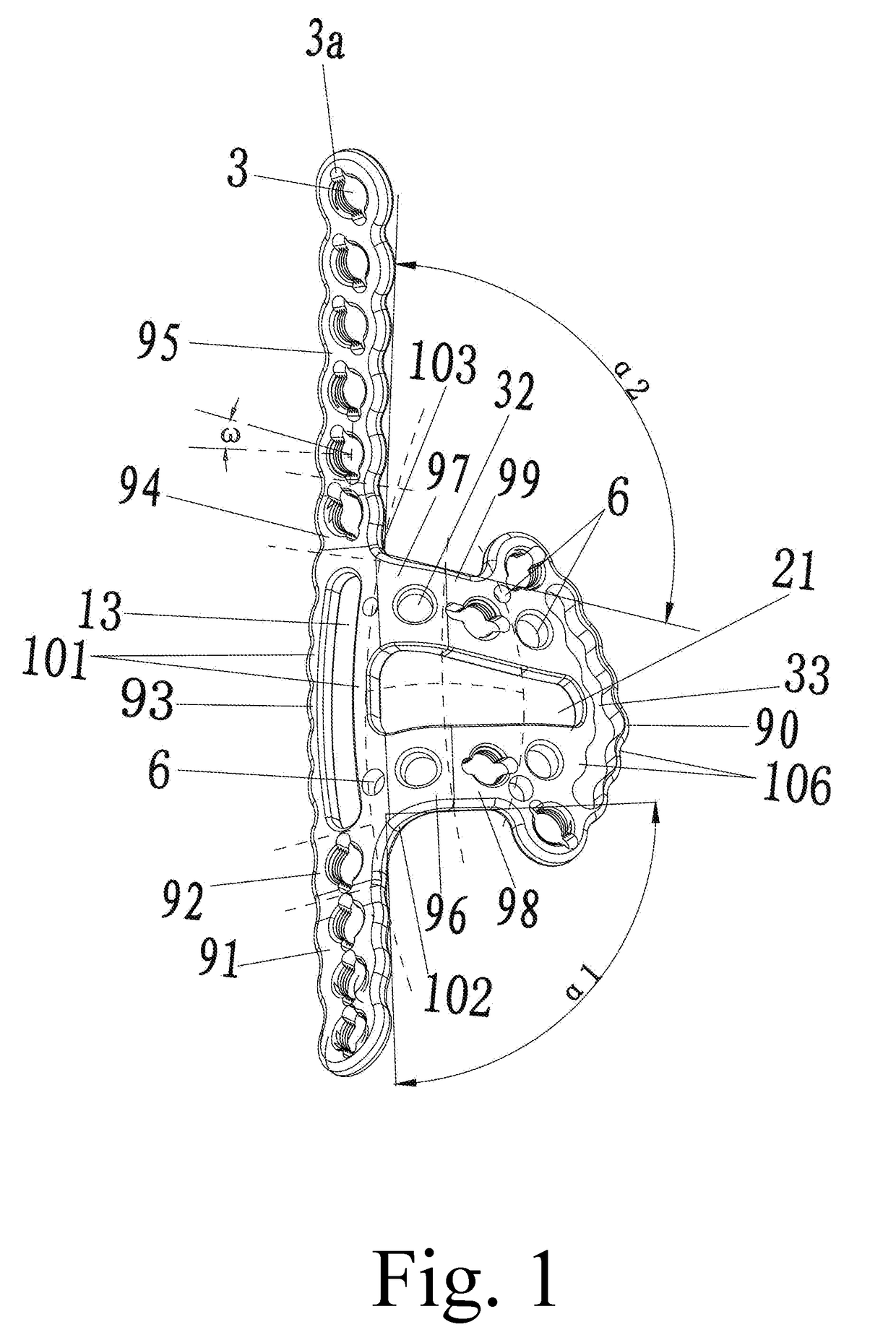
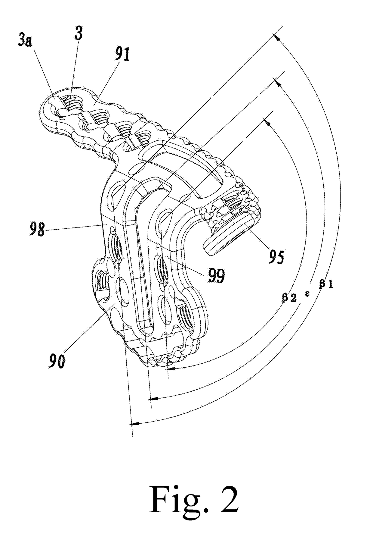
For the fracture fixation around the
quadrilateral area on the inner lateral sides of the acetabulum, the main problems are as follows: 1) two plates might be overlapped and fixed together, wherein the longitudinal plate is thinner and extends to the inner side of the acetabulum and therefore it doesn't work to fix comminuted fractures; moreover, since there are no locking sleeves, it is easy for screws to get into the joint or invade important vessels, nerves and organs around, and it thereby causes the secondary damage.
The longitudinal plate is of a long-strip shape and thus likely to stab internal organs and vessels, especially to aged
osteoporosis fractures.
2) The plates, screws, wires, steel needles used for pelvic and acetabular reconstruction are not stable enough, therefore some orthopaedic surgeons use two plates to form a “cross” in order to manually shape according to the different bone shapes of different patients during a model operation.
As a result, it is not guaranteed during the operation to make the plates and the bone fit each other perfectly.
During a model operation, shaping plates will prolong
operation time, increase
drug dosages, and cause more operation bleeding and different potential dangers.
If two plates do not match well, for example, if some uneven sheer force and sliding displacement exist between the plates, the risk of reduction losing will increase, and consequently it is uncertain for the patient to restore the joint function at the early stage.
In addition,
lying in
bed for a long period of time will also cause joint conglutination,
muscle atrophy, partially losing joint function and etc.
3) When plates shape or bend repeatedly, nicks and scratches will leave on the surface of the plates, it will further cause more focal points of
internal stress in the plates and accordingly cause implants to break more likely.
4) Shaping plates and matching bones unwell both will cause the
fracture site to loosen, displace, pain and cause the stress shelter of the bones, and may further cause the
fracture site to be
nonunion or even malunion.
With little
carelessness, those important structures or organs are very likely to be injured.
Therefore, it is improper to strip and
expose these structures during a model operation.
Exposure is even more difficult to the fat patients; in order to leave a certain space and place the plates, it is needed to strip soft tissues, it however will cause bigger operation injury, e.g., more operation hemorrhage, longer
anesthetic time, and longer
operation time.
6) because it is pretty difficult to fix the fractures around the
quadrilateral area on the inner lateral sides of the acetabulum, the incidence of complications is quite high, e.g., malreduction, unstable fixation,
nonunion, malunion, slow union, traumatic
arthritis, injuries of important vessels and nerves, breakage of internal implants, invalid or inefficient fixation, or failing operations.
7) There is a common
weakness for the internal
implant around the quadrilateral area on the inner lateral sides of acetabulum, that is inconformity with the
transmission system of
biomechanics of the
pelvis and the acetabulum, i.e., the
mechanics transmission
route of the bone, which causes the stress transmission in the bone to disorder, generates negative effects on the
fracture union, and it finally allows patients not to start the exercise at the early stage after the operation.
Because the anatomic shape of the pelvis and the acetabulum is irregular, and there are lots of important vessels, nerves, organs and soft tissues around, many aspects are limited, e.g., the
exposure range of the
surgical incision, and the size and strength of the internal
implant, which bring a huge difficulty and risk to the operative reduction and
internal fixation.
1) The positioning baffle around the quadrilateral area is of a U shape and no any locking holes are set therein, which is unable to effectively fix the smashed fracture fragments in a
free state, or even fixable but not firm enough, thus it will cause higher incidence of complications such as the
nonunion of model bone fractures, slow union or malunion, or traumatic
arthritis, or injury of important vessels and nerves, breakage of internal implants, or invalid or inefficient fixation, or failing operation, and make the applicable range of the plates limited.
2) The plates need to be shaped manually during the operation according to different bone shapes of every patient. However, it is not guaranteed to fit the shaped plates in a model operation and the bone perfectly. In addition, shaping the plates in a model operation will lead to longer
operation time, more anaesthetic dosage, more operative hemorrhage, and etc. Moreover, shaping or bending plates repeatedly will leave nicks and scratches on the surface of the plates, cause more focal points of
internal stress in the plates, and accordingly cause implants to break more likely.
3) The quadrilateral area on the inside of the acetabulum is located in the pelvis, with a deep position, a difficult
exposure and a complicated
local structure, being adjacent to important structures such as
uterus, ovaries, bladders, internal and external iliac arteries and veins, femoral arteries and veins, obturator nerves, obturator arteries and veins. During fixing the plates, it is easy for screws to get into the joint or damage important vessels, nerves and organs around, and causes the secondary damage. Therefore, the plate has lower safety and reduces the operative efficiency.
4) The inconformity with the
transmission system of
biomechanics of the pelvis and the acetabulum, i.e., the
mechanics transmission
route of the bone causes the stress transmission in the bone to disorder, generates negative effects on the
fracture union, which finally allows patients not to start the exercise at the early stage after the operation.
5) The screws in the plate can't reach and fix the
posterior wall of the acetabulum.
6) The baffle of the quadrilateral area of the plates is vertical to the plate body, which doesn't comply with the
normal shape of the surface of the bones of the pelvis.
7) The baffle of the quadrilateral area of the plates doesn't comply with the
normal shape of the surface of the bones of the acetabulum, because it just roughly shows the position of the quadrilateral area, but it doesn't actually fit the bone. The anterior and posterior edge of a normal acetabulum quadrilateral area have different torsion curvatures and radians, usually the difference being 10 degrees to 20 degrees, no matter for adults, children, for male or female.
8) There is no specific quantitative description with the plates to tell the specific angle and direction of the locking screw hole, but only roughly mentioning it with the locking hole. However, this should be one of the key processes in a model operation to internally fix the acetabulum fractures. If there is merely 3 degrees of error for a screw inserting, it might get into joints, or injure important vascular structures like obturator
artery and
vein, superior gluteal
artery and
vein, superior gluteal nurves.


