Predicting Weight Loss and Fat Metabolism Using Optical Signal Changes in Fat

a technology of optical signal and fat, applied in the field of medical diagnostic and biomedical optics, can solve the problems of inability to assess the structure or function of fat (adipose tissue), no widely used clinical methods for physiology in humans, and no non-invasive tools available to measure changes in fat (adipose tissue) structure or function in vivo, so as to reduce the incidence of obesity-related complications, prolong life span, and enhance white at sirto expression

Active Publication Date: 2017-07-27
RGT UNIV OF CALIFORNIA
View PDF8 Cites 6 Cited by
  • Summary
  • Abstract
  • Description
  • Claims
  • Application Information

AI Technical Summary

Benefits of technology

[0010]Recently, we utilized DOSI to obtain quantitative measures of near infrared (NIR) AT optical and physiological properties in 10 overweight adults during a three-month calorie-restricted diet. Calorie restriction, or CR, has been shown to extend life span and reduce incidence of obesity-related complications in mammals, and these effects are mediated in part by changes in AT. In mice, three months of CR can lead to nitric oxide (NO) mediated increases in white AT mitochondrial DNA, peroxisome proliferator-activated receptor-γ coactivator 1α (PGC-1α), and markers of mitochondrial biogenesis. It is suggested that enhanced white AT SIRTO expression contributes to the pro-longevity effects of CR in mammals. Specifically, studies of CR in humans have reported morphological changes in AT, such as adpipocyte size changes and altered profile of inflammatory cells. Weight reduction by CR leads to adipocyte shrinkage and it has been shown that a 10% weight loss results in a 16% decrease in adipocyte volume, whereas a high-fat diet increases adipocyte volume in a matter of weeks. In humans, a strong positive correlation has been found between adipocyte size and the presence of type 2 Diabetes or its associated risk factors in bariatric surgery candidates. Another recent study revealed a correlation between adipocyte hypertrophy and both insulin resistance and AT inflammation.
[0011]It is also possible that adipocyte size changes influence cellular metabolism. For example, some data suggest a relationship between adipocyte size and AT angiogenesis. More important is the hypothesized relationship between adipocyte size and O2 delivery. The diameters of hypertrophic adipocytes are thought to exceed the diffusion distance of O2, causing lower cellular PO2. This cellular hypoxia has been observed as a feature of AT in obesity in most studies, but there are some conflicting reports. Most importantly, there are data to suggest that it is increased AT PO2 that is associated with inflammation and insulin resistance in obesity. A recent investigation in mice found that expression of hypoxia-inducible factor (HIF)-1 alpha increased in animals fed a high-fat diet, in response to increased AT O2 consumption. Other lines of investigation have focused on the characterization of “beige” adipocytes, which tend to consume more O2 and are thought to correlate with enhanced metabolic health.
[0012]DOSI derived tissue concentrations of hemoglobin, water, and lipid and the wavelength-dependent scattering amplitude (A) and slope (b) obtained from 30 abdominal locations and three time points were calculated and analyzed using linear mixed effects models, and were also used to form three dimensional surface images. Subjects lost a mean of 11.7±3.4% of starting weight, while significant changes in A, b, tissue water fraction and deoxyhemoglobin [HbR] were observed. Optical scattering signals reveal alterations in tissue structure consistent with reductions in adipose cell volume, while water and hemoglobin dynamics suggest improved AT perfusion and oxygen extraction, consistent with possible “browning” of AT. These results suggest that DOSI measurements of NIR optical properties could be used to enhance understanding of the role of AT in disease progression, and to advance diagnostics and monitoring of metabolic disorders.
[0018]In yet another embodiment, the improved method also includes correlating the determined tissue absorption coefficient (μa) and reduced scattering coefficient (μs′) to a higher density and a reduced volume of adipose tissue within the patient. Here, correlating the determined tissue absorption coefficient (μa) and reduced scattering coefficient (μs′) to a higher density and a reduced volume of adipose tissue within the patient may include guiding therapeutic interventions including diet, exercise, and medication by assessing their impact on the adipose tissue within the patient. Alternatively, correlating the determined tissue absorption coefficient (μa) and reduced scattering coefficient (μs′) to a higher density and a reduced volume of adipose tissue within the patient may include correlating the tissue absorption coefficient (μa) and reduced scattering coefficient (μs′) with improvements in vascular disease risk observed in the patient.
[0025]In another embodiment, the improved method also includes correlating the created heat map representing a concentration of one of a plurality chromophores obtained from the tissue absorption coefficient and reduced scattering coefficient to a higher density and a reduced volume of adipose tissue within the patient. Here, correlating the created heat map representing a concentration of one of a plurality chromophores obtained from the tissue absorption coefficient and reduced scattering coefficient to a higher density and a reduced volume of adipose tissue within the patient may include guiding therapeutic interventions including diet, exercise, and medication by assessing their impact on the adipose tissue within the patient. Alternatively, correlating the created heat map representing a concentration of one of a plurality chromophores obtained from the tissue absorption coefficient and reduced scattering coefficient to a higher density and a reduced volume of adipose tissue within the patient may include correlating the created heat map with improvements in vascular disease risk observed in the patient.

Problems solved by technology

Despite the importance of AT in disease, there are no widely-used clinical methods for assessing AT physiology in humans.
In other words, there are no easily accessible, non-invasive tools available to measure changes in fat (adipose tissue) structure or function, which can provide indications of the progress of weight loss intervention and other pathologies associated with adipocyte size and metabolism such as insulin resistance and vascular dysfunction.
However, there are no diagnostic tools available to assess tissue structure or function in fat tissue in vivo.
These techniques have certain disadvantages and limitations, namely existing technologies are sensitive only to changes in macro-structure of adipose tissue layers, such as the thickness.
None of them are capable of measuring changes reflective of cell size, metabolism, or extracellular milieu, which occur prior to changes in the macro-structure.
Diffuse optical techniques are increasingly used in the study of cerebral and muscular hemodynamics and metabolism, as well as in cancer biology, but to this date they have not been explored for measuring AT.

Method used


Image

  • Predicting Weight Loss and Fat Metabolism Using Optical Signal Changes in Fat
  • Predicting Weight Loss and Fat Metabolism Using Optical Signal Changes in Fat
  • Predicting Weight Loss and Fat Metabolism Using Optical Signal Changes in Fat

Examples


Embodiment Construction

Method Overview

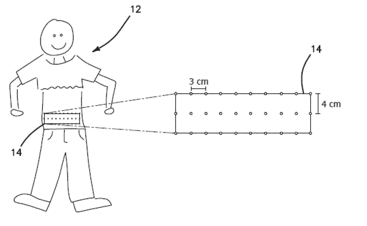
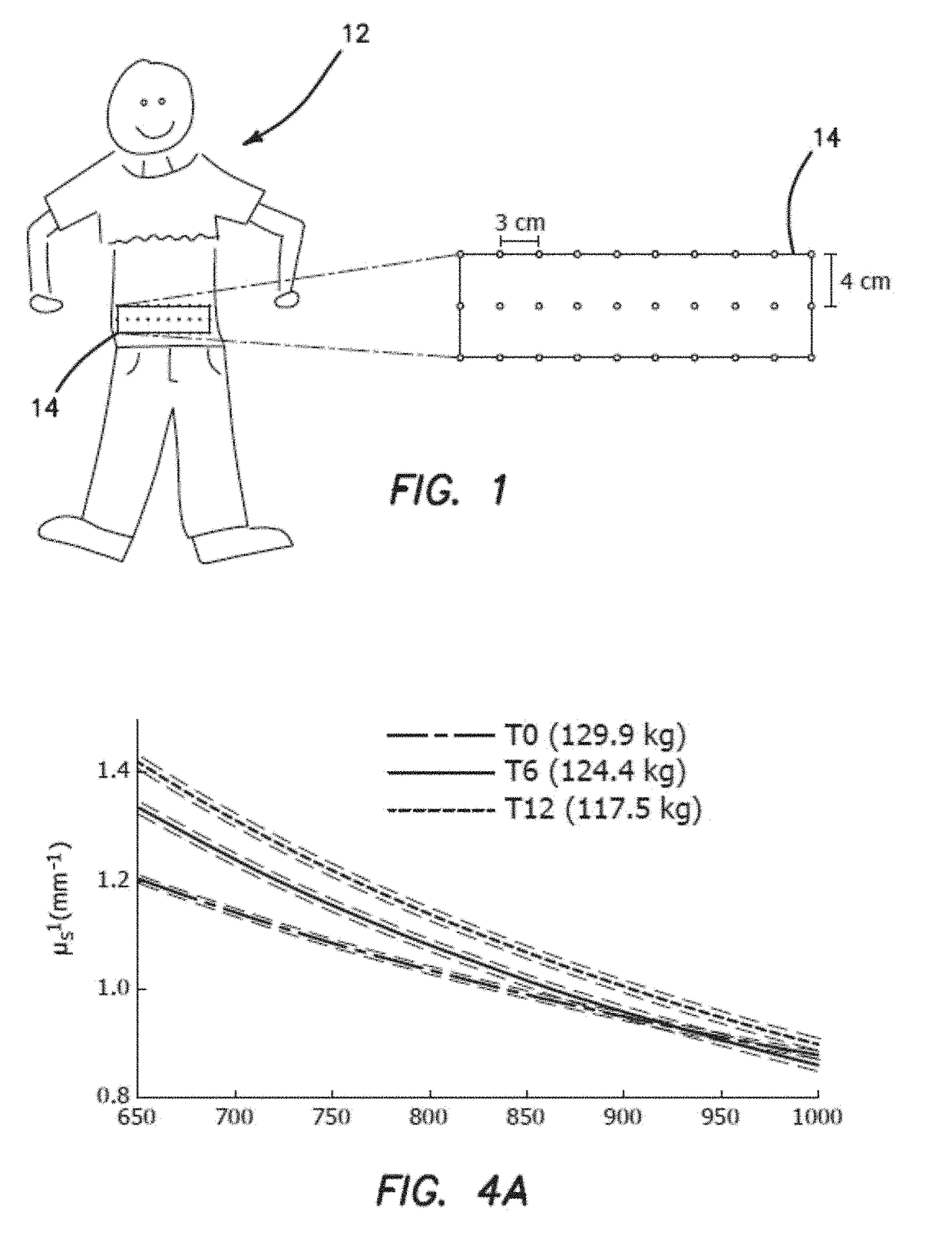
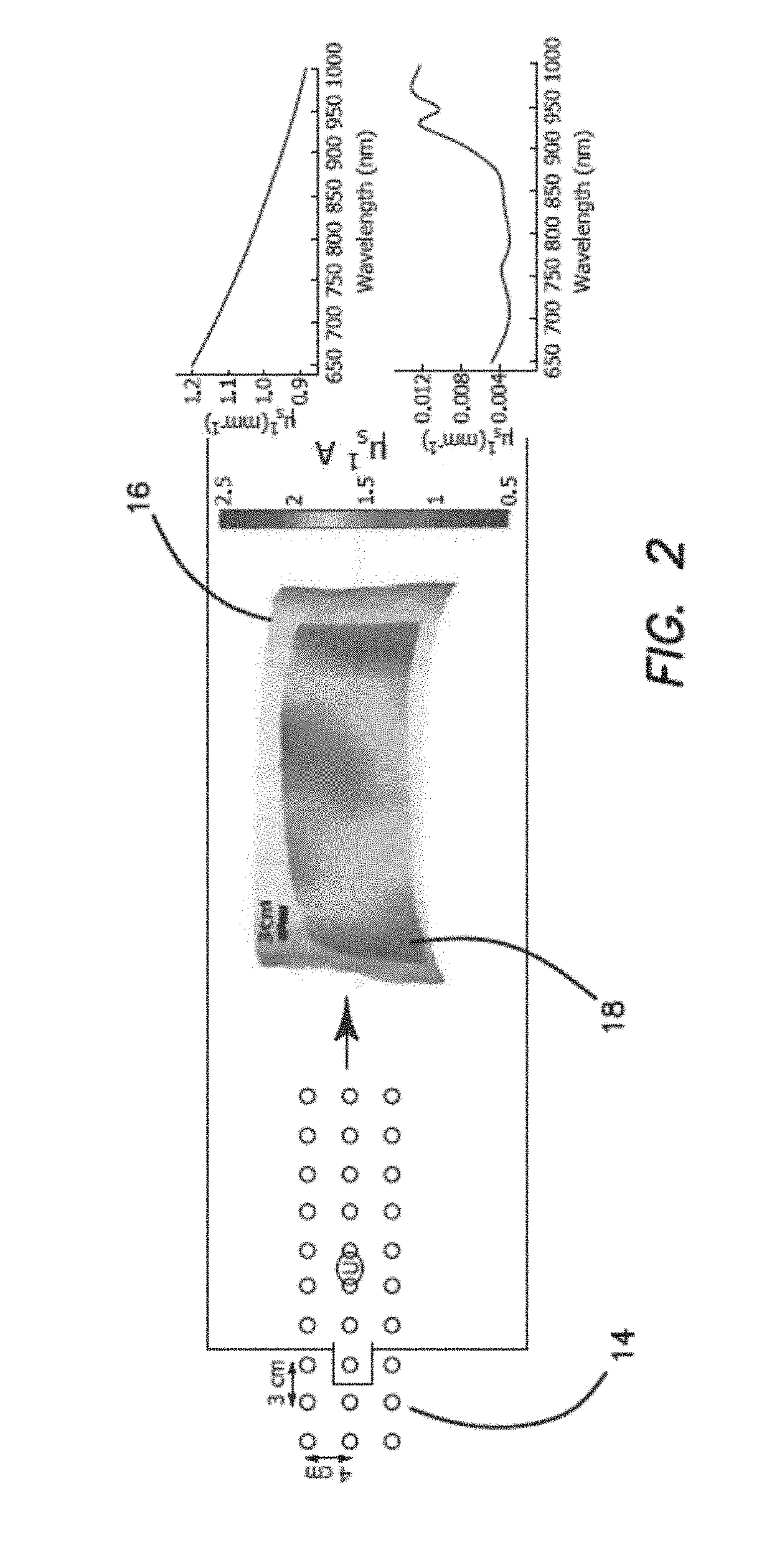
[0040]We are presenting for the first time the use of a quantitative, non-invasive optical spectroscopy technique for measuring dynamic changes in AT structure and metabolism in vivo. Our technique requires multiple wavelengths of light in the near-infrared (650-1000 nm). Using these wavelengths of light, we illuminate adipose tissue and analyze light that returns to a photodetector. Our analysis of the return signals allows for the calculation of absorption and reduced scattering coefficients (μa and μs′) at each wavelength. The obtained μa and μs′ values allow for quantification of biomarkers and indices which allow us to measure fat composition and metabolism. For example, we are able to determine the concentration of oxy- and deoxy-hemoglobin, the fractional water and lipid content, and information about the size distribution of light scatterers in the adipose tissue. We are not limited to this information, however, and our technique is sensitive to any unique abs...


PUM


Abstract

A quantitative, non-invasive optical spectroscopy technique for measuring dynamic changes in adipose tissue structure and metabolism in vivo. The technique requires multiple wavelengths of light in the near-infrared (650-1000 nm). Using these wavelengths of light, adipose tissue is illuminated and light that returns to a photodetector is analyzed. From the return signals, the absorption and reduced scattering coefficients (μa and μs′) at each wavelength are calculated. The obtained pa and μs′ values allow for quantification of biomarkers and indices which allow for measurement of fat composition and metabolism. The concentration of oxy- and deoxy-hemoglobin, the fractional water and lipid content, and information about the size distribution of light scatterers in the adipose tissue are also determined. A detailed and quantitative understanding of fat composition and metabolism is thereby provided which describes the effectiveness of interventions to improve the health of a patient.

Description

GOVERNMENT SUPPORT[0001]This invention was made with government support under grants EB015890 and TR000153 awarded by the National Institutes of Health. The government has certain rights in the invention.BACKGROUND[0002]Field of the Technology[0003]The invention relates to the field of medical diagnostic and biomedical optics and specifically relates to weight loss: a non-invasive, optical method to monitor and predict the effectiveness of weight loss interventions by measuring fat composition and metabolism.[0004]Description of the Prior Art[0005]Recent physiological studies have revealed the potentially critical role of adipose tissue (AT) in the development of metabolic disorders. Both visceral and subcutaneous AT have been shown to be subject to various pathophysiological processes, such as inflammation, dysregulated oxygenation, and disrupted endocrine signaling. Despite the importance of AT in disease, there are no widely-used clinical methods for assessing AT physiology in hu...

Claims


Application Information

Patent Timeline
Patent Type & Authority Applications(United States)
IPC IPC(8): A61B5/00A61B5/145A61B5/1455
CPCA61B5/4872A61B5/14551A61B5/14546A61B5/4875A61B5/7275A61B2090/373A61B5/7246A61B5/486A61B5/684A61B2090/306A61B2090/366A61B5/743A61B5/6823G16H50/20
Inventor WARREN, ROBERTGANESAN, GOUTHAMO'SULLIVAN, THOMASMALIK, SHAISTATROMBERG, BRUCE J.GALASSETTI, PIETRO
Owner RGT UNIV OF CALIFORNIA
Why Eureka
Try Eureka