
MULTIPLEX TP53/CEN17/B CELL GENE-PROTEIN CO-DETECTION ASSAY AND UNIQUELY SPECIFIC PROBES FOR 19q12, INSR, ATM, DLEU2, TP53, AND 13q12

a tp53 and cen17 technology, applied in the field of immunohistochemistry and in situ hybridization, can solve the problems of large capital investment, high diagnostic cost, and fish that does not incorporate the context of tissue for the reader/corer, and achieve the effect of quick identification of patients

Inactive Publication Date: 2017-07-27
VENTANA MEDICAL SYST INC
View PDF0 Cites 3 Cited by
  • Summary
  • Abstract
  • Description
  • Claims
  • Application Information

AI Technical Summary

Benefits of technology

The patent describes a way to quickly identify patients with a specific genetic abnormality (17p-aberration) by staining B cells with a combination of proteins, DNA markers, and a chemical treatment. The stained cells are then analyzed using a special technology, called bright field detection. This method allows for the efficient and simultaneous detection of multiple DNA markers and proteins on a single slide, which makes it easier to identify patients with this abnormality.

Problems solved by technology

Until most recently, patients testing 17p-deleted were evaluated for novel agents or stem cell transplantation, with limited results.
Some laboratories have begun incorporating CGH array testing or next generation sequencing technology to analyze for these CLL subtypes, but all of these diagnostic technology options are very costly, require a large capital investment in platforms and lab-specific requirements.
Further, FISH does not incorporate the context of tissue for the reader / scorer.
One of the reasons is because scoring a deleted entity (e.g., 17p) using bright field staining was thought to be not possible or at most would result in inaccurate scores.
Another reason it was thought not to be possible to perform such a gene-protein assay was because the protease treatment used in in situ hybridization was thought to destroy tissue morphology, and thus also destroy the staining of the B cells.

Method used


Image

  • MULTIPLEX TP53/CEN17/B CELL GENE-PROTEIN CO-DETECTION ASSAY AND UNIQUELY SPECIFIC PROBES FOR 19q12, INSR, ATM, DLEU2, TP53, AND 13q12
  • MULTIPLEX TP53/CEN17/B CELL GENE-PROTEIN CO-DETECTION ASSAY AND UNIQUELY SPECIFIC PROBES FOR 19q12, INSR, ATM, DLEU2, TP53, AND 13q12
  • MULTIPLEX TP53/CEN17/B CELL GENE-PROTEIN CO-DETECTION ASSAY AND UNIQUELY SPECIFIC PROBES FOR 19q12, INSR, ATM, DLEU2, TP53, AND 13q12

Examples


example 1

sis

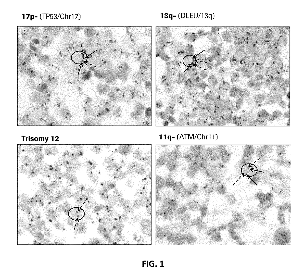
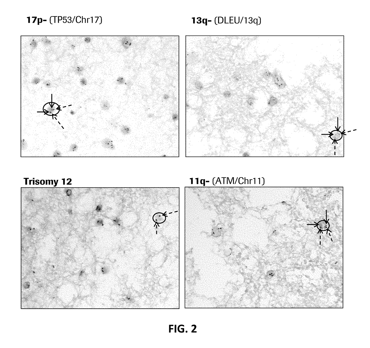
[0207]Abnormal cytogenetics are found in the majority of patients with CLL, and each subtype is associated with differentiated frequency, outcome, and suggested treatments (see Table 3).

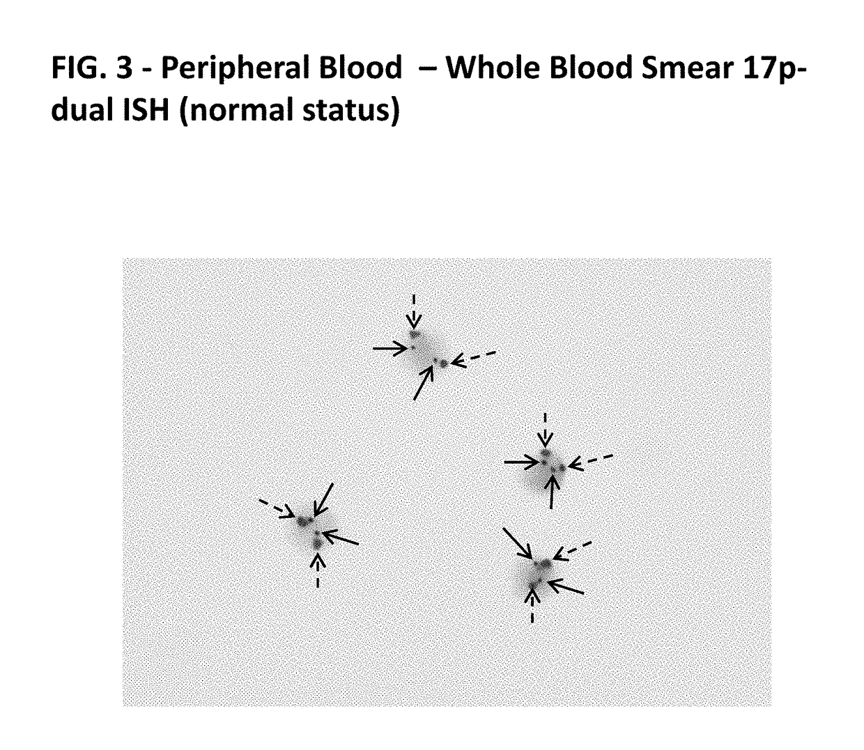
[0208]Example 1 demonstrates that probes of the present disclosure can detect such abnormalities via in situ hybridization (ISH) on formalin fixed paraffin embedded bone marrow / trephine samples.

[0209]A. Preparation of Reagents:

[0210]DNA probes were labeled with either DNP or DIG using standard reaction conditions. Specificity of each probe was verified using CGH Target Metaphase spread. Functionality of each probe was determined by standard dual ISH using the U Dual Color ISH Open Probe procedure on standard cancer tissues supplied by Ventana Medical Systems, Inc.

[0211]B. Reagents

(if commercially available from Ventana Medical Systems Inc., reagents are listed with the associated part number).

[0212]TP53 / Chr17:[0213]TP53-DNP [30 ug / ml] (mixture of 10 probes according to SEQ ID NOs: 51-60);[0214]AS...

example 2

Gene-Protein Assay for CD79a / TP53 / CEN17

[0264]This example describes a multiplex gene-protein assay for detection of CD79a protein, TP53 DNA, and chromosome 17 centromere DNA in a single sample. The staining strategy consists of essentially three staining steps: (1) CD79a protein; (2) TP53 Gene; and (3) CEN17. An overview of the staining strategy is illustrated at FIG. 6.

[0265]In the first detection, an antibody specific for CD79a 1 is contacted with the sample. A species-specific secondary antibody 2 conjugated to a first antibody-reactive group (such as a hapten) is then applied to the sample, which binds to the primary antibody 1. A tertiary antibody 3 is then applied to the sample. The tertiary antibody 3 is specific for the antibody-reactive subunit of the secondary antibody 2. The tertiary antibody 3 also is labeled with a detectable label. After reaction to visualize the detectable label, regions at which the primary antibody has bound appear as a first color. In the example i...

embodiment 1

[0270]A multiplex method for co-detecting a B cell marker protein, TP53 genomic DNA, and chromosome 17 centromere DNA in a sample on a single slide, said method comprising:

[0271]staining the B cell marker protein by contacting the sample with a B cell marker protein-specific antibody and contacting the sample with a first chromogen component for the B cell marker protein-specific antibody, the first chromogen component is adapted to emit or make visible a first color, wherein the presence of the first color indicates the presence of the B cell marker protein; and

[0272]staining TP53 genomic DNA and staining chromosome 17 centromere DNA by contacting the sample with a TP53 genomic DNA-specific nucleic acid probe and with a chromosome 17 centromere DNA-specific nucleic acid probe, and contacting the sample with a second chromogen component for the TP53 genomic DNA-specific nucleic acid probe and with a third chromogen component for the chromosome 17 centromere DNA-specific nucleic acid...


PUM

PropertyMeasurementUnit
Fractionaaaaaaaaaa
Fractionaaaaaaaaaa
Fractionaaaaaaaaaa

Abstract

Disclosed herein are multiplex methods for co-detecting a B cell marker, TP53 nucleic acid, and Chromosome 17 centromere DNA in a single sample. Samples stained for the B cell marker (e.g., CD79a protein), TP53 nucleic acid, and Chromosome 17 centromere DNA may allow for the identification of the subtype of chronic lymphocytic leukemia (CLL) with the 17p deletion. The methods feature staining the B cell marker (e.g., CD79a) a first distinct color, TP53 in a second distinct color, and chromosome 17 centromere DNA in a third distinct color. Further disclosed are nucleic acid probes specific for 19q12, INSR, ATM, DLEU2, TP53, and 13q12.

Description

CROSS-REFERENCE TO RELATED APPLICATIONS[0001]This patent application is a continuation of International Patent Application No. PCT / EP2015 / 072231 filed Sep. 28, 2015, which claims priority to and the benefit of U.S. Provisional Application No. 62 / 057,164 filed Sep. 29, 2014. Each of the above patent applications is incorporated herein by reference as if set forth in its entirety.BACKGROUND OF THE INVENTION[0002]Field[0003]This disclosure relates to immunohistochemistry and in situ hybridization, particularly to the detection of CD79a protein, TP53 nucleic acid, and chromosome 17 centromere DNA in a single sample. The disclosure also relates to nucleic acid probes, particularly useful for the in situ hybridization detection of 19q12, INSR, ATM, DLEU2, TP53, and 13q12.[0004]Background[0005]Many cancers are characterized by genetic changes that lead to aberrant control of cellular processes, or to uncontrolled growth and proliferation of cells. These genetic changes include gain or loss...

Claims


Application Information

Patent Timeline
IPC IPC(8): G01N33/574G01N1/30C12Q1/68
CPCG01N33/57492C12Q1/6886G01N2001/302C12Q2600/158G01N2333/70503G01N1/30G01N33/56972G01N33/57426G01N2333/70596C12Q1/6881
Inventor ALEXANDER, NELSON R.GROGAN, THOMAS M.HENRICKSEN, LEIGH A.KELLY, BRIAN D.NITTA, HIROSTANISLAW, STACEYTUBBS, ALISA
Owner VENTANA MEDICAL SYST INC
Learn More