
Systems and methods of disease modeling using static and time-dependent hydrogels

Inactive Publication Date: 2017-11-16
RGT UNIV OF CALIFORNIA
View PDF0 Cites 0 Cited by
  • Summary
  • Abstract
  • Description
  • Claims
  • Application Information

AI Technical Summary

Benefits of technology

The patent describes methods for creating and manipulating tissues on a chip using hydrogels and cross-linkers to mimic the stiffness of diseased tissues. These methods allow for the creation of disease models that can be used for disease modeling, dynamic sampling, and maintaining constant culture stiffness. The methods involve exposing the hydrogel to UV radiation and a photoinitiator to crosslink the hydrogel and create an elastic environment for cells. The resulting hydrogels can be used to mimic the progression of heart disease or breast cancer by inducing the differentiation of cells into specific cell types and then exposing them to additional UV radiation to further crosslink the hydrogel. The resulting hydrogels can also be used to induce hypoxia and maintain constant stiffness during the culture process. Overall, the methods provide a way to create and study disease models on a chip for research and potential treatment purposes.

Problems solved by technology

Identification and evaluation of new therapeutic agents or identification of suspected disease associated targets typically employ animal models which are expensive, time consuming, require skilled animal-trained staff and utilize large numbers of animals.
In vitro alternatives have relied on the use of conventional cell culture systems which are limited in that they do not allow the three-dimensional interactions that occur between cells and their surrounding tissue.
Normal tissue cells are generally not viable when suspended in a fluid.
Anchorage-dependent cells, therefore, are no longer viable if dissociated from the solid matrix and suspended in the culture media, even if soluble proteins are added to engage cell adhesion molecules, e.g., integrin-binding RGD peptide.
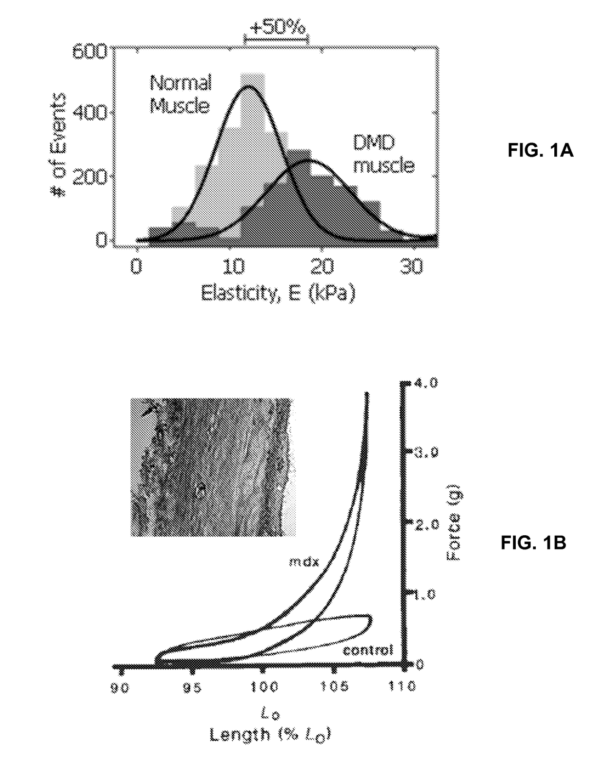
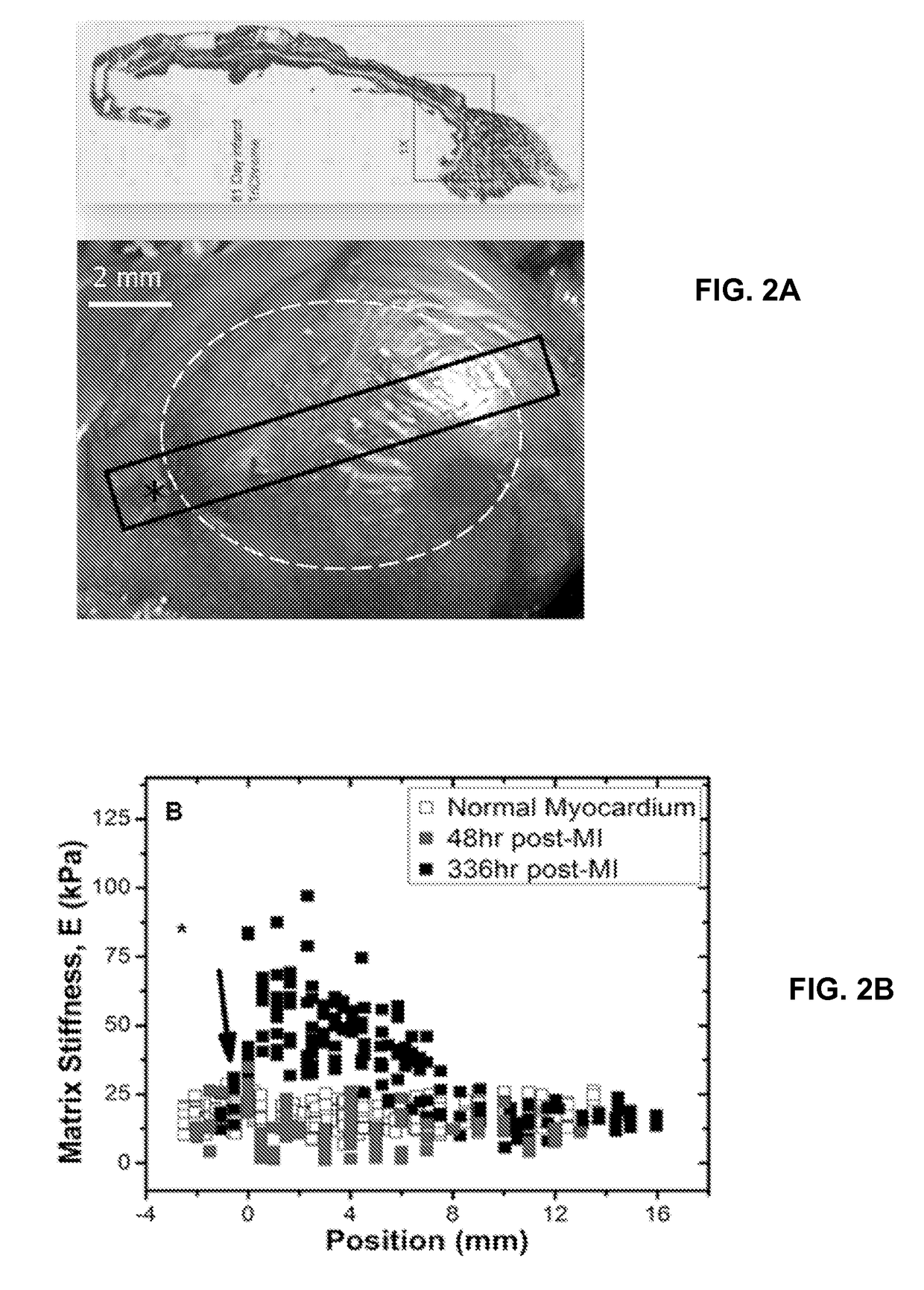
The heart then remodels itself to continue its pumping function, but with reduced efficiency.
Efforts to build biosynthetic materials or engineered tissues that recapitulate these structure-function relationships often fail because of the inability to replicate the in vivo conditions that coax this behavior from ensembles of cells.

Method used


Image

  • Systems and methods of disease modeling using static and time-dependent hydrogels
  • Systems and methods of disease modeling using static and time-dependent hydrogels
  • Systems and methods of disease modeling using static and time-dependent hydrogels

Examples


example 1

Polyacrylamide (PA) Hydrogel as a Static Substrate

[0070]PA gels are produced in this protocol by mixing various acrylamide and bis-acrylamide concentrations and inducing free radical polymerization. PA gel modulus of elasticity was quantified using atomic force microscopy (AFM), which is a nano-indentation method of calculating elasticity. This technique has been extensively detailed elsewhere (Rotsch et al., 1999; Rotsch and Radmacher, 2000).

[0071]Details of the PA hydrogel fabrication can be found in Tse and Engler, “Preparation of Hydrogel Substrates with Tunable Mechanical Properties,” Current Protocols in Cell Biology (2010), incorporated herein by reference.

[0072]Materials used are: 0.1 M NaOH; Distilled H2O; 3-Aminopropyltriethoxysilane (APES); 0.5% (v / v) glutaraldehyde in phosphate-buffered saline (PBS; Cellgro, cat. no. 46-013-CM); Dichlorodimethylsilane (DCDMS); 40% (w / v) acrylamide stock solution (Sigma-Aldrich, cat. no. A4058); 2% (w / v) bis-acrylamide stock solution (Sig...

example 2

Thiolated-Hyaluronic Acid (HA) Hydrogel as a Dynamic Substrate

[0075]The detailed methods for the HA hydrogels have been published for type one in Young and Engler. “Hydrogels with time-dependent material properties enhance cardiomyocyte differentiation in vitro,” Biomaterials (2011). Briefly, Hyaluronic Acid (HA) was obtained from Calbiotech (CA) and thiolated using a cleavable, carbohydrate selective, sulfhydryl-reactive crosslinker, PDPH (3-[2-Pyridyldithio]propionyl hydrazide) (Thermo Scientific-Pierce), MES Buffer (Thermo Scientific-Pierce), DMSO (Sigma), EDC (1-ethyl-3-[3-dimentylaminopropyl] carbodiimide hydrochloride) (Sigma), and DTT (dithiothreitol, Sigma). Alternatively, thiolated HA of similar functionality was also obtained from Glycosan Biosystems (UT). Poly(ethylene glycol) diacrylate (PEGDA) of different molecular weight was used as a crosslinker (Mw˜3400 Da from Glycosan Biosystems, UT and Mw˜258, 700 and 2000 Da from Sigma). For protein attachment on gels, EDC, NHS ...

example 3

Mimicking Fibrotic Stiffening Associated with Disease with Dynamic Methacrylated-Hyaluronic Acid (MeHA) Hydrogel

[0080]Genome-wide association studies have identified single nucleotide polymorphisms (SNPs) at the 9p21 gene locus as increasing the risk of coronary artery disease (CAD) and myocardial infarction susceptibility. Associations have implicated SNPs in enhancing smooth muscle cell proliferation and endothelial permeability but have not identified adverse effects in cardiomyocytes.

[0081]Using induced pluripotent stem cell-derived cardiomyocytes from patients that are homozygous risk / risk (R / R) and non-risk / non-risk (N / N) for 9p21 SNPs and either CAD positive (CAD+) or negative (CAD−), altered cardiomyocyte behavior was assessed when cultured on methacrylated-hyaluronic acid matrix (MeHA) capable of dynamically stiffening from healthy heart matrix stiffness, 11 kiloPascals (kPa), to that of fibrotic tissue, 50 kPa, to mimic the fibrotic stiffening associated with disease post-...


PUM


Abstract

Provided are methods and devices for the selection and regulation of the mechanical properties of substrates or tissue microenvironments as a technique to model disease progression in tissues. Substrate mechanical properties include elasticity, which is varied dynamically. Also provided are methods and devices for screening for compounds useful for treating such diseases.

Description

CROSS REFERENCE TO RELATED APPLICATION(S)[0001]This application claims the benefit of priority under 35 U.S.C. §119(e) of U.S. Ser. No. 62 / 088,405, filed Dec. 5, 2015, the entire content of which is incorporated herein by reference.GRANT INFORMATION[0002]This invention was made with government support under Grant Nos. DP02OD006460 and R21HL106529 awarded by The National Institutes of Health. The United States government has certain rights in the invention.BACKGROUND OF THE INVENTIONField of the Invention[0003]The invention relates generally to hydrogels and more specifically to altering the properties of certain hydrogels to mimic environmental changes of diseased tissue.Background Information[0004]Identification and evaluation of new therapeutic agents or identification of suspected disease associated targets typically employ animal models which are expensive, time consuming, require skilled animal-trained staff and utilize large numbers of animals. In vitro alternatives have relie...

Claims


Application Information

Patent Timeline
IPC IPC(8): G01N33/50
CPCG01N33/5044G01N33/5011G01N2800/325
Inventor ENGLER, ADAM
Owner RGT UNIV OF CALIFORNIA
Learn More