
Atherosclerosis imaging agents and methods of using the same

a technology of atherosclerosis and imaging agents, applied in the field of atherosclerosis imaging agents and methods of using the same, can solve the problems of insufficient mr angiograms using gadolinium contrast agents to show atheroma or plaque in the smaller vessels of patients, clinical events, myocardial infarction or stroke, etc., to achieve less leakage or accumulation, less mb leakage susceptibility, and more complex pharmacokinetics

Inactive Publication Date: 2018-03-01
THE GENERAL HOSPITAL CORP
View PDF1 Cites 5 Cited by
  • Summary
  • Abstract
  • Description
  • Claims
  • Application Information

AI Technical Summary

Benefits of technology

The present patent provides methods for using MB as a targeting imaging agent for diagnosing atherosclerosis, a disease in which plaque builds up in the arteries. Unlike other agents currently used for imaging atherosclerosis, MB has several advantages, including less susceptibility to leakage or accumulation, a unique binding profile, and a longer half-life. These advantages make MB a more effective tool for detecting atherosclerosis and potentially high-risk plaques.

Problems solved by technology

This build-up of lesions in the arteries and veins of a patient may lead to clinical events such as myocardial infarction or stroke.
However, MR angiograms using gadolinium contrast agents are inadequate to show atheroma or plaque in the smaller vessels of patients such as coronary arteries or veins, for example.
This inadequacy is due to the limitations on resolution and detail from MR imaging of gadolinium agents in the vascular system.
However, the usefulness of intimal medial thickness as an indicator of atherosclerosis is disputed.
Additionally, changes in the intimal medial thickness over time may also not be correlated with atherosclerosis development, as the systems of vascular response are complex.
Further, the differentiation between atherosclerotic plaque and normal thickening of the arteries and veins in particular can be very difficult based on current imaging methods.
Thus, there are problems with the current methods for diagnosing atherosclerosis.
Therefore, the diagnosis of atherosclerosis can not always be made with confidence based on intimal medial thickness measurements alone, and sometimes when the diagnosis of atherosclerosis is felt to be confident, it is incorrect.
Additionally, IVUS, like other structural imaging modalities, does not give information about the biology of plaques within the vessel wall.
X-ray, MR, and computed tomography (CT) angiography are likewise inadequate for confident distinction of atheroma or arterial thickening from atherosclerosis in a single imaging evaluation.
This is due to also due to the inability to distinguish between normal and abnormal structures indicated, as well as the background noise of blood in the imaged vessels.
Further, where ICG appears to have good sensitivity for binding to atheroma and macrophages, in larger plaques or where endothelial permeability is high, ICG may also accumulate in these interstitial gaps or even leak into intraplaque hemorrhage.

Method used


Image

  • Atherosclerosis imaging agents and methods of using the same
  • Atherosclerosis imaging agents and methods of using the same
  • Atherosclerosis imaging agents and methods of using the same

Examples


example 1

[0069]Using an atherosclerosis model in rabbits, it has been determined that a routine clinical-type intravenous dose of methylene blue (MB) (1 mg / kg tested) can produce deposition of MB in plaques of rabbits, and can be detected by near-infrared fluorescence (NIRF) imaging.

Methods:

[0070]New Zealand white rabbits (3-4 kg, Charles River Laboratories, n=2) were fed a high cholesterol diet (0.3% total cholesterol, 5% peanut oil; Research Diets) and were subjected to an infrarenal abdominal aorta injury using a 3F Fogarty embolectomy balloon (Edwards Lifesciences). The 3F balloon was inserted percutaneously via the femoral artery, inflated to nominal pressure, and withdrawn under tension and repeated. After recovery from the injury, the rabbits were continued on the 0.3% high cholesterol diet, and their total serum cholesterol levels were routinely measured (Hemagen Diagnostics).

Methylene Blue:

[0071]24 hours prior to atheroma imaging, 1 mg / kg concentration of MB in a phosphate buffer sa...

example 2

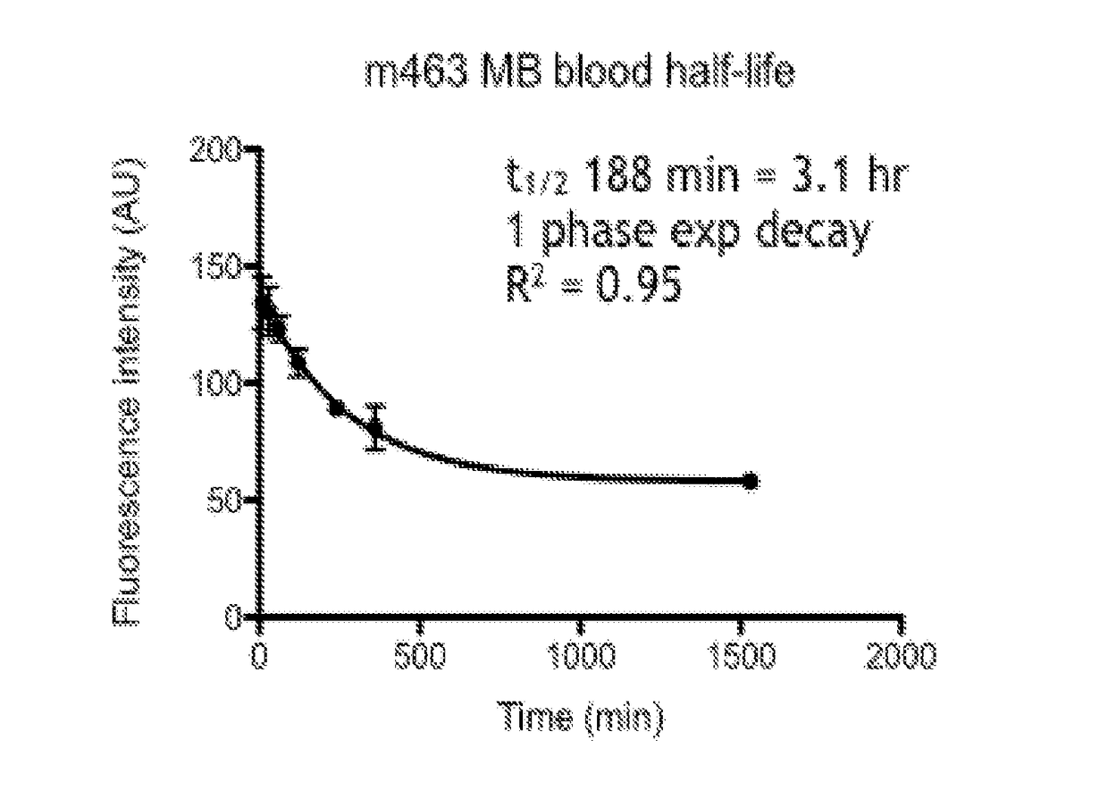
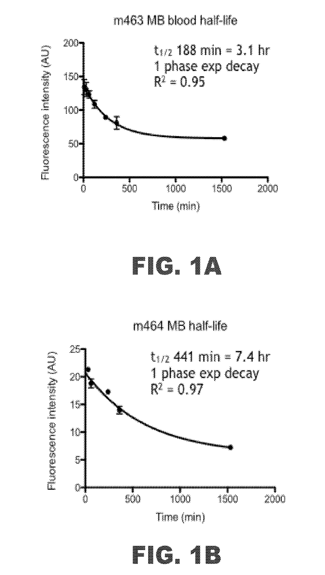
[0081]The methylene blue (MB) binding and uptake was evaluated in a rabbit model of atherosclerosis of the abdominal aorta. The half-life for MB in human bloodstreams has been documented as around 5 to 6.5 hours (Peter, Eur. J. Clin. Pharmacol., 2000 56(3):247-50). The half-life for MB in rabbit bloodstreams was measured in two subjects, m463 and m464 that had been subjected to a high cholesterol diet and the high inflammation protocol. FIG. 1A shows a graph of the fluorescence intensity of MB in the bloodstream of m463 over time. The fluorescence intensity had a 1 phase exponential decay. The half-life was found to be 188 minutes, or about 3 hours. FIG. 1B shows a graph of the fluorescence intensity of MB in the bloodstream of m464 over time. The fluorescence intensity similarly had a 1 phase exponential decay. The half-life was found to be 441 minutes, or about 7.4 hours. The average of the MB half-life in the rabbits tested was around 5.25 hours, which falls within the range foun...


PUM

PropertyMeasurementUnit
Timeaaaaaaaaaa
Timeaaaaaaaaaa
Dimensionless propertyaaaaaaaaaa

Abstract

Methods for detecting the presence of atherosclerotic structures in order to diagnose or prevent atherosclerosis are provided herein. In particular, it has been found that methylene blue injected intravenously acts as an excellent indicator because the compound targets high-risk plaque, atheroma, macrophages, and other atherosclerotic structures formed within the endothelial walls of a vessel of a subject. Because the compound provides a unique binding profile with uptake only in plaque or atheroma, and not the normal or healthy vascular interstitial tissue, methylene blue maintains a good plaque-to-background ratio for imaging purposes. This enables healthcare providers to determine the status of atherosclerosis development in vivo within a patient with higher certainty and at lower costs. The disclosed methods allow for high-resolution mapping of plaque build-up, plaque pathobiology, and other atherosclerotic structures within a vessel of a subject by using methylene blue as an imaging agent.

Description

CROSS-REFERENCES TO RELATED APPLICATIONS[0001]This application claims priority from U.S. Patent Application No. 62 / 129,243 filed Mar. 6, 2015.STATEMENT REGARDING FEDERALLY SPONSORED RESEARCH[0002]Not Applicable.BACKGROUND OF THE INVENTION1. Field of the Invention[0003]The present disclosure relates to a method for the targeted imaging and detection of characteristic structures or molecules indicative of atherosclerosis. In particular, intravenously injected methylene blue or derivatives thereof may be used as imaging agents targeting atheroma or related structures in the vascular system of a subject.2. Description of the Related Art[0004]Atherosclerosis is a vascular or chronic inflammatory disease associated with the development of atherosclerotic plaque or atheroma, made up of macrophages and lipids, within vessel walls. This build-up of lesions in the arteries and veins of a patient may lead to clinical events such as myocardial infarction or stroke. Atheroma in veins or arteries...

Claims


Application Information

Patent Timeline
IPC IPC(8): A61K49/00A61B5/00
CPCA61K49/006A61B5/0059A61K49/003A61B5/004A61K31/54A61K49/0021A61B5/0036
Inventor JAFFER, FAROUC A.MAUSKAPF, ADAMOSBORN, ERICTEARNEY, GUILLERMO J.
Owner THE GENERAL HOSPITAL CORP
Learn More