
Cell therapy with polarized macrophages for tissue regeneration

a cell therapy and macrophage technology, applied in the field of cell therapy with polarized macrophages for tissue regeneration, can solve the problems of limited success of physiotherapy for treating musculoskeletal injuries, inability to accelerate the healing process and reduce scar formation,

Active Publication Date: 2018-03-29
XCELL MEDICAL SOLUTIONS SL
View PDF0 Cites 1 Cited by
  • Summary
  • Abstract
  • Description
  • Claims
  • Application Information

AI Technical Summary

Benefits of technology

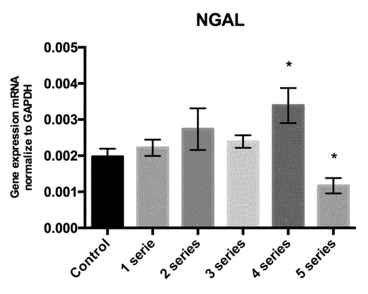
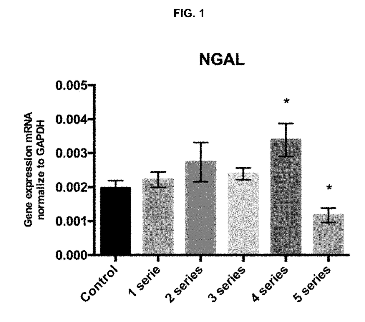
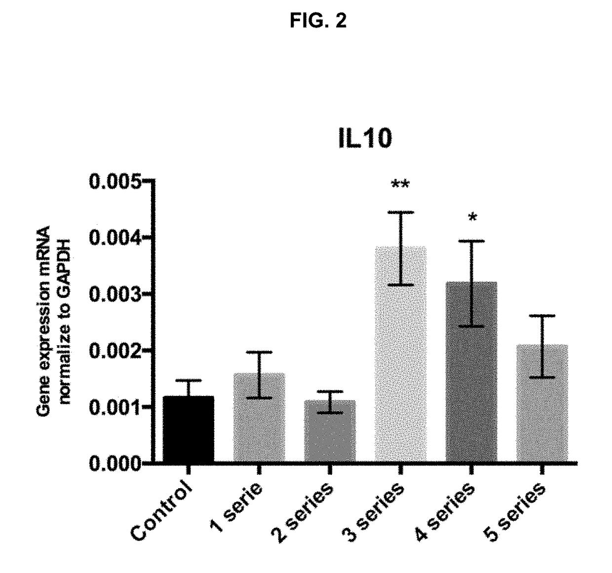
The patent is about a method for culturing and polarizing macrophages to promote tissue regeneration and reduce inflammation. The main advantage of this method is that the macrophages can be autologous, which reduces risks associated with adverse immunological reactions. The method also involves easily obtaining and isolating macrophages from the patient's bloodstream. The resulting M2 macrophages have been shown to promote tissue regeneration, reduce inflammation, and clear fibrosis. The overexpression of IL-10 and NGAL is important for these activities. The patent also describes a device that allows for the recovery of cells suitable for injection in tissue. Overall, this patent provides a technical solution for obtaining and manipulating macrophages for therapeutic purposes.

Problems solved by technology

However, macrophage polarization is a complex process and it has emerged that there is a broad set of signals that induce distinct macrophage phenotypes.
On the other hand, methods based on physiotherapy for treating musculoskeletal injuries, including injuries to muscle, tendon and fascial components, immobilization, and various physiotherapies, have limited success.
The use of drugs to reduce the pain is also commonly used, but none of these methods have been able to accelerate the healing process and reduce scar formation.
The use of platelet rich plasma and stem cells seems to be a promising method for the treatment of these injuries, and several approaches has been developed, but none of these methods has achieved a complete regeneration of the injured tissue.
Furthermore, the use of stem cell therapy has intrinsic risks given the loss of control on the administered cells once they have been implanted in the recipient with potential tumorigenic risk or inappropriate differentiation.
But the effect of this technique has not been previously used in treatment of musculoskeletal injuries in order to accelerate the wound healing and tissue regeneration, reducing scar formation.
None of these devices would be useful for clinical application since the processes are not carried out in aseptic conditions.
In all the systems previously described, the cell cultures are placed and removed from the hypoxic chamber to external environment, and therefore can be easily contaminated.

Method used


Image

  • Cell therapy with polarized macrophages for tissue regeneration
  • Cell therapy with polarized macrophages for tissue regeneration
  • Cell therapy with polarized macrophages for tissue regeneration

Examples


example 1

Effects of Short Periods of Anoxia-Reoxygenation on the Polarization of Peritoneal Macrophages

1.1. Objective.

[0089]The goal was to assess if the exposure of the macrophages to 1, 2, 3, 4 or 5 anoxia-reoxygenation series (3′anoxia—45″ reoxygenation) promotes the polarization of macrophages to an M2 phenotype, with an increase in NGAL and IL10 expression compared to the control subjected to standard incubation conditions.

1.2. Experimental Design.

[0090]a. Control group under oxygen standard conditions (CO2 5% plus atmospheric air).

[0091]Peritoneal macrophages were extracted and isolated from six mice and cultured during 24 h under standard conditions.

[0092]b. Groups subjected to 1, 2, 3, 4 or 5 anoxia (nitrogen 95%; CO2 5%; O2 0%).—reoxygenation series plus 1 h and 30 min of reoxygenation.

[0093]Peritoneal macrophages were extracted and isolated from six mice and cultured during 24 h under standard conditions. Then, macrophages were subjected to 1, 2, 3, 4 or 5 anoxia-reoxygenation seri...

example 2

Description of a Preferred Embodiment of the Device

[0109]In FIG. 3A it can be appreciated a diagram of the proposed device for inducing hypoxia and re-oxygenation conditions on isolated macrophages according to the method previously described. In FIG. 3B there is a perspective view of said device.

[0110]As can be seen in said figure, the essential elements of the device are a removable chamber (1) configured to house the isolated macrophages, a first gas conduction circuit (2) and a second gas conduction circuit (3).

[0111]The first gas conduction circuit (2) comprises a first removable connection (4), which is preferably a stopcock, to the removable chamber (1) and a first gas source (5) and a second gas source (6). The first and second gas sources (5, 6) are connected to an electrovalve (7) which is configured to select the gas source from which the gas passes to the removable chamber (1).

[0112]The first gas conduction circuit (2) can also comprise a press sensor (16) placed between...

example 3

In Vivo Therapy For Treating Injured Tissue

3.1. Experimental Design and Procedure.

[0133]a. Mice without injury (Control)

[0134]b. Mice with an injury not treated

[0135]c. Mice with an injury treated with an in vivo ischemia reperfusion protocol (PREC)

[0136]d. Mice with an injury treated with an in vivo ischemia reperfusion protocol (PREC)+M2 macrophages

[0137]An injury in the mice leg muscle was performed by laceration using a 5 mm diameter biopsy.

After 48 h

[0138]Peritoneal macrophages were extracted and isolated from mice (d) and polarized to an M2 phenotype using the procedure described in example 1.

[0139]The legs of mice (c) and (d) were surrounded with a plastic track above the twin. Once it is well caught, it begins to tighten to make it a restriction of the flow during 3 minutes. After this period the track is released and the blood flow was recovered during 45 seconds. Afterwards the same ischemia reperfusion process was repeated 2 times.

[0140]After the PREC protocol, M2 macroph...


PUM

PropertyMeasurementUnit
timeaaaaaaaaaa
timeaaaaaaaaaa
diameteraaaaaaaaaa

Abstract

The invention provides an in vitro method for inducing macrophage polarization to an M2 phenotype useful for tissue repair. The method described in the present invention comprises the in vitro exposure of macrophages to repeated series of hypoxia-reoxygenation. Activated M2 macrophages obtained by this method overexpress molecules important for tissue remodeling and amelioration of inflammation, such as NGAL and anti-inflammatory cytokines (IL-10). Thus, M2 macrophages obtained by this method are useful as cell therapy for tissue regeneration. The invention also provides pharmaceutical compositions and kits comprising the M2 macrophages obtained by the described method. The invention further refers to a device for inducing hypoxia and re-oxygenation conditions on isolated macrophages according to the described method.

Description

[0001]This invention belongs to the field of cell therapies for tissue repair. Specifically, it refers to an in vitro method for obtaining macrophages polarized to an M2 phenotype which are suitable for tissue regeneration. It also refers to the M2 macrophage populations obtained by this method and to medicaments and pharmaceutical compositions comprising the same for their use in cell therapy in the regeneration of damaged, injured or impaired tissue.BACKGROUND ART[0002]Macrophages are derived from circulating monocytes that exit the vasculature and invade into the surrounding tissues where they differentiate under the influence of local signals into resident tissue macrophages. Resident macrophages have a variety of roles; they patrol tissues for damaged or apoptotic cells, which they clear by phagocytosis, they identify and eliminate such invading pathogens as bacteria, fungi, and virally infected cells, they scavenge lipoproteins, and they are also responsible for regulating tis...

Claims


Application Information

Patent Timeline
Patent Type & Authority Applications(United States)
IPC IPC(8): C12N5/0786A61K35/15C12M1/00C12M1/04
CPCC12N5/0645A61K35/15C12M1/005C12M1/045C12N2506/115C12N2500/02A61P43/00A61K39/4622A61K39/4614A61K39/464
Inventor HOTTER CORRIPIO, MARIA GEORGINASOLA MART NEZ, ANA MARIAMART N CORDERO, JORGE VICENTEGARC A DE LA RIVA MESTRE, PABLODOM NGUEZ S NCHEZ, RUBENS NCHEZ MORENO, JAIMEGINESTA BUCH, XAVIERRODR GUEZ GARCIA, ANNACASTILLO GARC A, ADRIAN
Owner XCELL MEDICAL SOLUTIONS SL
Learn More