
Method for the Diagnosis of Gaucher's Disease

a gaucher's disease and diagnostic method technology, applied in the field of gaucher's disease diagnostic method, can solve the problems of gaucher's disease not being applicable as a biomarker, gb1 is not applicable, and the specificity and sensitivity of the method are low

Active Publication Date: 2018-09-13
CENTOGENE IP GMBH
View PDF0 Cites 0 Cited by
  • Summary
  • Abstract
  • Description
  • Claims
  • Application Information

AI Technical Summary

Benefits of technology

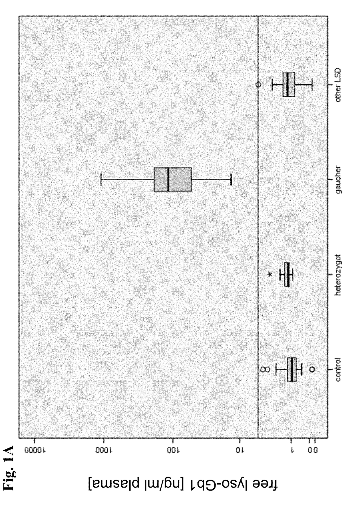
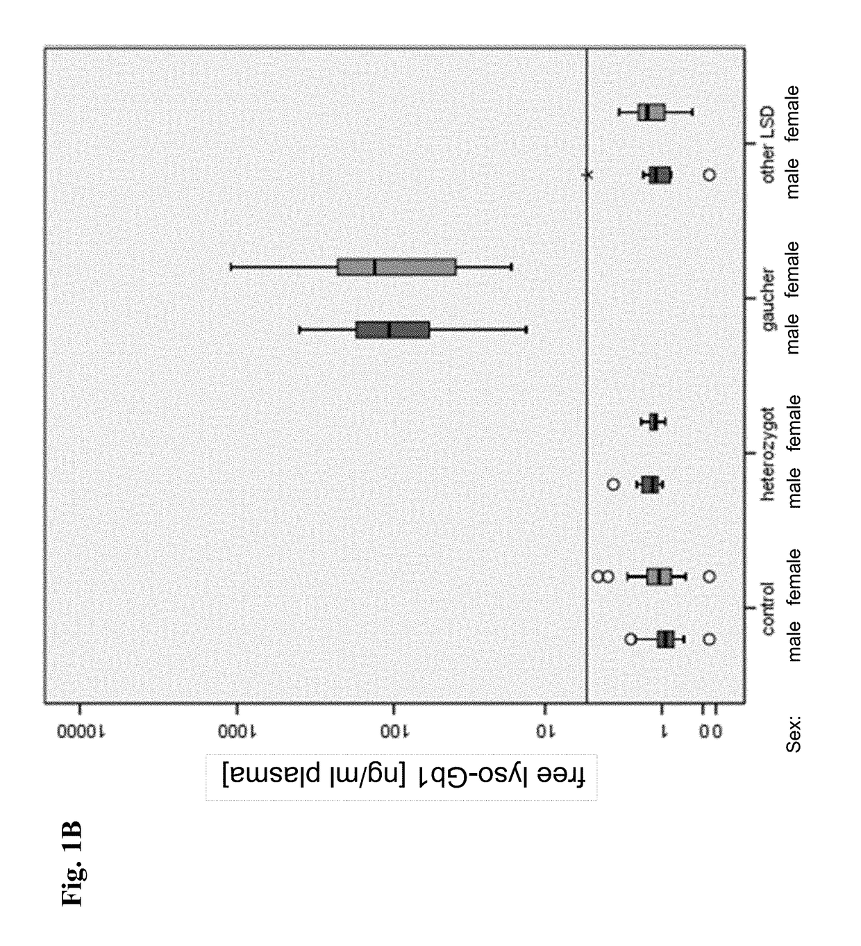
[0157]The present inventors have surprisingly found that free lyso-Gb1 constitutes a biomarker which allows for a method for diagnosing Gaucher's disease in a subject, more specifically diagnosing Gaucher's disease in a subject with high specificity and sensitivity using said free lyso-Gb1 as the biomarker.
[0158]The present inventors have also surprisingly found that free lyso-Gb1, which can be detected by the methods of the present invention, is circulating in the blood of a subject in a concentration of approximately 1 / 1000 of total Gb1. Moreover, the present inventors have surprisingly found that, unlike total Gb1, free lyso-Gb1 which is present in the blood of a subject is useful in a method for diagnosing Gaucher's disease in a subject comprising a step of detecting a biomarker in a sample from the subject, wherein the biomarker is free lyso-Gb1. The present inventors have also surprisingly found that the level of free lyso-Gb1 determined in the sample from a subject by the methods of the present invention allows for diagnosing Gaucher's disease with high sensitivity and high specificity.
[0159]In so far the present invention turns away from the teaching of the state of the art in that the method of the present invention comprises determining the level of a lyso-compound using said lyso-compound as a biomarker for diagnosis of a sphingolipidoses. More specifically, the present inventors have surprisingly found that determining the level of free lyso-Gb1 in a sample from a subject allows for diagnosing Gaucher's disease with high sensitivity and high specificity.
[0168]These subtypes have come under some criticism for not taking account of the full spectrum of observable symptoms. There are also compound heterozygous variations which considerably increase the complexity of predicting disease course.
[0172]ERT is based on chronic intravenous administration of a recombinant glucocerebrosidase (imiglucerase, Genzyme; velaglucerase, Shire; taliglucerase, Protalix) (G. A. Grabowski et al., Enzyme therapy in type I Gaucher's disease: comparative efficacy of mannose-terminated glucocerebrosidase from natural and recombinant sources, Ann. Intern. Med. 122 (1995) 33-39.). For type I and most type III patients, ERT with intravenous recombinant glucocerebrosidase (such as, e.g., imiglucerase) can significantly reduce liver and spleen size, reduce skeletal abnormalities, and reverse other manifestations.
[0174]Both ERT and SRT generally result in marked clinical improvements such as reduction in hepatosplenomegaly, corrections in hematological abnormalities, stabilization or improvement in skeletal deterioration.

Problems solved by technology

Lysosomal storage diseases affect mostly children and they often die at a young and unpredictable age, many within a few months or years of birth.
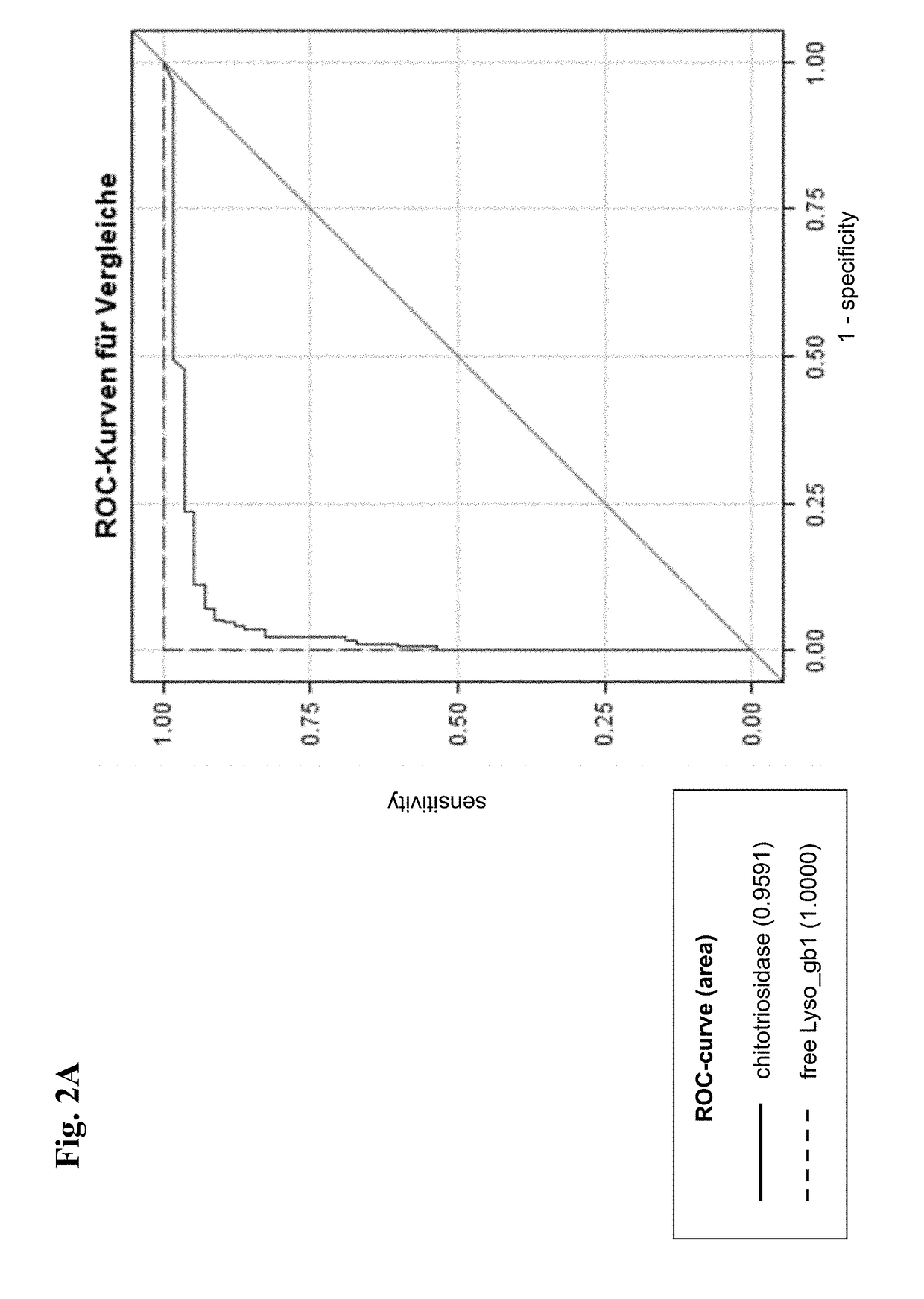
However, beside other disadvantages in the use of chitotriosidase as a biomarker for Gaucher's disease, said enzyme accumulates independent of a direct link to the pathology of Gaucher's disease.
Furthermore, up to 35% of given ethnicities demonstrate a defect of the gene coding for chitotriosidase resulting in an artificially reduced or non-measurable chitotriosidase activity.
Nevertheless, although Gb1 measured in said study was increased in plasma of said patients, said increase of Gb1 was not prominent and thus the specificity and the sensitivity of the method were low showing that Gb1 is not applicable as a biomarker for Gaucher's disease.
Thus, said lyso-compounds might not be suitable biomarkers for diagnosis of sphingolipidoses such as Gaucher's disease.
It is important to note that until today no use of a highly specific and highly sensitive biomarker and no method for the diagnosis of Gaucher's disease is available beside the methods described above, that exhibit an unsatisfactory limit of detection, sensitivity and / or specificity and thus proved to be unsuitable for clinical application.
This is because macrophages that clear these cells are unable to eliminate the waste product, which accumulates in fibrils, and turn into so called Gaucher cells, which appear on light microscopy to resemble crumpled-up paper.
Symptoms may begin early in life or in adulthood and include enlarged liver and grossly enlarged spleen (together hepatosplenomegaly); the spleen can rupture and cause additional complications.
Diseased subjects in this group usually bruise easily (due to low levels of platelets) and experience fatigue due to low numbers of red blood cells.
Symptoms include an enlarged liver and spleen, extensive and progressive brain damage, eye movement disorders, spasticity, seizures, limb rigidity, and a poor ability to suck and swallow.
These subtypes have come under some criticism for not taking account of the full spectrum of observable symptoms.
There are also compound heterozygous variations which considerably increase the complexity of predicting disease course.
Persons affected most seriously may also be more susceptible to infection.
It is important to mention that this procedure carries significant risk and is rarely performed in Gaucher's disease patients.
There is currently no effective treatment for the severe brain damage that may occur in patients with types II and III Gaucher's disease.
Although total Gb1 measured as lyso-Gb1 in said study of the prior art was increased in plasma of said patients, said increase in total Gb1 was not prominent and thus the specificity and the sensitivity of the method were low showing that Gb1 is not suitable as a biomarker for Gaucher's disease.
(Groener et al., supra) is prejudicial compared to the methods of the present invention in that the specificity and sensitivity of said method of the prior art is lower and diagnosing of Gaucher's disease based on such method of the prior art using total Gb1 rather than free lyso-Gb1 is not suitable for reliable clinical application thereof, i.e. the method has no sensitivity and specificity sufficient to diagnose Gaucher's disease by a reliable statistically secured prediction.
Additionally, after treatment the level of chitotriosidase changes extremely slowly making chitotriosidase unsuitable for assessing quickly the efficacy of treatment to which the patient is or has been subjected as well as a relapse of the disease independent form the cause of the disease.
Furthermore, the use of plasma chitotriosidase as a Gaucher cell marker is hampered by the fact that patients, including those with Gaucher's disease, may be deficient in chitotriosidase activity due to a 24-base pair (bp) duplication in the chitotriosidase gene.
Obviously these individuals cannot be monitored by the measurement of plasma chitotriosidase activity (Hollak et al.
Furthermore, in these cases the marker cannot further be used as a follow-up marker for monitoring, e.g., ERT.
The person skilled in the art will acknowledge that Gb1 present in a sample from a subject which is subjected to a step of deacetylation, e.g. by hydrolysis in methanolic sodium hydroxide, will result in cleavage of the fatty acid moiety from the Gb1 and thus will undesirably result in a chemically converted, transformed or derivatized form of Gb1 which cannot be differentiated from free lyso-Gb1.
Conversely, the closer the curve comes to the 45-degree diagonal of the ROC graph, the less accurate the test.
A skilled person will immediately understand that a high limit of detection, e.g. higher than an ideal cut-off level would possibly result in a low sensitivity of the method since the percentage of true positives that are predicted by a test to be positive also depends on whether a level of the biomarker may be determined for said true positives.
In connection therewith is important to note that a level of chitotriosidase from 200 to 1000 nmolMU / h / ml may also be found in a sample from a subject suffering from another LSD such as Niemann-Pick type C or Krabbe's disease, thereby rendering the use of chitotriosidase for diagnosing Gaucher's disease unsuitable.

Method used


Image

  • Method for the Diagnosis of Gaucher's Disease
  • Method for the Diagnosis of Gaucher's Disease
  • Method for the Diagnosis of Gaucher's Disease

Examples


example 1

r the Detection of Free Lyso-Gb1 in Human Serum

[0258]Equipment

[0259]For detecting free lysoGb-1 in a sample of plasma from a subject the following equipment was used.

Apparatus / Piece ofEquipmentType / ProducerHPLC pumpSeries 200, Perkin Elmer, USASample injectorSeries 200, Perkin Elmer, USAColumn ovenSeries 200, Perkin Elmer, USAMass selective detectorAPI 4000 Q TRAP, AB SCIEX, USA / CanadaMulti-tube vortexerHenry Troemner LLC, USADVX-2500Vortex mixerVortex Genie 2; Scientific Industries, USACentrifugeMegafuge 1.0; Heraeus, GermanyMultipette(s), pipette(s)Eppendorf, GermanyWater bathSW21-C, Julabo, Germany

[0260]Reagents

[0261]For detecting free lysoGb-1 in a sample of plasma from a subject the following reagents were used.

[0262]To that extent that values depend on temperature (e.g. the pH value) such values were determined at a temperature of 25° C.

ReagentPurityAcetonitrile (ACN)HPLC-grade or Gradient gradeAcetone99.5%Dimethylsulfoxide (DMSO)HPLC gradeEthanol (EtOH)p.a., 96%Formic acid (F...

example 2

esting and Classification of Study Participants

[0303]After consenting of patients to participation in the study, patients were subjected to a genetic testing for mutations of the glucocerebrosidase gene. Accordingly, 5 to 10 ml of EDTA blood were sequenced according to Seeman et al. (Seeman et al., 1995). Were appropriate other genes beside the glucocerebrosidase gene were sequenced in addition, particularly in controls. Furthermore the chitotriosidase gene was sequenced for detection of the 24 bp duplication as mentioned above. Said genetic testing was controlled using test samples of age and sex matched control patients.

253 subjects were tested.

[0304]According to the result of the above described genetic testing, patients participating in the study were classified into the following groups:

1.) Patients having Gaucher's disease: gold standard for the diagnosis was the detection of two pathogenic mutations within the glucocerebrosidase gene, either homozygous or compound heterozygou...

example 3

of Gaucher's Disease Using Free Lyso-Gb1 as a Biomarker

[0311]The protocols described in Example 1 above were used to generate HPLC-mass spectrometric chromatograms of 485 blood samples derived from the 253 subjects. Exemplary HPLC-mass spectrometric chromatograms displaying peak intensity of free lyso-Gb1 and IS of three samples from two Gaucher's disease patients and one healthy control person are depicted in FIG. 5A, FIG. 5B and FIG. 5C.

[0312]Gold standard for the classification of patients into the group “Gaucher”, was based on the sequencing of the entire coding area as well as the the intron-exon-boundaries of the glucocerebrosidase gene according to the genetic testing as described in Example 2 resulting in the detection of either a homozygous mutation or a compound heterozygosity.

[0313]The results of a determination of the levels of Chitotriosidase or CCL18 in samples from patients were available in 58 or 44 Gaucher's disease patients, respectively. Said results were obtained...


PUM

PropertyMeasurementUnit
diameteraaaaaaaaaa
timeaaaaaaaaaa
temperatureaaaaaaaaaa

Abstract

The present invention is related to an in vitro method for diagnosing Gaucher's disease in a subject comprising a step ofa) detecting a biomarker in a sample from the subject, wherein the biomarker is free lyso-Gb1.

Description

FIELD OF THE INVENTION[0001]The present invention is related to a method for diagnosing Gaucher's disease in a subject, a method for determining the course of Gaucher's disease in a subject, a method for determining the effectiveness of at least one treatment applied to a subject being positively tested for suffering from or being at risk for developing Gaucher's disease, a method of determining the effectiveness of a compound for the treatment of Gaucher's disease, use of mass spectrometry for the detection of a biomarker, use of a biomarker for Gaucher's disease, a kit for determining the presence of a biomarker in a sample from a subject and a software product, wherein the biomarker is free lyso-Gb1.BACKGROUND OF THE INVENTION[0002]Lysosomal storage diseases, also referred to herein as lysosomal storage disorders or LSDs, are a group of rare inherited metabolic disorders that result from defects in lysosomal function. LSDs result when a specific organelle in the body's cells—the ...

Claims


Application Information

Patent Timeline
Patent Type & Authority Applications(United States)
IPC IPC(8): G01N33/68G01N33/92
CPCG01N33/92G01N33/6893G01N2405/10G01N2800/044G01N33/50C12N9/2442
Inventor ROLFS, ARNDTMASCHER, HERMANN
Owner CENTOGENE IP GMBH
Learn More