Method for separating the fraction bound to glycosaminoglycans and applications thereof

Active Publication Date: 2018-11-15
SERVIZO GALEGO DE SAUDESERGAS +3
View PDF0 Cites 1 Cited by
  • Summary
  • Abstract
  • Description
  • Claims
  • Application Information

AI Technical Summary

Benefits of technology

[0013]The authors of the present invention have developed a new method for separating the fraction bound to sulfated glycosaminoglycans (GAGs) in biological samples. This method allows studying alterations in the glycosylation of GAGs in a manner that is simple, quick, and cost-effective and requires a very small amount of sample (microliters). This

Problems solved by technology

Although interferences in measurements due to other molecules that were invariably purified together with GAGs have already been descr

Method used


Image

  • Method for separating the fraction bound to glycosaminoglycans and applications thereof
  • Method for separating the fraction bound to glycosaminoglycans and applications thereof
  • Method for separating the fraction bound to glycosaminoglycans and applications thereof

Examples


example 1

on of Sample Processing Before the Isolation of the Fraction Associated with Glycosaminoglycans and / or Exosomes

[0409]In the case of urine, the process started with the second morning urine (discarding the first stream to prevent contaminations of the external genitourinary system) collected in protease-free containers, and urine with the visible presence of hematuria and urinary tract infection were discarded. The urine samples must be stored at −20° C. until use if they are not used on the same day.

[0410]In the case of blood samples, they must be collected in a suitable tube depending on whether serum or plasma is to be obtained. In the case of plasma, the blood sample must be collected in a tube with anticoagulant, for example heparin or EDTA, and centrifuged at a maximum speed for 10 minutes. In the case of serum, the sample must be collected in an STII or biochemical tube, left to sit for at least 30 minutes, and centrifuged at a maximum speed for 10 minutes. Both the plasma and...

example 2

on of the Exosome Isolation Method

[0411]The samples were processed following a modification of the method that is well established in the literature (Christianson et al., Proc Natl Acad Sci USA 2013, 110:17380-5; Hogan et al., J Am Soc Nephrol 2009, 20:278-288). In the case of urine, the method started with the urine supernatant obtained by means of centrifuging urine at 1000 g for 5 minutes. Briefly, the urine supernatants were centrifuged at 5,000 g for 20 minutes, subjected to filtration through low protein adsorption filters having a pore size of 0.22 μm, and ultracentrifuged at 100,000 g for 2 hours. The exosome pellets were resuspended in a suitable volume of buffer (for example, PBS 1×) and kept at −20° C. until use.

example 3

on of the Separation Method for Separating the Fraction Bound to Glycosaminoglycans

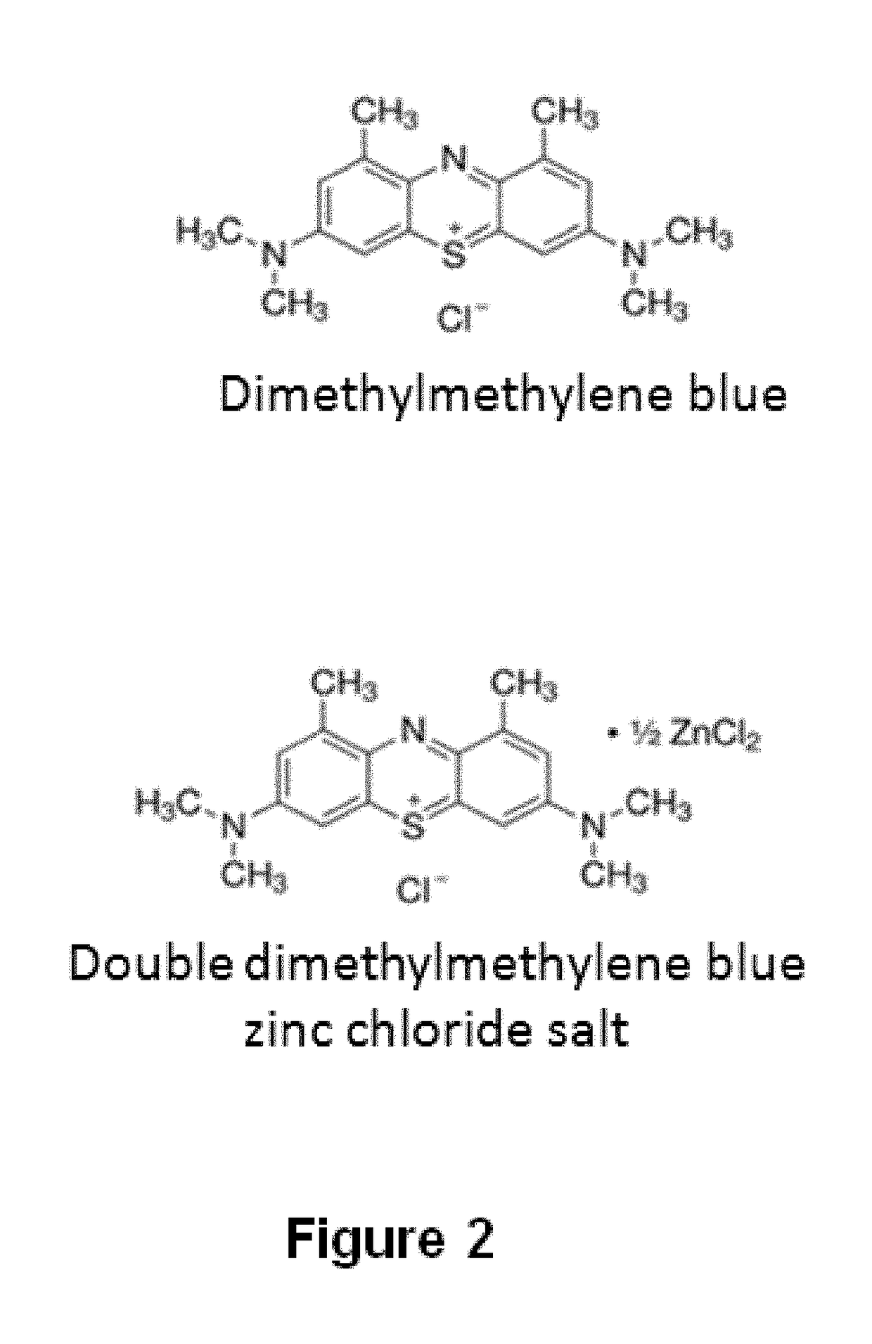
[0412]Dimethylmethylene blue (Serva) at a concentration of 0.29 mM dissolved in ethanol was used, and 0.2 M formate buffer pH 3.5 was added at a 1:99 ratio. It was then mixed with the biological sample at a 1:2 ratio. It was subsequently incubated for 15 minutes at room temperature. Finally, it was centrifuged at 10,000 g for 10 minutes at 4° C. The supernatant was removed and the precipitate containing the fraction bound to glycosaminoglycans was recovered.


PUM

PropertyMeasurementUnit
Temperatureaaaaaaaaaa
Molar densityaaaaaaaaaa

Abstract

The present invention is comprised in the field of glycobiology. In particular, it relates to a method for separating, in biological samples, the fraction bound to or associated with sulfated glycosaminoglycans (GAGs), and the applications thereof in biomedicine, such as for identifying the profile of glycoproteins or the profile of lipids bound to or associated with sulfated GAGs, detecting an alteration in the pattern of glycosylation by sulfated GAGs, identifying biomarkers for the diagnosis, for the prognosis, for monitoring the progression of a disease or of the effect of a therapy, or for identifying compounds suitable for the treatment of a disease. The invention also relates to methods for diagnosing mucopolysaccharidosis and for diagnosing and determining the prognosis of a kidney disease.

Description

FIELD OF THE INVENTION[0001]The present invention is comprised in the field of glycobiology. In particular, it relates to a method for separating, in biological samples, the fraction bound to or associated with sulfated glycosaminoglycans and the applications thereof in biomedicine.BACKGROUND OF THE INVENTION[0002]Glycosaminoglycans (GAGs) are large unbranched polysaccharides formed by repeating disaccharide sequences, in which one of the components is always an amino sugar (D-galactosamine or D-glucosamine) and the other component is a uronic acid, such as L-glucuronic or L-iduronic, with the exception of keratan sulfate. Except for hyaluronic acid, all GAGs have sulfate groups, such as O-esters or N-sulfate.[0003]GAGs are part of proteoglycans and lipids. Proteoglycans are a specific type of glycoproteins having at least one GAG chain bound to the protein, and they are classified based on the GAG chain present.[0004]The glycosylation of molecules is a post-translational enzymatic ...

Claims


Application Information

Patent Timeline
IPC IPC(8): G01N33/68G01N33/58G01N33/50
CPCG01N33/6839G01N33/585G01N33/5091G01N2400/40G01N2800/04G01N2800/52G01N2800/50G01N33/6893G01N33/92G01N2800/347G01N2800/042G01N2800/56G01N2333/765G01N2333/47G01N33/53
Inventor ALONSO SAMPEDRO, MANUELALVAREZ GONZ LEZ, VICTORCOLON MEJERAS, CRISTOBALGARC A GONZ LEZ, MIGUEL A.LAMAS GONZ LEZ, OLAYA
Owner SERVIZO GALEGO DE SAUDESERGAS
Why Eureka
Try Eureka