
Rod-shaped plant virus nanoparticles as imaging agent platforms

Active Publication Date: 2019-01-31
CASE WESTERN RESERVE UNIV
View PDF0 Cites 0 Cited by
  • Summary
  • Abstract
  • Description
  • Claims
  • Application Information

AI Technical Summary

Benefits of technology

This patent describes the use of tobacco mosaic virus (TMV) as a scaffold to display para magnetic resonance imaging (MRI) contrast agents. TMV can form spherical nanoparticles that can be tailored in size. The inventors have loaded TMV nanorods and spheres with contrast agents and have shown that they can undergo thermal re-shaping to form high-relaxivity nanospheres. They have also used TMV to target and image atherosclerotic plaques in vivo using a dual-modal imaging platform. The technical effects of this patent include the creation of a unique platform to study rod-shaped and spherical nanomaterials side-by-side, the efficient loading of cargos, and the development of a novel class of MRI contrast agents with high relaxivities.

Problems solved by technology

However, they have not previously been used as a drug delivery vehicle or contrast agent.

Method used


Image

  • Rod-shaped plant virus nanoparticles as imaging agent platforms
  • Rod-shaped plant virus nanoparticles as imaging agent platforms
  • Rod-shaped plant virus nanoparticles as imaging agent platforms

Examples


example 1

osaic Virus Rods and Spheres as Supramolecular High-Relaxivity MRI Contrast Agents

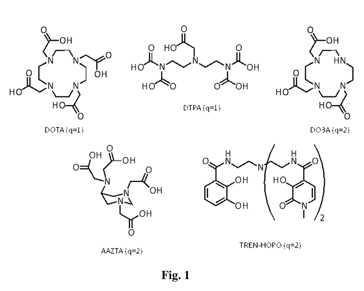
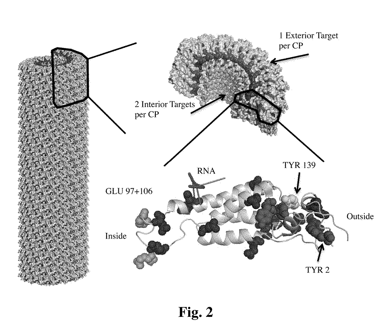
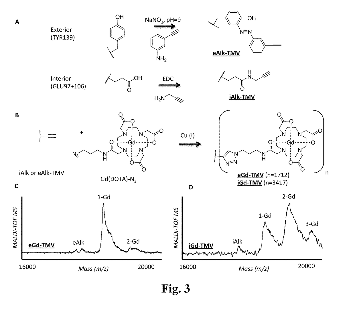
[0073]To compensate for the low sensitivity of magnetic resonance imaging (MRI), nanoparticles have been developed to deliver high payloads of contrast agents to sites of disease. The inventors have developed supramolecular MRI contrast agents using the plant viral nanoparticle tobacco mosaic virus (TMV). Rod-shaped TMV nanoparticles measuring 300×18 nm were loaded with up to 3,500 or 2,000 chelated paramagnetic gadolinium (III) ions selectively at the interior (iGd-TMV) or exterior (eGd-TMV) surface, respectively. Spatial control is achieved through targeting either tyrosine or carboxylic acid side chains on the solvent exposed exterior or interior TMV surface. The ionic T1 relaxivity per Gd ion (at 60 MHz) increases from 4.9 mM−1s−1 for free Gd(DOTA) to 18.4 mM−1s−1 for eGd-TMV and 10.7 mM−1s−1 for iGd-TMV. This equates to T1 values of ˜30,000 mM−1s−1 and ˜35,000 mM−1s−1 per eGd-TMV and iGd-TMV nanop...

example 2

l MRI and Fluorescence Imaging of Atherosclerotic Plaques In Vivo Using VCAM Targeted Tobacco Mosaic Virus

[0111]The nanoparticles formed by plant viruses are emerging tools for molecular imaging in medicine. Here, the rod-shaped tobacco mosaic virus was used to target and image atherosclerotic plaques in vivo. TMV was loaded with magnetic resonance (MR) and fluorescence contrast agents to provide a dual-modal imaging platform. Targeting to atherosclerotic plaques was achieved with vascular cell adhesion molecule (VCAM) receptors present on activated endothelial cells. Dual, molecular imaging was confirmed using a mouse model of atherosclerosis.

Methods

[0112]Isolation of TMV. TMV particles were isolated from Nicotiana benthamiana or N. rusitca plants using a previously established protocol. Boedtker H, Simmons N S. JACS, 80:2550-6 (1958). The TMV concentration was determined based on UV-Vis absorbance at 260 nm with an extinction coefficient of 3.0 mL mg−1 cm−1.

[0113]Bioconjugation of...


PUM


Abstract

A rod-shaped plant virus having an interior surface and an exterior surface, and at least one imaging agent that is linked to the interior and / or exterior surface is described. The rod-shaped viruses can be combined into larger spherical nanoparticles. A rod-shaped plant virus or spherical nanoparticles including an imaging agent can be used in a method of generating an image of a tissue region of a subject such as a tumor or atherosclerotic tissue by administering the virus particle to the subject and generating an image of the tissue region of the subject to which the virus particle has been distributed.

Description

CROSS-REFERENCE TO RELATED APPLICATIONS[0001]This application claims priority to U.S. Provisional Patent Application Ser. No. 61 / 711,492, filed Oct. 9, 2012, which is incorporated herein by reference.GOVERNMENT FUNDING[0002]The present invention was supported by NIH / NIBIB grant P30 EB011317 (to NFS)), Mt. Sinai Foundation (to NSF), and NIH / NHLBI training grant T32 HL105338. The Government has certain rights in this invention.BACKGROUND[0003]Magnetic resonance imaging (MRI) is an emerging technology used for detection of disease and for follow-up diagnosis after surgery or treatment. While MRI shows great potential because of its high spatial resolution, deep soft tissue contrast, and use of non-ionizing radiation, its low sensitivity remains a drawback. To overcome this shortcoming, paramagnetic contrast agents, such as Magnevist® (an FDA approved chelated gadolinium reagent), can be used to enhance the detection sensitivity of MRI. The conjugation of contrast agents to a macromolec...

Claims


Application Information

Patent Timeline
IPC IPC(8): A61K49/18C12N7/00
CPCC12N7/00A61K49/1896A61K49/1884C12N2770/00031
Inventor STEINMETZ, NICOLEBRUCKMAN, MICHAEL
Owner CASE WESTERN RESERVE UNIV
Learn More