Implantable medical devices for optogenetics

a technology of optogenetics and medical devices, applied in the field of optogenetics, can solve problems such as heightened risk of adverse events, and achieve the effect of precise monitoring and control of biologic function

Active Publication Date: 2019-02-21
THE BOARD OF TRUSTEES OF THE UNIV OF ILLINOIS +1
View PDF1 Cites 11 Cited by
  • Summary
  • Abstract
  • Description
  • Claims
  • Application Information

AI Technical Summary

Benefits of technology

[0005]Provided herein are electronic devices specially configured for implantation, injection or surface mount into or onto various soft tissues, such as biological soft tissues in living animals. Ultrathin and mechanically compliant electronic device components, for example, permit access to the interior of living tissues without unduly impacting biologic function. Because the physical devices provided herein are minimally invasive, they can be used even for long-term implantation to interface with tissue that is not normally physically accessible. For example, devices provided herein may be injected into a tissue with an attendant impact that is no more than that caused by a micro-needle. Furthermore, the electronic devices can be sized to a cellular and sub-cellular scale, thereby providing precise monitoring and control of biologic function on a cell-by-cell basis. This provides unique and technologically sophisticated applications that are not achieved with conventional systems that are confined to tissue surfaces.

Problems solved by technology

For example, tissue trauma is associated with a robust immune response along with heightened risk of adverse events ranging from device rejection requiring device removal, to thrombi, lesions and the like that can affect the tissue.

Method used


Image

  • Implantable medical devices for optogenetics
  • Implantable medical devices for optogenetics
  • Implantable medical devices for optogenetics

Examples


example 1

t, Fully Implantable Optoelectronics Technology with Near-Field Wireless Operation for Broad Application in Optogenetics

[0224]Abstract

[0225]Optogenetics, which enables light-induced, area-confined stimulation or inhibition of genetically targeted neurons, is now an essential tool for modern research in neuroscience. Implantable optoelectronic technologies offer the most powerful and versatile capabilities in optogenetics experiments, but their specialized designs are not easily adapted to the type of mass manufacturing approaches that are needed for distribution to the broader community. Furthermore, the associated radio frequency schemes for power delivery and control depend sensitively on details associated with the sizes, shapes and positions of metallic components and / or water features in and around the experimental environment. In this example, we present a fully implantable device platform compatible with near field communication hardware, similar to that used for RFID tags an...

example 2

e and Implantable Photometry Devices

[0266]This example relates to the development of a robust, minimally invasive wireless photometry system for in vivo calcium measurements in freely moving behavior. To achieve this, a miniaturized, wireless, ‘injectable’ photometry platform (˜300 mm wide, ˜100 mm thick and several mm long) that enables quantitative measurements of fluorescence stimulated using a high performance microscale inorganic light emitting diode (μ-ILED) and captured using a co-located, sensitive microscale inorganic photodetector (μ-IPD) is described. These devices directly address current limitations in measuring calcium transient activity within any environment and facilitate sensing of genetically defined neural networks in more ethologically relevant behaviors. These small-scale device components will mount on thin, flexible filaments with overall dimensions significantly smaller than fiber optic cables. The resulting systems will greatly reduce motion artifacts, due ...

example 3

aracterization

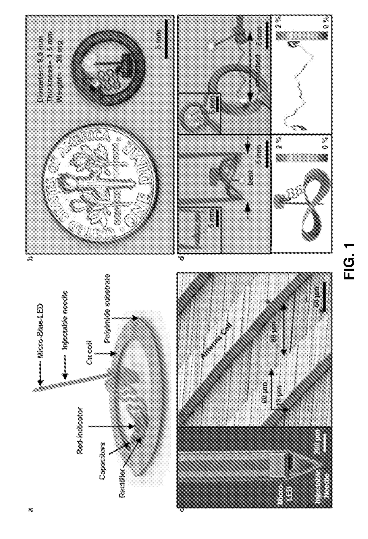
[0308]FIGS. 32-55 further illustrate the biocompatibility of the devices and in vivo operation. For example, FIG. 32 illustrates a wireless photometry system for deep brain Ca2+ measurement. Panel (a) is a schemetic exploded-view illustration of a wireless, injectable, ultrathin photometry probe with a μ-ILED and a μ-IPD at the tip end. (b) Optical micrograph of the injectable photometry probe. The tip has a total width of ˜350 μm and a thickness of ˜150 μm. The weight is 29 mg. (Inset) Magnified colorized SEM image of the tip (orange: polyimide; yellow: interconnection; blue: μ-ILED; green: μ-IPD with an optical filter). Scale bar, 200 μm. (c) Schematic illustration of a GaAs μ-IPD. (Bottom) SEM image of a representative μ-IPD (lateral dimensions of 100×100 mm2, and thickness of 5 μm). Metal electrodes are colorized in yellow. Scale bar, 50 μm. (d) Schemetic exploded-view illustration of a transponder. (e) Photographic image of the wireless detachable transponder on f...


PUM


Abstract

Provided are implantable biomedical devices and related methods for interfacing with a target tissue. The devices comprise a substrate, an electronic device supported by the substrate and a freely positionable injectable needle electronically connected to the electronic device by a deformable interconnect, where the injectable needle has one or more optical sources provided on a distal tip end. The injectable needle may further comprise a photodetector.

Description

CROSS-REFERENCE TO RELATED APPLICATIONS[0001]This application claims the benefit of and priority to U.S. provisional patent app. No. 62 / 316,708, filed Apr. 1, 2016, which is incorporated by reference herein in its entirety, except to the extent inconsistent herewith.STATEMENT REGARDING FEDERALLY SPONSORED RESEARCH OR DEVELOPMENT[0002]This invention was made with government support under TR01 NS081707 and EUREKA DA037152 awarded by the National Institutes of Health. The government has certain rights in the invention.BACKGROUND OF INVENTION[0003]Provided herein are devices for implantation into biological tissues and related methods. The devices and methods utilize electronic devices arranged in ultrathin functional layers, along with stacking of those functional layers in a special geometric configuration to achieve device implantation that is minimally invasive while providing the ability to interface with tissues on a cellular-scale resolution. Minimal disturbance of the tissue mak...

Claims


Application Information

Patent Timeline
Patent Type & Authority Applications(United States)
IPC IPC(8): A61B5/00
CPCA61B5/0084A61B5/0031A61B5/6848A61B5/6868A61B5/0071A61B5/076A61N2005/0662A61N2005/0661A61N5/0601A61N2005/0659A61N5/0622A61B2560/0219A61B17/04
Inventor ROGERS, JOHN A.SHIN, GUNCHULBANKS, ANTHONY R.BRUCHAS, MICHAELGEREAU, ROBERT
Owner THE BOARD OF TRUSTEES OF THE UNIV OF ILLINOIS
Why Eureka
Try Eureka