

Methods of modulating melanosome ph and melanin level in cells
a melanosome and cell technology, applied in the field of modulating melanosome ph and melanin level in cells, can solve the problems of unclear mechanism by which melanocytes regulate melanosome ph and remain elusive signaling pathways that control pigmentation
- Summary
- Abstract
- Description
- Claims
- Application Information
AI Technical Summary
Benefits of technology
Problems solved by technology
Method used
Image

Examples
example 1
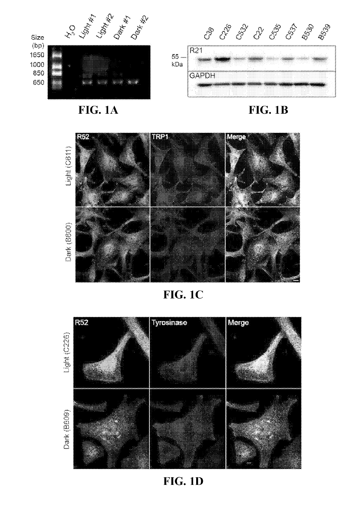
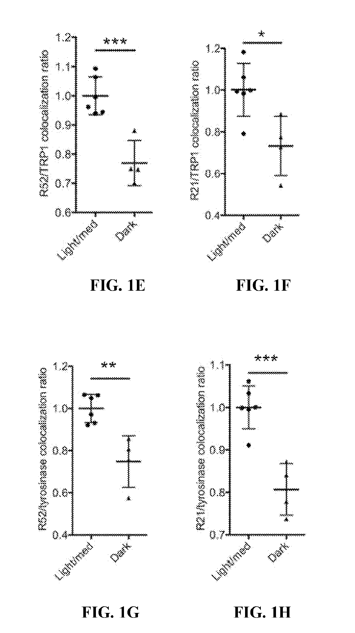
[0124]This example demonstrates that sAC is expressed in human melanocytes and localizes to melanosomes.
[0125]To determine the expression pattern of sAC in melanocytes, sAC expression and localization was determined in isolated human primary melanocytes. Briefly, primary human melanocytes derived from neonatal foreskins were obtained from the Yale Dermatology Cell Culture Facility (New Haven, Conn., USA) and grown in Opti-MEM medium supplemented with 5% fetal bovine serum (FBS), 1% penicillin-streptomycin, 10 ng / ml of fibroblast growth factor-2, 1 ng / ml of heparin, 0.1 μM dibutyryl cAMP (dbcAMP), and 0.1 mM 3-isobutyl-1-methylxanthine (IBMX). Prior to experiments, melanocytes were cultured in“cAMP starvation media” without dbcAMP and without IBMX for 24 hours. The expression of sAC mRNA was measured by RT-PCR using the primers 5′-GAGCCCACCTCCAGGGAAGAAGAGGC-3′ (SEQ ID NO: 2) and 5′-GGAGGAGTCCACTGTGGAACTTGAGG-3′ (SEQ ID NO: 3) which are directed against exons 25 and 29, respectively. ...
example 2
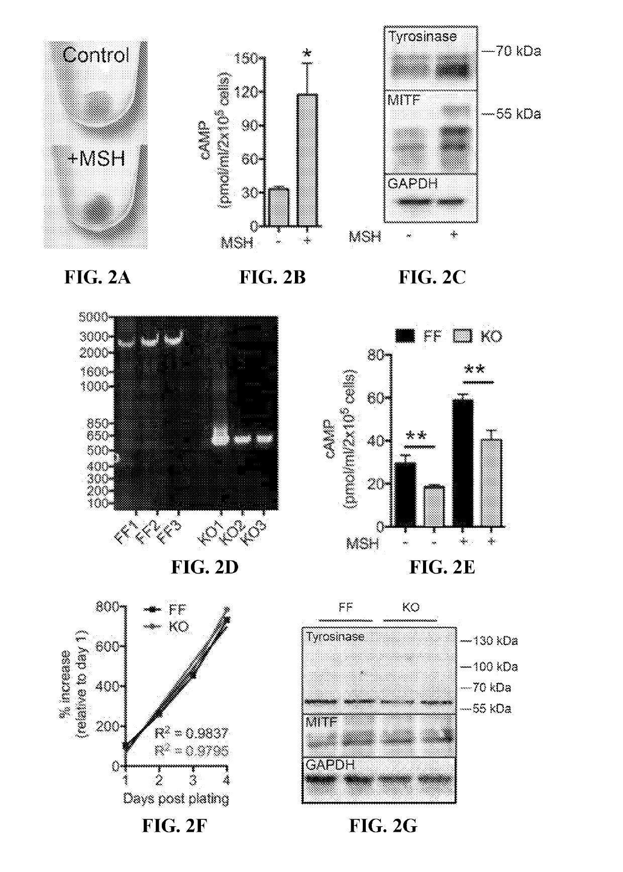
[0130]This example demonstrates that the loss of sAC activity leads to an alkalization of melanosome pH.
[0131]To genetically evaluate the role of sAC in melanosome biology, a strain of mice with three exons encoding the second of two catalytic domains of the ADCY10 gene flanked by loxP sites (ADCY10fl / fl) (Chen et al., Brain Res., 1518: 1-8 (2013); Watson et al. Journal of Experimental Medicine, 212(7): 1021-1041 (2015)) was utilized to generate immortalized mouse melanocytes by serial passage (Tamura et al., In Vitro Cell Dev Biol., 23(7): 519-522 (1987)). Briefly, newborn mice were euthanized and skin was removed from the back, placed in a Petri dish epidermis side up, and incubated in 2.5 ml Dispase in MEMS overnight at 4° C. The next day the dermis was discarded and the epidermis was incubated in trypsin solution until cells became dissociated. Cells were washed to remove the trypsin solution then cultured in TAV medium [Ham's F12 plus glutamine, Penn / Strep, horse serum 7%, feta...
example 3
[0139]This example demonstrates that pharmacologic inhibition of sAC activity leads to an alkalization of melanosome pH.
[0140]The effect of pharmacologic inhibition of sAC was examined using the pH-sensitive vial dye LysoSensor Yellow / Blue DND-160 assay, described above. Briefly, cultured sACFF melanocytes were treated for 4 hours with 30 μM KH7, 30 μM LRE1, or control (untreated). The cells were incubated with 1 μM LysoSensor DND-160 (Invitrogen) for 5 minutes at 37° C. Lysosensor was excited at 405 nm and its emission detected at 417-483 nm (W1) and 490-530 nm (W2). The ratio of emissions (W1 / W2) in Lysosensor-stained puncta was assigned to a pH value based on a calibration curve generated for each experiment using solutions containing 125 mM KCl, 25 mM NaCl, 24 μM monensin, and varying concentrations of MES to adjust the pH to 4, 4.5, 5.0, 5.5, 6.0, 6.5, 7.0, 7.5. The fluorescence ratio was linear for pH 5.0-7.0.
[0141]Similar to the genetic loss of sAC, pharmacologic inhibition o...
PUM
| Property | Measurement | Unit |
|---|---|---|
| pH | aaaaa | aaaaa |
| pH | aaaaa | aaaaa |
| pH | aaaaa | aaaaa |
Abstract
Description
Claims
Application Information