Method and device for non-invasively classifying a tumorous modification of a tissue

a tumorous modification and non-invasive technology, applied in the field of non-invasive imaging modalities for oncologic imaging, can solve the problems of only properly characterized tissue, nephrogenic systemic fibrosis, and nephrogenic fibrosis, and achieve the effects of shortening the examination time, increasing the cost of the imaging method, and reducing the risk of allergic reactions

Pending Publication Date: 2020-06-04
DEUTES KREBSFORSCHUNGSZENT STIFTUNG DES OFFENTLICHEN RECHTS
View PDF1 Cites 1 Cited by
  • Summary
  • Abstract
  • Description
  • Claims
  • Application Information

AI Technical Summary

Benefits of technology

[0062]Long examination times in MRI may significantly increase the cost of the imaging method and shorten the availability to a broader patient audience, in particular, hinder an introduction of such methods and devices into clinical routine. In contrast hereto, the present method and device offer examination times of 0.5 min to 35 min, preferably of 5 min to 15 min, which considerably differ from examination times as known from experimental or scientific studies which, typically, exceed 30 min, 1 hour, or even more. Examination times can further be reduced since conventional imaging protoc

Problems solved by technology

Tissue characterization in the field of oncologic imaging by using non-invasive imaging modalities is still a challenging task.
Using MRI for detecting and characterizing suspicious change of a tissue commonly includes sophisticated examination protocols as well as an intravenous application of a contrast agent, in particular, a compound comprising gadolinium, which may, however, imply risks for allergic reactions, nephrogenic systemic fibrosis, and gadolinium deposition in the brain [2-7].
The

Method used


Image

  • Method and device for non-invasively classifying a tumorous modification of a tissue
  • Method and device for non-invasively classifying a tumorous modification of a tissue
  • Method and device for non-invasively classifying a tumorous modification of a tissue

Examples


Embodiment Construction

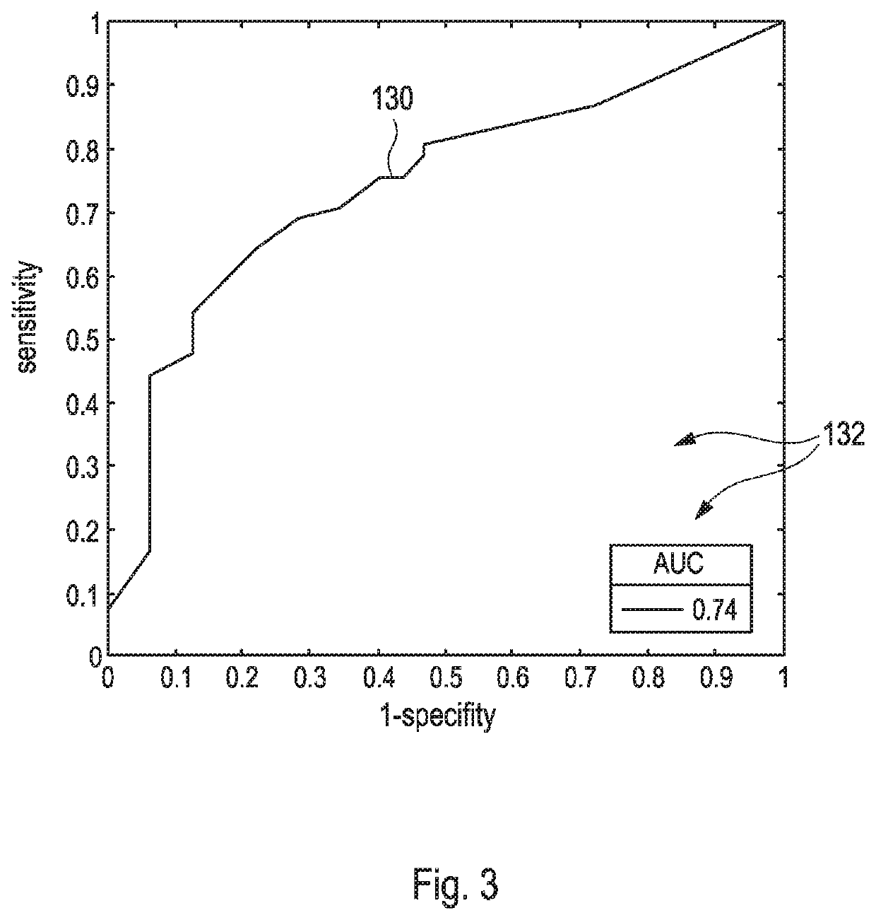
[0071]For comparison purposes, FIGS. 1 to 3 demonstrate that currently available solitary quantitative parameters based on diffusion weighted imaging (DWI) sequences for differentiating between benign and malignant tissue in magnetic resonance imaging (MRI) only exhibit of a rather limited diagnostic value.

[0072]In particular, FIG. 1 illustrates a scheme 110 which comprises three different receiver operating curves (ROC) 112, 114, 116 for three different types of solitary quantification schemes, wherein each of the solitary quantification schemes was applied for determining microstructural tissue correlates in benign and malignant breast lesions as extracted from diffusion weighted imaging (DWI). Hereby, each of the curves 112, 114, 116 renders values of a sensitivity as depicted versus values of 1-specify, wherein each of the curves 112, 114, 116 represents one of the following different solitary quantification schemes, wherein[0073]the curve 112 refers to ‘diffusional kurtosis ima...


PUM


Abstract

A method for non-invasively classifying a tumorous modification of a tissue according to different stages of the tumorous modification comprises the steps of: a) receiving raw magnetic resonance imaging (MRI) data that has been recorded by applying at least one diffusion weighted imaging (DWI) sequence using three to nine different b-values to a tissue being suspicious to a tumorous modification without application of a contrast agent; b) extracting at least two quantification scheme parameters from the raw MRI data by using at least one quantification scheme, wherein each of the quantification scheme parameters is related to a microstructural property of the tissue; c) applying a weight to each quantification scheme parameter, wherein the weight is dependent on a kind of the tissue and on the quantification scheme, whereby a set of weighted quantification scheme parameters is obtained; d) determining a scoring value by combining the weighted quantification scheme parameters within the set, wherein each of the weighted quantification scheme parameters is used only once for determining the scoring value; and e) classifying the tumorous modification of the tissue into one of at least two classes according to the scoring value. The method and a corresponding classification device are capable of performing non-invasive tissue characterization without contrast agent administration in a highly accurate manner while supplementary information related to conventional imaging properties and clinical information can further increase the high diagnostic accuracy. They are used in their entirety for classifying the tumorous modification of the tissue.

Description

TECHNICAL FIELD OF THE INVENTION[0001]The invention relates to a method and a device for non-invasively classifying a tumorous modification of a tissue, in particular of a human tissue, preferably without an administration of contrast agents or ionizing irradiation. The method and the device according to the present invention specifically may be used in the field of oncologic imaging. However, other applications are possible.RELATED ART[0002]Tissue characterization in the field of oncologic imaging by using non-invasive imaging modalities is still a challenging task. Modalities which are currently available for detecting and characterizing suspicious changes of a tissue include ultrasound, x-ray imaging, computer tomography, positron emission tomography (PET), and magnetic resonance imaging (MRI), whereby MRI is increasingly used in oncologic imaging [1]. Using MRI for detecting and characterizing suspicious change of a tissue commonly includes sophisticated examination protocols as...

Claims


Application Information

Patent Timeline
IPC IPC(8): A61B5/00A61B5/055A61B6/03A61B6/00A61B8/08G06T7/00G16H50/20G16H30/40
CPCA61B5/055A61B5/4312A61B6/032G06T2207/10081A61B8/5261G06T2207/30081G06T2207/30068G06T2207/10104A61B5/4381G16H30/40G16H50/20A61B6/037G06T7/0016G06T2207/10132A61B6/5247G06T2207/30096A61B5/7267A61B5/4331A61B2576/02A61B5/7264G01R33/5608G01R33/56341
Inventor BICKELHAUPT, SEBASTIANKONIG, FRANZISKA
Owner DEUTES KREBSFORSCHUNGSZENT STIFTUNG DES OFFENTLICHEN RECHTS
Why Eureka
Try Eureka