Treatment agent for epidermolysis bullosa
a treatment agent and epidermolysis technology, applied in the field of cell product in regenerative therapy, can solve the problems of limiting the daily living of patients, the risk of complicated fungal or bacterial infections, and the use of ointments containing antibiotics that are not positively and necessarily used except, so as to improve or heal skin symptoms, easy and viable treatment methods, and excellent safety
- Summary
- Abstract
- Description
- Claims
- Application Information
AI Technical Summary
Benefits of technology
Problems solved by technology
Method used
Image
Examples
example 1
n with Full-Thickness Wound Model Mouse
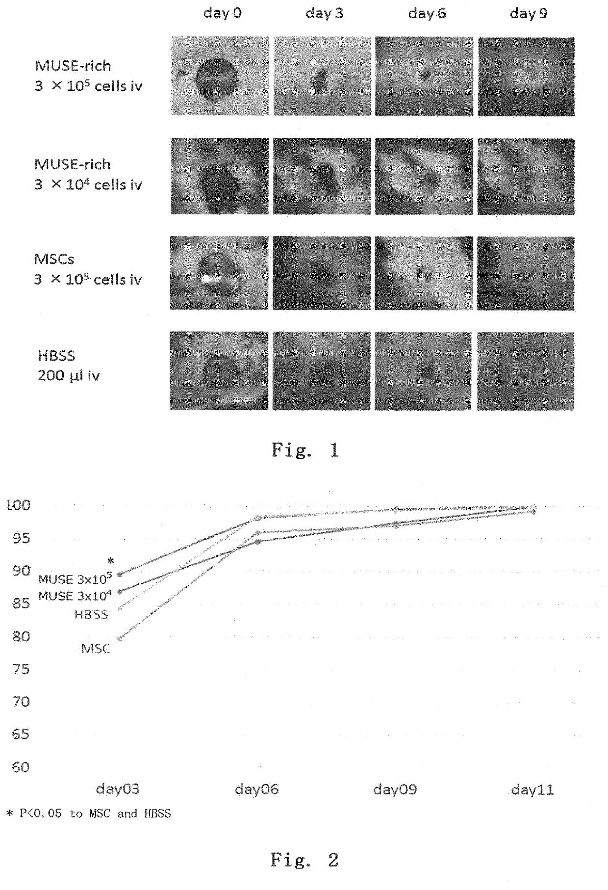
[0107]A full-thickness wound was made on the back of an adult C57BL / 6 mouse, and the mouse was used as a full-thickness wound model. Within 30 minutes after full-thickness wounding, the above-prepared Muse cells (3×10⁵ per mouse or 3×10⁴ per mouse), MSCs (3×10⁵ per mouse), or 200 μl of HBSS was injected into the tail vein, and then the therapeutic effect was investigated. The epithelization rate was calculated as below.
[0108]The back skins at the time of wounding and at day 3, 6, 9, and 11 after wounding were photographed with a digital camera together with a ruler, and then the skin ulcer areas (mm2) were determined using Image J software (version 1.50i). Using the area at the time of wounding as a reference value, the percent area reduction was calculated.
Epithelizationrate(%)=Areaatthetimeofwounding(mm2)-Ulcerareaateachday(mm2)Areaatthetimeofwounding(mm2)×100
[0109]The results are shown in FIGS. 1 and 2. In all groups, the wounds were healed ...
example 2
n with COL17-Knockout Epidermolysis Bullosa Model Mouse
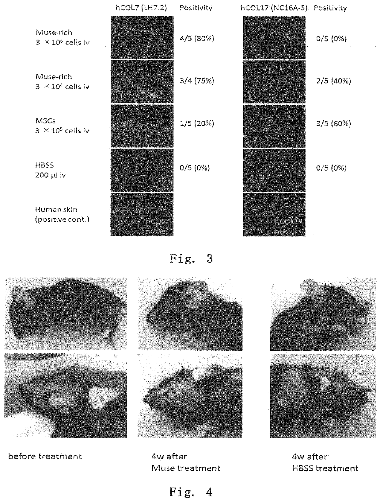
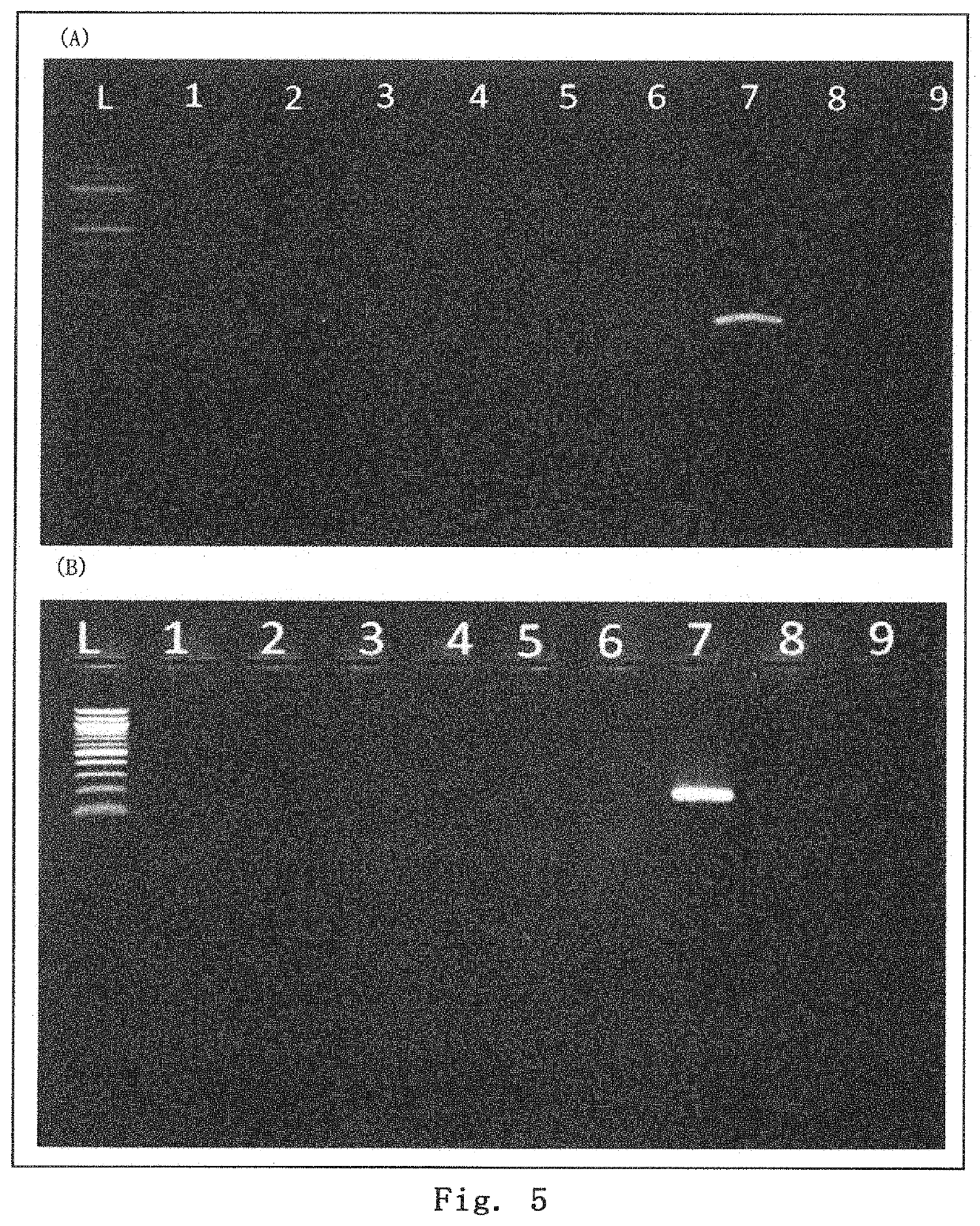
[0111]Using a 3 to 4-week COL17 gene-knockout mouse (see Nat Med. 2007 Mar; 13 (3): 378-83.), the epidermis was chafed to form a blister. Then, within 30 minutes after the blister formation, Muse cells (3×10⁵ per mouse) were injected into the tail vein. Observing the skin state after one month, as shown in FIG. 4, the control mouse treated with HBSS had a poor hair coat, showing extensive formation of wounds and mucosal erosion, while the mouse treated with Muse cells gave mild results for both hair coat and wound formation. In addition, the skin tissue one month after the administration was removed, and RNA was extracted from the skin tissue. RT-PCR was performed to examine the expression of human-derived COL7 and COL17 genes. The results are shown in FIG. 5. As can be seen from the results, the Muse cell-administered mice showed the presence of human COL7 and human COL17, and thus the administered Muse cells provided the adhes...
example 3
iation of Muse Cells into Keratinocytes
[0112]Differentiation of Muse cells into keratinocytes was made by culturing them according to the following procedure:
[0113]Day 0: plating Muse cells;
[0114]Day 1: culturing them in a DMEM low glucose medium supplemented with 10% FBS, KGF (10 ng / ml), and EGF (20 to 30 ng / ml) for three days; and
[0115]Day 4: culturing them in a DMEM low glucose medium supplemented with 10% FBS, KGF (10 ng / ml), EGF (20 to 30 ng / ml), HGF (10 ng / ml), and IGF2 (60 ng / ml) for 8 to 14 days with the culture medium exchanged every other day.
[0116]The results are shown in FIGS. 7 to 9. As shown in FIG. 7, cells at day 8 of differentiation showed keratinocyte-like morphology. In addition, as shown in FIGS. 8 and 9, differentiated cells expressed keratinocyte markers at protein and mRNA levels.
PUM
 Abstract
Description
Claims
Application Information
 - R&D
- Intellectual Property
- Life Sciences
- Materials
- Tech Scout
- Unparalleled Data Quality
- Higher Quality Content
- 60% Fewer Hallucinations
Browse by: Latest US Patents, China's latest patents, Technical Efficacy Thesaurus, Application Domain, Technology Topic, Popular Technical Reports.
© 2025 PatSnap. All rights reserved.Legal|Privacy policy|Modern Slavery Act Transparency Statement|Sitemap|About US| Contact US: help@patsnap.com


