
Method for diagnosing anomalies in the coagulation blood

a technology of coagulation blood and anomalies, applied in the direction of material analysis, biological material analysis, instruments, etc., can solve the problems of lack of specificity, limited to experienced laboratories, lack of sensitivity for moderate diseases on the one hand, and the inability to carry out. , to achieve the effect of reducing the required sampling volum

Active Publication Date: 2020-08-13
HOSPICES CIVILS DE LYON
View PDF0 Cites 0 Cited by
  • Summary
  • Abstract
  • Description
  • Claims
  • Application Information

AI Technical Summary

Benefits of technology

The patent describes a method for quickly and accurately detecting if a person has a clotting disorder and if it is severe or moderate. The method can be performed using small samples of blood, which makes it useful for children. This method can help diagnose disorders of secondary hemostasis, like hemophilias, with a single test.

Problems solved by technology

This test, which is interesting because it is carried out under close to physiological conditions, is difficult to carry out because of the specific pre-test conditions required and a lack of standardization, and is thus limited to experienced laboratories.
The major drawbacks of these tests are their lack of sensitivity for moderate diseases on the one hand and their lack of specificity on the other; it is often necessary to carry out additional tests (determination of factors, assaying of circulating anticoagulant, etc.) in order to identify the origin of the clotting disorder observed.
This entails increased expenditure, both in terms of the reagents and material used and with respect to working time.
Moreover, a major drawback of the tests currently in use is that they must be carried out on platelet-poor plasma obtained from a whole blood sample after said sample has been centrifuged for 10 to 15 minutes.
This limits the use of these tests in emergencies because of the additional time required for this centrifugation step and imposes an additional cost due to pre-treatment of the blood sample.
The drawback of this method is that it is necessary to use numerous reagents and carry out several tests in parallel in order to get an overall view of the various characteristics of clotting.
Moreover, the sensitivity of this method for certain clotting anomalies, in particular hemophilia, is mediocre.
At present, there is no test that allows the identification of a subject suffering from a coagulopathy, and in particular a subject suffering from a hemophilic disorder, based on a single test.
Moreover, there is no test that allows identification of a subject having a deficiency of another clotting factor based on a single test.
More generally, there is no comprehensive, rapid test that may be conducted with a small volume of whole blood without requiring an initial step of centrifugation and uses a single reagent that is capable of predicting hemorrhagic risk and detecting congenital or acquired clotting anomalies.

Method used


Image

  • Method for diagnosing anomalies in the coagulation blood
  • Method for diagnosing anomalies in the coagulation blood
  • Method for diagnosing anomalies in the coagulation blood

Examples


example 1

tal Protocol

[0185]300 μl of a solution of NaCl / CaCl2 (Roche Diagnostic) are placed in the cup before adding 300 μl taken from a citrated tube with a concentration of 0.107 M or 0.109 M. The cup is equipped with a magnetic bar for agitation and two pairs of silver electrodes connected to the Multiplate® unit (Roche Diagnostic).

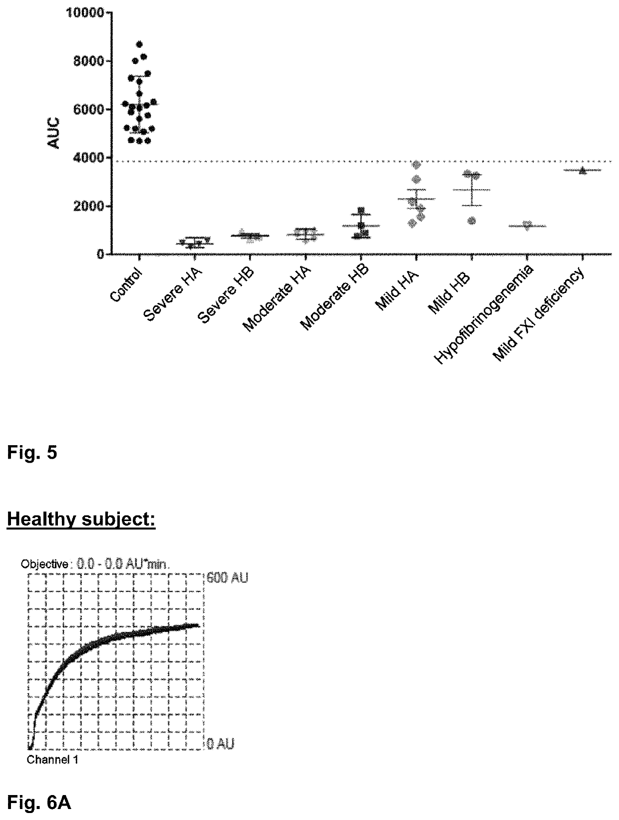
[0186]After incubation for 120 seconds, clotting is initiated by adding a solution of tissue factor diluted in HEPES buffer; the final concentration in the sample is 2 pM (FIGS. 1 through 4), 100 pM (FIGS. 6A, 6B), or 1 pM (FIGS. 7A, 7B).

[0187]Measurement of the impedance variation between the electrodes (expressed in arbitrary units—AU) is carried out for a period of 20 minutes. A curve is thus generated as a function of time, and the curve thus obtained defines an area under the curve measured in AU / min (indicated in the figures as ‘AUC’).

example 2

btained by Adding Tissue Factor in a Concentration of 2 pM

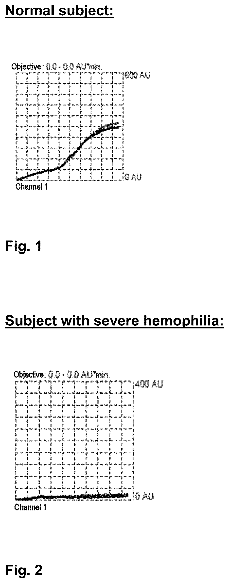
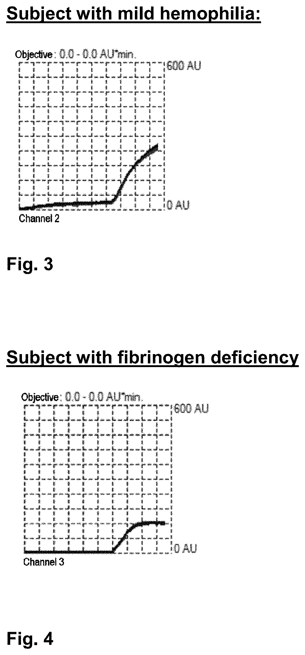
[0188]FIGS. 1 through 4 respectively show the following:[0189]the curve obtained for a blood sample from a subject with no clotting disorders; the area under the curve is 4703 AU / min (FIG. 1.)[0190]the curve obtained for a blood sample from a subject diagnosed with severe hemophilia; the area under the curve is 301 AU / min. (FIG. 2.)[0191]the curve obtained for a blood sample from a subject diagnosed with mild hemophilia; the area under the curve is 2180 AU / min. (FIG. 3.)[0192]the curve obtained for a blood sample from a subject diagnosed with a fibrinogen deficiency; the area under the curve is 1173 AU / min. (FIG. 4.)

[0193]It may therefore be deduced from these curves that when the area under the curve is less than a threshold value referred to as a “reference value,” a clotting anomaly is present.

[0194]FIG. 5 shows the distribution of values for area under the curve as a function of the characteristics of the subjects tested....

example 3

t of Concentration of Tissue Factor Required for Initiating Clotting

[0201]Experiments were conducted according to the same protocol as that described in example 1 in order to determine the optimum concentration range of tissue factor sufficient to initiate blood clotting in vitro in the test described in the present application.

[0202]Two concentrations were tested: 100 pM and 1 pM, in two types of blood samples: one from a healthy subject without a clotting anomaly (A) and the other from a subject with moderate hemophilia.

[0203]When clotting is initiated with a concentration of 100 pM of tissue factor, the values for area under the curve are elevated and distinguishable between the two subjects:[0204]17,191 AU / min for a healthy subject with no clotting disorder;[0205]11,638 AU / min for a subject with hemophilia. (FIG. 6A, 6B)

[0206]When clotting is initiated with a concentration of 1 pM of tissue factor, the values for area under the curve are less elevated and are more sharply distin...


PUM

PropertyMeasurementUnit
volumeaaaaaaaaaa
volumeaaaaaaaaaa
temperatureaaaaaaaaaa

Abstract

The present invention relates to a method for diagnosing anomalies in the coagulation of blood, comprising the following successive steps: a) placing a sample of total blood in a holder containing two pairs of electrodes connected to an apparatus generating an electrical current; b) incubating this sample for 60 to 180 seconds in the presence of calcium; c) adding to this sample tissue factor in a concentration high enough to trigger the coagulation of the blood; d) measuring the impedance variation between the electrodes as a function of time, for a period comprised between 10 and 30 minutes from step (c), and generating a curve of the impedance values as a function of time; e) comparing the value of the area under the curve generated in step (d) with the value of an area under a reference curve.

Description

[0001]The present invention relates to a novel method for ex vivo evaluation of the process of blood clotting, specifically in order to diagnose coagulation anomalies such as hemophilic states or other clotting factor deficiencies based on a blood sample from a subject.[0002]Blood clotting is a complex process that makes it possible to plug a gap in the endothelial wall of blood vessels in mammals.[0003]Coagulation, or hemostasis, begins almost instantly after a lesion of the blood vessel wall. It is composed of three main steps:[0004]1) Formation of a “platelet plug” by platelet aggregation in order to stop the bleeding: this is primary hemostasis.[0005]2) Induction of a complex cascade of events during which plasma proteins referred to as “clotting factors” react to form fibrin fibers that reinforce the platelet plug: this is secondary hemostasis.[0006]3) Destruction of the primary clot or fibrinolysis.[0007]Numerous clotting disorders that lead to the risk of more extensive bleed...

Claims


Application Information

Patent Timeline
Patent Type & Authority Applications(United States)
IPC IPC(8): G01N33/49G01N27/07
CPCG01N2800/52G01N33/4905G01N2800/224G01N27/07G01N27/06
Inventor NOUGIER, CHRISTOPHE
Owner HOSPICES CIVILS DE LYON
Learn More