Method and apparatus for diagnostic analysis of the function and morphology of microcirculation alterations

a technology of microcirculation and diagnostic analysis, applied in the direction of diagnostic recording/measuring, instruments, image enhancement, etc., can solve the problems of not directly imaging but rather measuring, and none of these clinical technologies can image the capillaries, etc., to achieve easy measurement, difficult diagnosis, and great precision

Active Publication Date: 2020-10-01
ACTIVE MEDICAL BV
View PDF3 Cites 7 Cited by
  • Summary
  • Abstract
  • Description
  • Claims
  • Application Information

AI Technical Summary

Benefits of technology

[0029]According to an optional example, the learning processor may, based on the MC analysis, identify heart failure of a patient, which carries with it a risk for myocardial infarction (MI). Currently, as well as being a risk for MI, there is no known mechanism for evaluating a success of therapy for the clinician or even in a home-care environment. Employing the IVM device herein described enables an observation sublingually on the MC image to identify if the heart is functioning well enough to achieve adequate microcirculatory perfusion. Abnormalities in blood flow kinetics might indicate myocardial instability. In addition analysis of microcirculatory patterns may also provide sensitive information regarding the functioning of the failing heart being supported either by extracorporeal assist device or using medication. Identifying the success of such therapy is very difficult, which is greatly eased by using the IVM device described herein, as it can provide a tool for evaluating heart function at the level of the microcirculation and identify a success (or failure) of therapeutic support of the failing heart.
[0030]According to an optional example, the learning processor may, based on the MC analysis, identify a presence of infectious disease and a potential burden of infectious agents, which can otherwise only be shown by blood withdrawal and analysis. There exists a real need for bloodless diagnostics especially when presented with potential harmful pathogens, such as viral and bacterial agents. In addition an inexpensive methodology, allowing easy measurement in large populations, is needed. This is especially needed in a setting of tropical disease, such as malaria and denge. The claimed IVM device has been shown to be sensitive in identifying microcirculatory alterations in malaria and denge. Furthermore, the inventors of the present invention have recognised and appreciated a need for a point-of-care methodology, for identifying infected people the infection itself and possibly then identify the infectious agent without having to withdraw blood.
[0031]There is a need in surgery for a surgical diagnostic hand tool that would allow precise examination needed for making surgical decisions (e.g. to resect or not). According to an optional example, the IVM device herein described may identify abnormal microvascular structures on tumour surfaces as well ischemic boundaries associated with surgical reconstructions, which may lead to anastomotic leakage with great precision. Thus, the IVM device can be used to identify boundaries of pathology and healthy tissue or the nature of pathology of tissues, such as a tumour (or absence thereof) or ischemia (or absence thereof). Thus, the IVM device may allow direct diagnosis and identification of abnormal tissue and provide the surgeon important information in support of clinical decision making during surgery.
[0032]There is a need to identify diabetes, which is presently and predominantly performed by analysis of blood samples requiring blood withdrawal. One of the main targets of diabetes, however, are the blood vessels especially that of the microcirculation. HVM devices have proven successful in identifying morphological changes in vessel structures and subcellular components associated with the severity of diabetes. Differentiation and quantification of the severity of disease in diabetes may be achieved by the IVM device using, say, AI analysis of the different components of the microcirculation, such as the morphology, leucocytes kinetics, subcellular structures glycocalyx. In some examples, the IVM device may provide / output this information in a point-of-care manner, as well as potentially providing a self-care type of IVM device.
[0033]Hypovolemia is a condition where there is insufficient volume of blood in the circulation to allow the heart to sufficiently pump blood to perfuse the microcirculation with oxygen rich blood. It can occur as a result of blood loss or dehydration (which is the primary cause of death amongst children worldwide). Hypovolemia is difficult to diagnose. However, in accordance with some optional examples of the invention, the IVM device allows precise identification and also enables a distinguishing between blood loss and dehydration.
[0034]Currently there is no direct; non-invasive imaging modality for assessment of a vascular health of an individual. According to examples of the present invention, the IVM device described herein is able to image and interpret the properties of the vasculture that would allow an individual (and also across a population) to track as a vascular bioprint the development of the vasculuture. Since the microcirculation is that part of the cardiovascular system that is closest to the tissues, it can be regarded as the most relevant to follow in this respect. It can be visualized by the IVM device, but needs interpretation and quantification, which can also be provided by the described IVM device. Being able to measure the microcirculation and interpret its health and response to lifestyle would allow one to track ones vascular health over time. Since it is known that exercise benefits vascular health, the IVM device can allow an individual (or patient) to assess the benefit of exercise for the development of the (micro) vasculture needed for cardiovascular health.

Problems solved by technology

However, these clinical technologies, without exception, do not directly image but rather measure surrogates of microcirculatory flow and / or oxygenation.
Most importantly none of these clinical technologies can image the capillaries of the microcirculation (main physiological compartment of the MC) where oxygen is transported to the tissues.
This is a severe limitation of monitoring the microcirculation, since with these clinical techniques it is neither possible to measure quantitatively parameters, nor do they provide quantitative information regarding the function of the capillaries.
However, the inventors of the present invention have recognised and appreciated that this has limited value in practical scenarios.
However, the suggested device only yields image sequences, and the inventors of the present invention have recognised and appreciated that this has limited value in practical scenarios where identification of MC abnormalities coupled to a specific disease state, advice on the optimal type therapy and the prognosis of the state of disease is required.

Method used


Image

  • Method and apparatus for diagnostic analysis of the function and morphology of microcirculation alterations
  • Method and apparatus for diagnostic analysis of the function and morphology of microcirculation alterations
  • Method and apparatus for diagnostic analysis of the function and morphology of microcirculation alterations

Examples


first embodiment

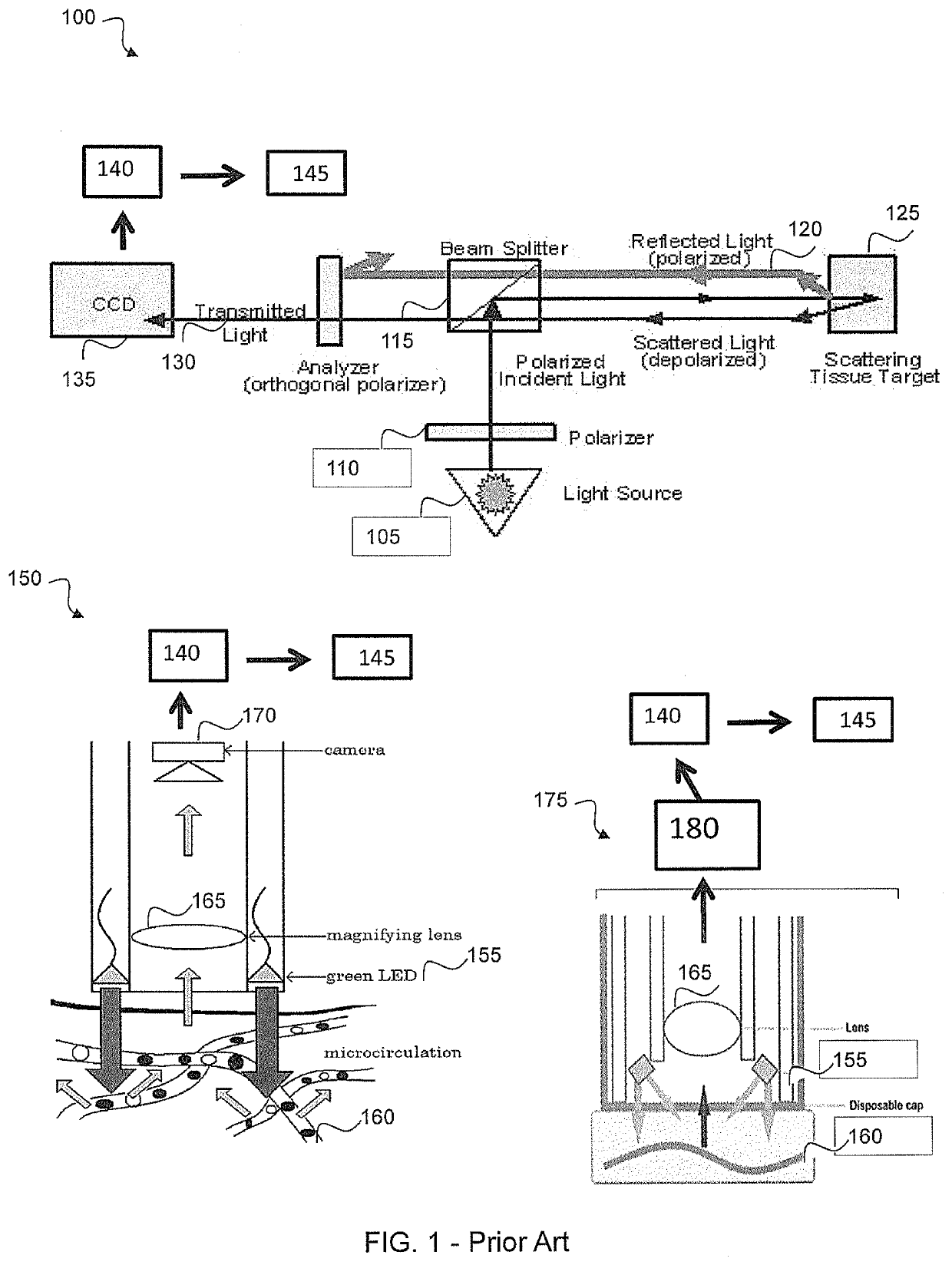
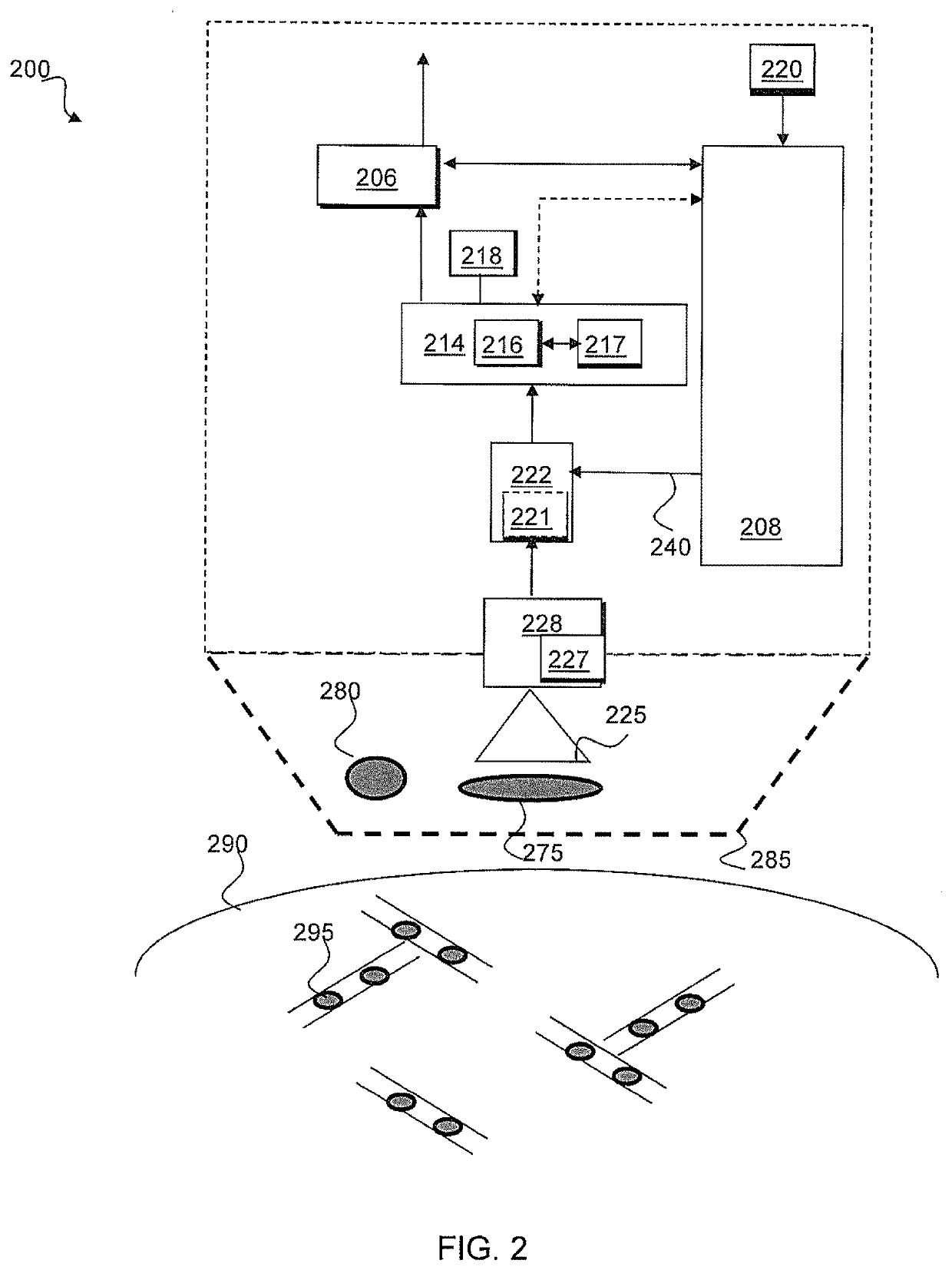
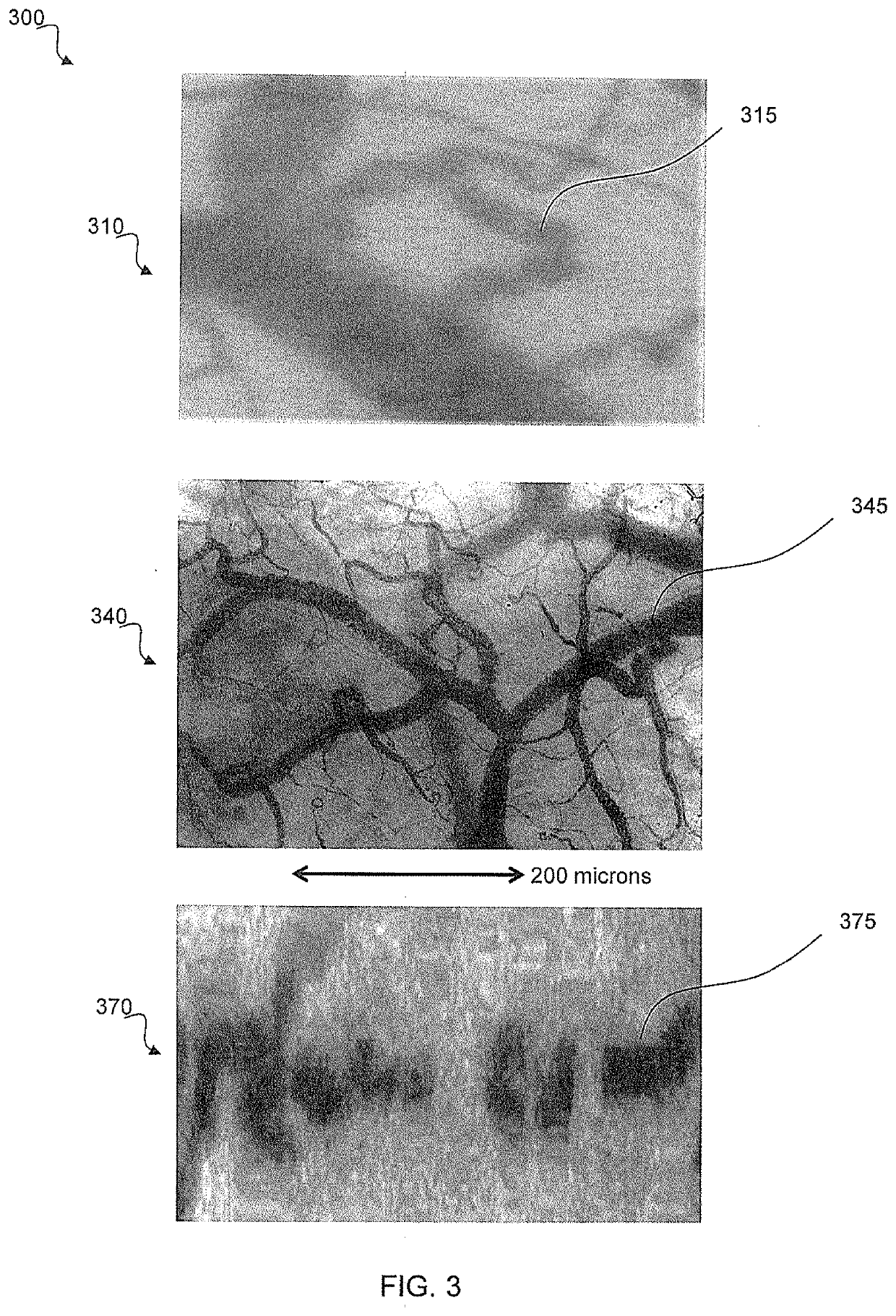
[0167]A first embodiment is the method wherein IVM analysis of the MC and quantification of morphological and functional parameters of the microcirculation and changes therein over time is used to evaluate health, disease and in response to therapy. The input variable parameters in this AI analysis include but not limited to movie clips and images of the MC and derived parameters including visual inspection obtained by IVM and variables using other MC techniques described in the Date Set section of all organ surfaces including but not limited to the sublingual and oral region. Examples of the Input variable parameters include but are not limited functional parameters of the MC including: Microcirculatory hemodynamic values: capillary, venule and arteriolar blood flow and blood volume, identification of types of vessels (e.g. capillaries, capillary loops, arterioles, venules), Proportion of perfused vessel density or functional capillary density (PVD [mm / mm2]. FCD (density of functio...

seventh embodiment

[0175]A seventh embodiment is the method wherein IVM analysis of MC is used to show changes in microcirculatory function and morphological improvement related to health as a result of exercise and therapy. It has been reported that exercise (>150 min / week) in hypertension patients results in improved functional capillary density and red blood cell flow.

[0176]Improvement of the MC can also be achieved by therapy, for example, by use of cardiac assist devices. It has been shown that sustained improvement of the MC using such Impella technology can be used for bridge to treat following myocardial infarction. Following session of the Impella treatment MC function which had improved during Impella therapy remained improved following withdrawal of the Impella device. Similarly, it has been shown that weaning from an ECOS can be judged as a result of sustained improved sublingual red blood cell flow upon reduction of ECOS pump flow. It has been shown that such devices or cardiotonics in he...


PUM


Abstract

An intelligent vital microscopy, IVM, device is described. The IVM device includes: a receiver configured to receive at least one IVM image of a human microcirculation, MC, of an organ surface; a learning processor coupled to the receiver and configured to: process the at least one IVM image and extract at least one MC variable therefrom, and identify from the extracted at least one MC variable of the at least one IVM image at least one of: an underlying cause for an observed abnormality, an intervention, a disease state, a disease diagnosis, a medical state of the human; a presence of a pathogen; and an output coupled to the learning processor and configured to output the identification.

Description

FIELD OF THE INVENTION[0001]The field of this invention relates to a method and apparatus for processing at least one or sequence of vital microscopy image(s) and performing a diagnostic analysis of a function and morphology of microcirculation alterations of humans related to health and disease, for example for identifying a disease of a subject and an associated therapeutic resolutionBACKGROUND OF THE INVENTION[0002]Several types of clinical technologies have been described in the literature that purports to monitor the human microcirculation (MC). These have included laser Doppler imaging, laser speckle imaging, near-infrared spectroscopy, contrast enhanced ultrasound, spectrophotometry and oxygen electrodes. However, these clinical technologies, without exception, do not directly image but rather measure surrogates of microcirculatory flow and / or oxygenation. Most importantly none of these clinical technologies can image the capillaries of the microcirculation (main physiologica...

Claims


Application Information

Patent Timeline
Patent Type & Authority Applications(United States)
IPC IPC(8): G02B21/36G02B21/00G06N3/063G06N3/08G06T7/00G16H50/20
CPCG02B21/36G06T7/0012G06N3/063G06T2207/10056G06T2207/10016G06N3/08G16H50/20G02B21/0012G06T2207/20084G06T2207/30004A61B5/0261A61B5/7264A61B5/201Y02A90/10
Inventor INCE, CANHILTY, MATTHIAS PETERINCE, YASINAHISKA, YAVUZ
Owner ACTIVE MEDICAL BV
Why Eureka
Try Eureka