Tissue Engineered Intestine
a technology of intestine and construct, which is applied in the direction of tissue regeneration, prosthesis, pharmaceutical delivery mechanism, etc., can solve the problems of limited cell source, limited application range, and inability to reach adults, etc., to achieve accurate mechanical properties, facilitate normal tissue function, and promote proper stem cell differentiation
- Summary
- Abstract
- Description
- Claims
- Application Information
AI Technical Summary
Benefits of technology
Problems solved by technology
Method used
Image

Examples
example 1
Analysis of Scaffold Materials
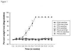
[0120]This detailed evaluation of the numerous potential scaffold materials was carried out to determine potential scaffold materials for the construction of a multilayer nanofiber scaffold for use in generating engineered intestine constructs. The purpose of this study was to characterize seven different scaffold materials according to degradation rates, histologic changes, and tensile strength to determine which would be best suited for the production of the engineered intestine constructs.
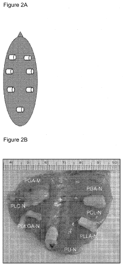
[0121]Initially, the seven different single tube scaffolds were fabricated using electrospinning as described in Example 2 and above. These scaffolds were comprised of poly(glycolic acid)(PGA) nanofibers, Poly(-caprolactone) (PCL) nanofibers, Poly(-caprolactone-co-lactic acid) (PLC) nanofibers, Poly(L-lactic acid) (PLLA), Poly(D-lactic acid-co-glycolic acid) (PDLGA), Polyurethane (PU) nanofibers and PGA macrofibers. The physical and chemical characteristics of the n...
example 2
Scaffold Fabrication
[0147]A multilayer nanofiber scaffold was fabricated for generation of an engineered intestine constructs. Multilayer scaffolds were constructed in order to facilitate the delivery of cells of different sizes, e.g. neural stem cells, smooth muscle cells and crypt cells. In addition, the multilayer scaffold allows for different mechanical properties within the scaffold, allows for a smooth lumen and allows for separation of different cell types which allowed for the generation of different types of tissue.
[0148]To seed the scaffolds with cells, the scaffolds are coated onto a cell culture plate for three dimensional cell culture. Human smooth muscle cells are plated and within these cultures the cells migrated along the nanofibers after 5 days in culture. The migration of the smooth muscle cells (SMC) demonstrates that upon seeding of smooth muscle cells into a circumferentially or longitudinally aligned nanofiber tubular layer, the SMCs will align, orientate, mig...
example 3
In Vitro Characterization of the Nanofiber Scaffolds
[0153]A total of seven nanofiber scaffolds (PCL, PLC, PCL-FHB-EGF, PLC-FHB-EGF) are fabricated as described in Example 1 and each of these scaffolds are analyzed using the following in vitro characterizations. Any nanofiber scaffold of the invention will be characterized using one or more of the following analyses.
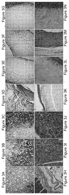
Scanning Electron Microscopy (SEM)
[0154]Scaffolds are sputter coated with gold and then observed under a scanning electron microscope at an accelerated voltage of 15 kV. Fiber and pore size of the inner and outer layers are measured using Image J software and continuity between pores are assessed. Fiber and pore size between HB-EGF-coated and noncoated scaffolds are compared using Student t-test, with p<0.05 considered statistically significant. The scanning electron microscopy studies provide guidance on how modify the fiber size and pore size to best accommodate the cells to be seeded into the scaffold.
Modulus Determina...
PUM
| Property | Measurement | Unit |
|---|---|---|
| relative porosity | aaaaa | aaaaa |
| temperature | aaaaa | aaaaa |
| voltage | aaaaa | aaaaa |
Abstract
Description
Claims
Application Information