Systems, methods, and devices for medical image analysis, diagnosis, risk stratification, decision making and/or disease tracking

a technology of medical image analysis and decision-making, applied in the field of systems, methods and devices for medical image analysis, diagnosis, risk stratification, decision-making and/or disease tracking, can solve the problems of blood clots, most effective treatment, and considered more life-threatening

Active Publication Date: 2021-08-19
CLEERLY INC
View PDF0 Cites 0 Cited by
  • Summary
  • Abstract
  • Description
  • Claims
  • Application Information

AI Technical Summary

Benefits of technology

The patent text is about a computer-implemented method that improves the accuracy of algorithms used for analyzing medical images. This is done by using a normalization device that can adjust the first and second medical images based on different parameters, such as variable acquisition parameters, image capture technology, or contrast agents. This helps to make the images more consistent and reliable for the algorithm-based analysis, which can be more accurate and reliable. In simple terms, the method improves the quality of medical images for analysis by using a normalization device to adjust them for better accuracy.

Problems solved by technology

However, a recent federally funded $100 million study calls into question whether the current trends in treating cardiovascular disease are the most effective treatment for all types of patients.
However, if the plaque build-up is significant enough to cause severe stenosis or narrowing of the arterial vessel such that blood flow to heart muscle might be blocked, then an invasive angioplasty procedure to implant a stent may likely be required because heart attack or sudden cardiac death (SCD) could occur in such patients without the implantation of a stent to enlarge the vessel opening.
For example, in some embodiments of systems, devices, and methods disclosed herein, arteries with “good” or stable plaque or plaque comprising hardened calcified content are considered non-life threatening to patients whereas arteries containing “bad” or unstable plaque or plaque comprising fatty material are considered more life threatening because such bad plaque may rupture within arteries thereby releasing such fatty material into the arteries.
Such a fatty material release in the blood stream can cause inflammation that may result in a blood clot.
Further, in some instances, it is generally more difficult for blood to flow through fatty plaque buildup than it is for blood to flow through calcified plaque build-up.
Further, while blood tests and drug treatment regimens are helpful in reducing cardiovascular health issues and mitigating against cardiovascular events (for example, heart attacks), such treatment methodologies are not complete or perfect in that such treatments can misidentify and / or fail to pinpoint or diagnose significant cardiovascular risk areas.
For example, the mere analysis of the blood chemistry of a patient will not likely identify that a patient has artery vessels having significant amounts of fatty deposit material bad plaque buildup along a vessel wall.
Similarly, an angiogram, while helpful in identifying areas of stenosis or vessel narrowing, may not be able to clearly identify areas of the artery vessel wall where there is significant buildup of bad plaque.
In certain circumstances, areas where there exist areas of bad plaque can lead to a rupture wherein there is a release of the fatty materials into the bloodstream of the artery, which in turn can cause a clot to develop in the artery.
A blood clot in the artery can cause a stoppage of blood flow to the heart tissue, which can result in a heart attack.
This is stark contrast to existing measures today which do not produce reproducible prognosis or assessment, take extensive amounts of time, and / or require invasive procedures.
As a result, even if two different scans were taken of the same subject, the brightness and / or darkness of the resulting medical image may be different, which can result in less than accurate analysis results processed from that image.
However, in some embodiments, such CT image can comprise a non-contrast CT image, in which case it can be more difficult to identify and / or determine non-calcified plaque due to its low radiodensity value and overlap with other low radiodensity values components, such as blood for example.

Method used


Image

  • Systems, methods, and devices for medical image analysis, diagnosis, risk stratification, decision making and/or disease tracking
  • Systems, methods, and devices for medical image analysis, diagnosis, risk stratification, decision making and/or disease tracking
  • Systems, methods, and devices for medical image analysis, diagnosis, risk stratification, decision making and/or disease tracking

Examples


example embodiments

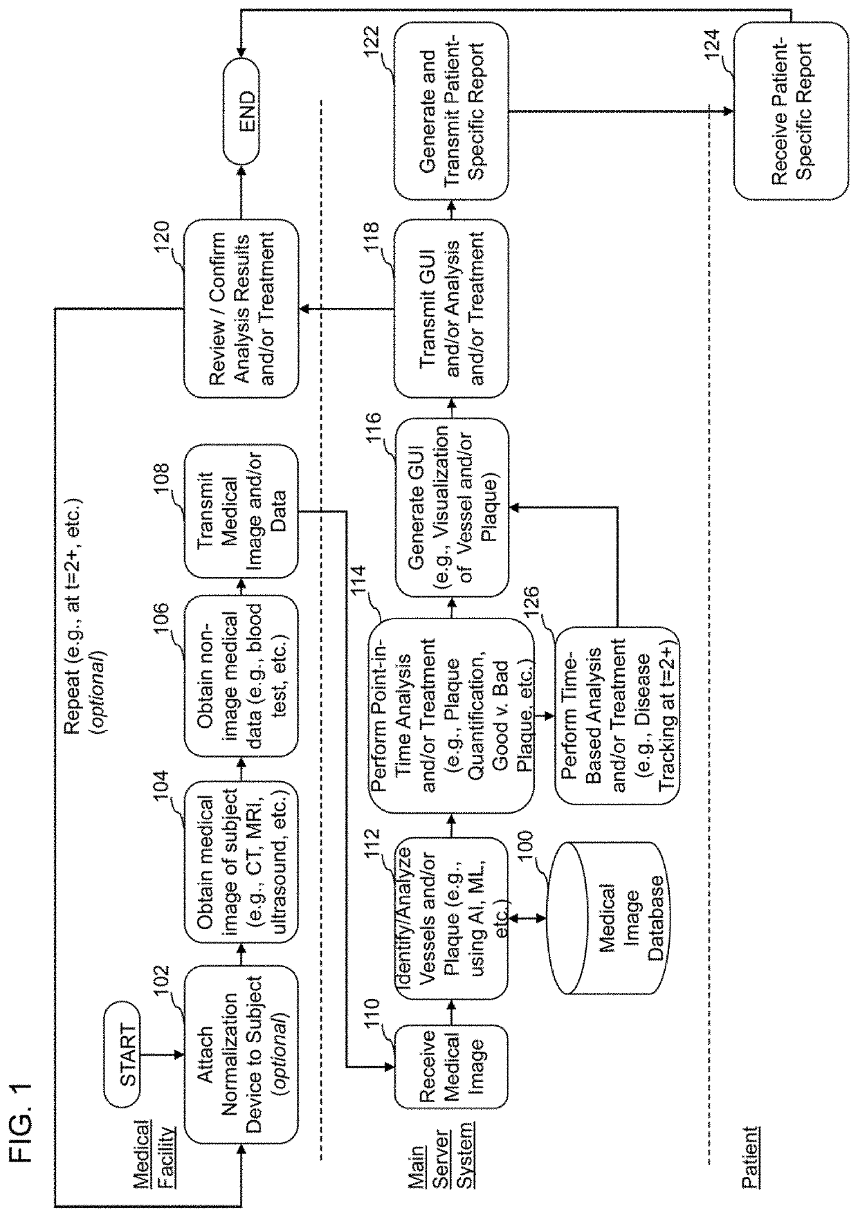
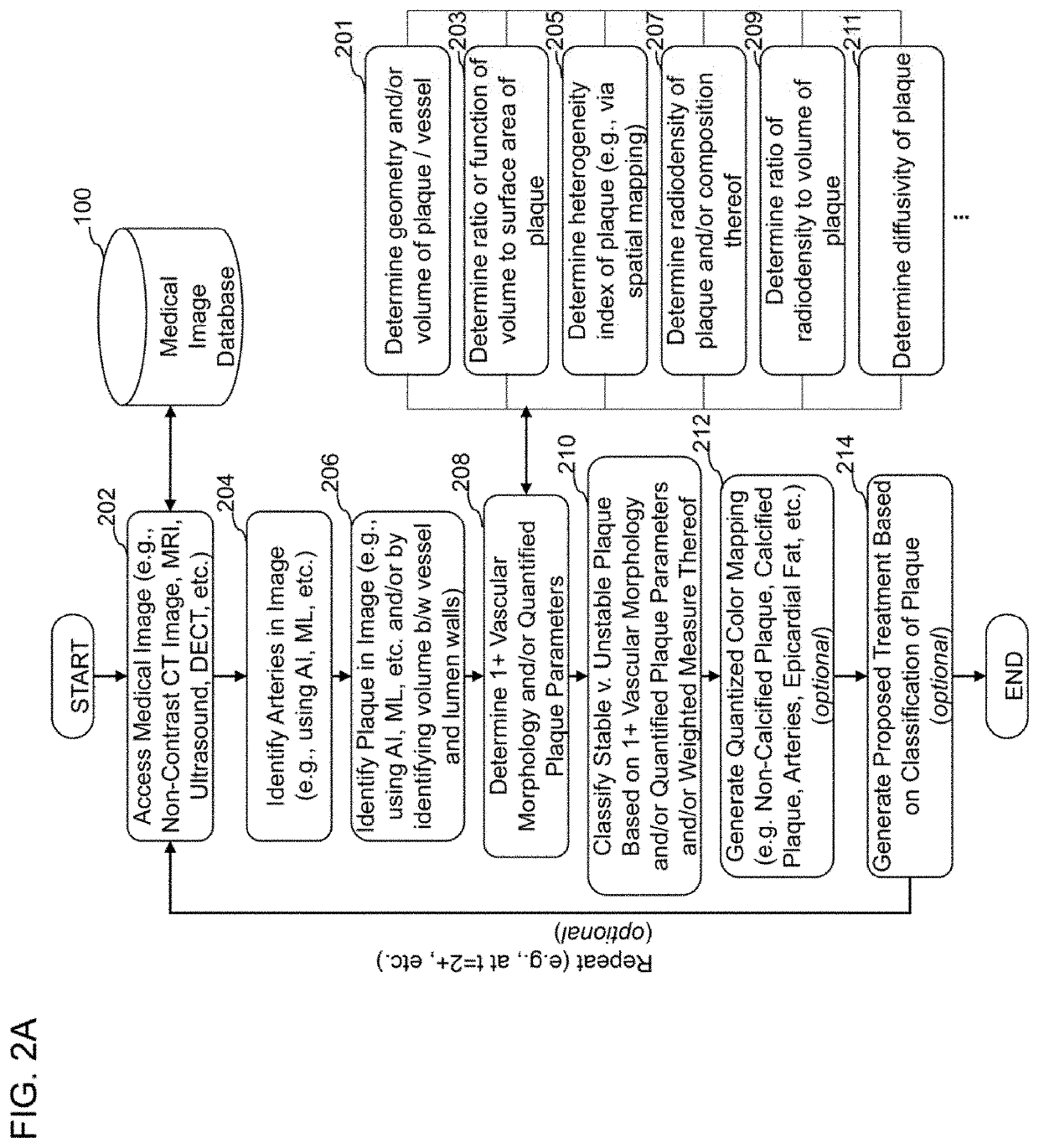
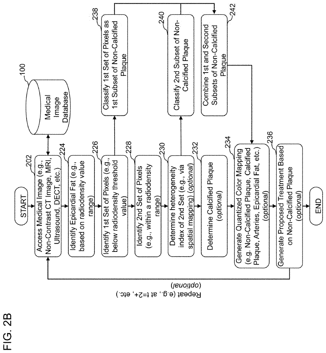
[0459]The following are non-limiting examples of certain embodiments of systems and methods of characterizing coronary plaque. Other embodiments may include one or more other features, or different features, that are discussed herein.

embodiment 1

[0460]A computer-implemented method of quantifying and classifying coronary plaque within a coronary region of a subject based on non-invasive medical image analysis, the method comprising: accessing, by a computer system, a medical image of a coronary region of a subject, wherein the medical image of the coronary region of the subject is obtained non-invasively; identifying, by the computer system utilizing a coronary artery identification algorithm, one or more coronary arteries within the medical image of the coronary region of the subject, wherein the coronary artery identification algorithm is configured to utilize raw medical images as input; identifying, by the computer system utilizing a plaque identification algorithm, one or more regions of plaque within the one or more coronary arteries identified from the medical image of the coronary region of the subject, wherein the plaque identification algorithm is configured to utilize raw medical images as input; determining, by t...

embodiment 2

[0461]The computer-implemented method of Embodiment 1, wherein one or more of the coronary artery identification algorithm or the plaque identification algorithm comprises an artificial intelligence or machine learning algorithm.


PUM


Abstract

The disclosure herein relates to systems, methods, and devices for medical image analysis, diagnosis, risk stratification, decision making and / or disease tracking. In some embodiments, the systems, devices, and methods described herein are configured to analyze non-invasive medical images of a subject to automatically and / or dynamically identify one or more features, such as plaque and vessels, and / or derive one or more quantified plaque parameters, such as radiodensity, radiodensity composition, volume, radiodensity heterogeneity, geometry, location, and / or the like. In some embodiments, the systems, devices, and methods described herein are further configured to generate one or more assessments of plaque-based diseases from raw medical images using one or more of the identified features and / or quantified parameters.

Description

CROSS-REFERENCE TO RELATED APPLICATIONS[0001]This application is a continuation of U.S. application Ser. No. 17 / 142,120, filed Jan. 5, 2021, which claims the benefit of U.S. Provisional No. 62 / 958,032, filed Jan. 7, 2020, and titled SYSTEMS, METHODS, AND DEVICES FOR CARDIOVASCULAR IMAGE ANALYSIS, DIAGNOSIS, RISK STRATIFICATION, DECISION MAKING AND / OR DISEASE TRACKING, and these disclosures are incorporated herein by reference in their entirety. Any and all applications for which a foreign or domestic priority claim is identified in the Application Data Sheet as filed with the present application are hereby incorporated by reference under 37 C.F.R. § 1.57.BACKGROUNDField[0002]The present application relates to systems, methods, and devices for medical image analysis, diagnosis, risk stratification, decision making and / or disease tracking.Description[0003]Coronary heart disease affects over 17.6 million Americans. The current trend in treating cardiovascular health issues is generally...

Claims


Application Information

Patent Timeline
Patent Type & Authority Applications(United States)
IPC IPC(8): G06T7/00A61B5/00A61B6/00A61B6/03G06K9/62A61K49/04A61B8/14A61B8/12A61B5/055G06V10/20G06V10/764G06V10/82
CPCG06T7/0012G06T2207/20081A61B6/504A61B6/5205A61B6/481A61B5/7475A61B6/037A61B5/7267G06K9/6298A61K49/04A61B5/742A61B6/032A61B8/14A61B5/0066A61B8/12A61B5/055G06T2207/10088G06T2207/30101G06T2207/10132G06T2207/10081G06T2207/30048G06T2207/10101A61B5/0075A61B6/5217A61B6/583A61B8/0891A61B8/587A61B8/5223G06T2207/10104G06T2207/10108G06T2207/10116G06T2207/20101A61B5/02007A61B5/7264A61B5/1076A61B5/004G06V10/245G06V10/247G06V10/20G06V2201/03G06V10/82G06T2207/20084G06V10/764G06V10/761G06F18/22G06F18/2413Y02A90/10G06F18/10A61B6/463A61B6/467
Inventor MIN, JAMES K.EARLS, JAMES P.RODRIGUES MARQUES, HUGO MIGUEL
Owner CLEERLY INC
Why Eureka
Try Eureka