Biomembrane-covered nanoparticles (bionps) for delivering active agents to stem cells

a technology of biomembrane-covered nanoparticles and stem cells, which is applied in the direction of antibacterial agents, drug compositions, immunological disorders, etc., can solve the problems of poor dna insertion, limited loading capacity of viral vectors, and long-standing problem of cargo delivery to hspcs

Pending Publication Date: 2021-09-09
UNIVERSITY OF DELAWARE
View PDF0 Cites 0 Cited by
  • Summary
  • Abstract
  • Description
  • Claims
  • Application Information

AI Technical Summary

Benefits of technology

[0031]The delivery method may further comprise preventing a disease or condition in the second subject. The disease or condition may be selected from the group consisting of bone marrow failure disorder, leukemia, lymphoma, multiple myeloma, aplastic anemia, sickle cell disease, thalassemia, autoimmune disorders, HIV, multiple sclerosis, myeloproliferative disorder and myelodysplastic syndrome. In one embodiment, the disease or condition is cancer.
[0032]A method for treating a disease or condition in a subject in need thereof is provided. The treatment method may comprise administering to the subject an effective amount of the bio-nanoparticles or the composition of the present invention. The disease or condition may be selected from the group consisting of bone marrow failure disorder, leukemia, lymphoma, multiple myeloma, ap

Problems solved by technology

. . that may transform medical practice.” However, cargo delivery to HSPCs is a long-standing problem.
Current delivery methods in the form of viral vectors (lentivirus, adeno-associated virus) have limited loading capacity, poor DNA insertion, and produce too much cytotoxicity.
Although nanoparticles wrapped in membranes derived from red blood cells, platelet

Method used


Image

  • Biomembrane-covered nanoparticles (bionps) for delivering active agents to stem cells
  • Biomembrane-covered nanoparticles (bionps) for delivering active agents to stem cells
  • Biomembrane-covered nanoparticles (bionps) for delivering active agents to stem cells

Examples


examples 1-4

D CHARACTERIZATION OF BIONPS CONTAINING HYDROPHOBIC CARGO

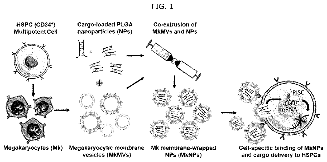
[0082]To demonstrate the synthesis of BioNPs containing hydrophobic molecules, we used DiD fluorophores as model cargo, as this allows visualization of cargo delivery to HSPCs by fluorescence microscopy. In short, we synthesized PLGA NPs encapsulating DiD by the method described above method by dissolving 50:50 PLGA with an inherent viscosity of 0.67 dL / g in acetone along with DiD fluorophores and adding this mixture dropwise to water in a 1:3 ratio. We have adjusted the concentration of PLGA used in this synthesis from 1 to 4 mg / mL, resulting in particles ranging from 50 to 120 nm diameter. Likewise, we have used the above lysis and homogenization method to produce MkMVs approximately 150 nm in diameter, and we have co-extruded these MkMVs with DiD-loaded PLGA NPs to produce Mk membrane-wrapped BioNPs. The resultant BioNPs were characterized by several techniques, summarized below.

example 1

e Successfully Wrapped with Mk-Derived Membranes

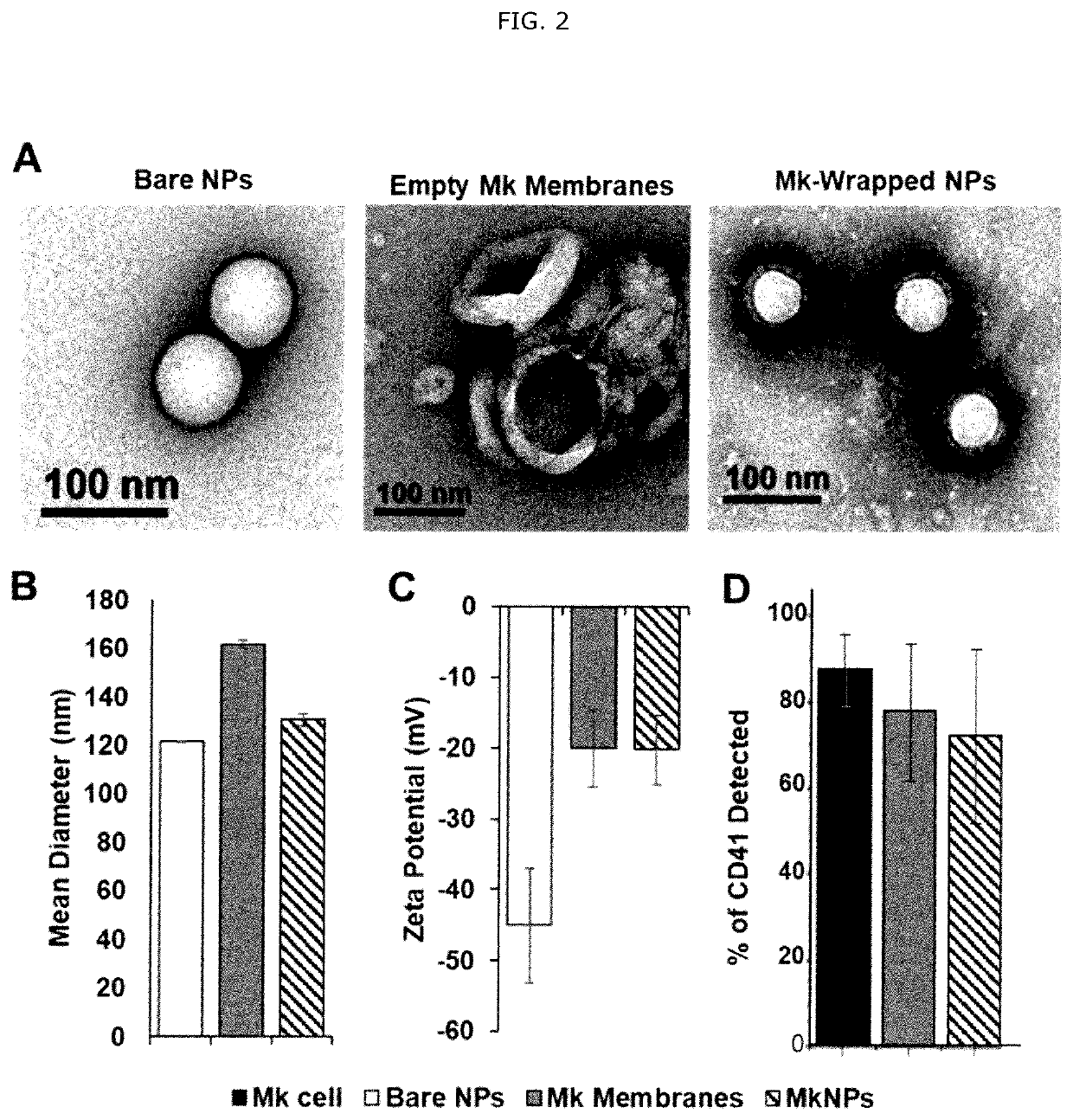
[0083]The successful production of BioNPs was confirmed by using transmission electron microscopy (TEM) of uranyl-acetate stained samples to visualize unwrapped (bare) PLGA NPs, empty MkMVs, and Mk membrane-wrapped BioNPs (FIG. 2A). As seen in these images, bare PLGA NPs have a homogenous spherical shape and MkMVs appear as hollow shells. By comparison, Mk membrane-wrapped BioNPs have core / shell structure indicative of PLGA NPs (brighter interior) surrounded by Mk-derived biological membranes (darker exterior).

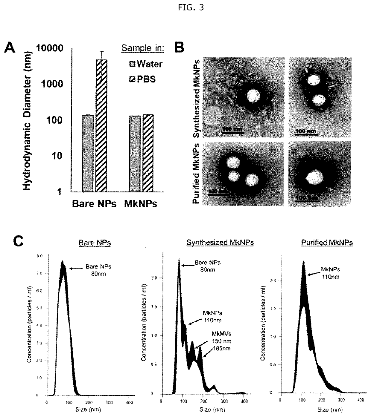
[0084]The hydrodynamic diameter and zeta potential of bare NPs, MkMVs, and BioNPs were also measured to corroborate the TEM findings and confirm successful membrane wrapping. As shown in FIG. 2B, BioNPs are slightly larger than bare PLGA NPs, but smaller than empty MkMVs (which typically have a mean diameter ranging from 140-160 nm). In general, we have found by TEM and nanoparticle tracking analysis (NTA) that BioNPs are 10-20 n...

example 2

intain the Membrane Composition of Their Source Cells

[0085]For BioNPs to maintain the unique HSPC-specific targeting capabilities of Mk cells and MkMPs, they must retain the characteristic membrane proteins. To confirm membrane composition is preserved after wrapping, we synthesized BioNPs as above, and then incubated the samples with a solution of 1-μm streptavidin beads decorated with antibodies against CD41, a surface marker of Mks. The antibodies on the beads can bind CD41 found on BioNPs, whole Mk cells, and empty Mk membrane vesicles. The samples can then be incubated with FITC-labeled anti-CD41 antibodies and analyzed by flow cytometry to determine the relative amount of CD41 present in each group. As shown in FIG. 2D, using this technique we determined that the fraction of streptavidin beads exhibiting positive CD41 signal in the case of whole Mk cells was approximately 85%, which reduced to approximately 75% for empty MkMVs and fully wrapped BioNPs. This indicates that CD41...


PUM

PropertyMeasurementUnit
Biological propertiesaaaaaaaaaa
Hydrophobicityaaaaaaaaaa
Electrostatic interactionaaaaaaaaaa

Abstract

The present invention provides bio-nanoparticles (BioNPs) for delivering an active agent into hematopoietic stem & progenitor cells (HSPCs). Each BioNP comprises a core and a biological membrane covering the core, which comprises the active agent and a polymer. The biological membrane comprises a phospholipid bilayer and one or more surface proteins of a megakaryocyte (Mk). The active agent remains active after being delivered into the HSPC. Also provided are methods for preparing the BioNPs and uses of the BioNPs for targeted delivery of an active agent into HSPCs and/or treating or preventing a disease or condition in a subject in need thereof.

Description

CROSS-REFERENCE TO RELATED APPLICATION[0001]This application is a Continuation Application of International Application No. PCT / US2019 / 063685, filed Nov. 27, 2019, claiming priority to United States Provisional Application No. 62 / 772,311, filed Nov. 28, 2018, the contents of which are incorporated herein by reference in their entireties for all purposes.REFERENCE TO U.S. GOVERNMENT SUPPORT[0002]This invention was made with government support under Grant No. 1752009 from the National Science Foundation. The United States has certain rights in the invention.FIELD OF THE INVENTION[0003]The invention relates generally to biomembrane-covered nanoparticles (BioNPs) comprising active agents and uses thereof for targeted delivery of the active agents into hematopoietic stem & progenitor cells (HSPCs) with high specificity and controlled release of the active agents from the BioNPs in the HSPCs.BACKGROUND OF THE INVENTION[0004]Hematopoietic stem & progenitor cells (HSPCs) are located in the ...

Claims


Application Information

Patent Timeline
IPC IPC(8): A61K9/51A61K45/06
CPCA61K9/5184B82Y5/00A61K45/06A61K9/5153A61P31/10A61P31/04A61P35/00A61P37/04B01J13/04B01J13/22
Inventor PAPOUTSAKIS, ELEFTHERIOS T.DAY, EMILYWINTER, ERICAHARRIS, JENNAKAO, CHEN-YUANDAS, SAMIK
Owner UNIVERSITY OF DELAWARE
Why Eureka
Try Eureka