Utilization of nuclear structural proteins for targeted therapy and detection of proliferative and differentiation disorders

Inactive Publication Date: 2001-09-11
RGT UNIV OF CALIFORNIA
View PDF1 Cites 41 Cited by
  • Summary
  • Abstract
  • Description
  • Claims
  • Application Information

AI Technical Summary

Benefits of technology

Using a 3D model of human mammary epithelial cell culture, we have shown that the distribution of the structural protein NuMA within the nucleus depends on the cell status, and that the distribution

Problems solved by technology

Mutation of the predicted p34cdc2 phosphorylation sites in NuMA

Method used


Image

  • Utilization of nuclear structural proteins for targeted therapy and detection of proliferative and differentiation disorders
  • Utilization of nuclear structural proteins for targeted therapy and detection of proliferative and differentiation disorders
  • Utilization of nuclear structural proteins for targeted therapy and detection of proliferative and differentiation disorders

Examples


Example

Example 1

Internal Nuclear Organization Is Remodeled When HMECS Are Cultured Within 3D rBM.

HMT-3522 HMECs, like primary HMECs, undergo morphogenesis to form tissue-like acini when cultured in 3D rBM. Neither cell type undergoes acinar differentiation when cultured as 2D monolayers.

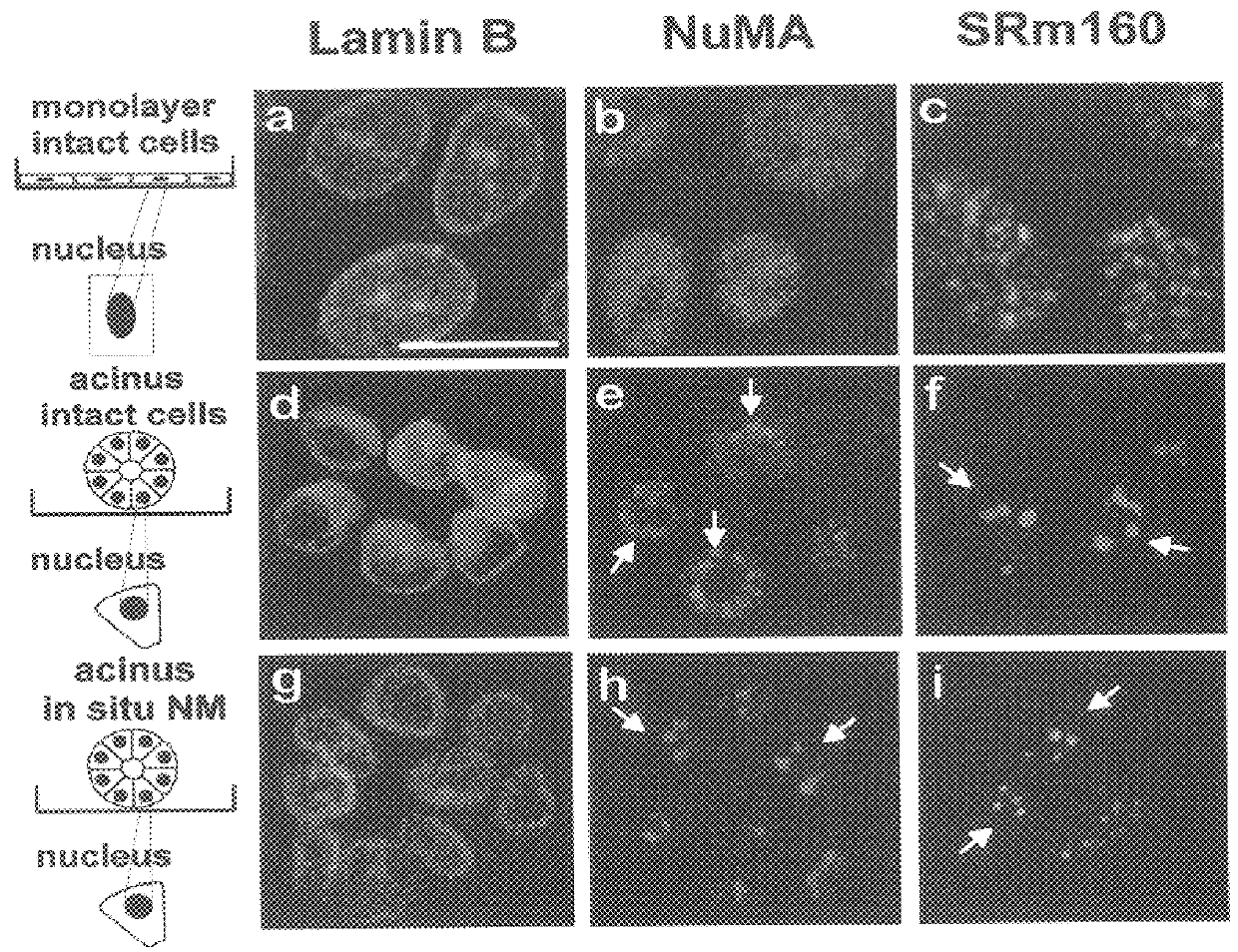
Referring now to FIG. 1, confocal fluorescence irnages (0.2.mu. optical sections) of lamin B, NuMA and splicing factor SRml60 in cells grown as monolayers (2D),( a-c) and within rBMs (3D), (d-i) are shown. NuMA was diffusely distributed in the nuclei of cells grown as monolayers (b), but reorganized into large nuclear foci in cells induced to undergo morphogenesis (acini formation) in response to a rBM (e). SRm160 was distributed as multiple nuclear speckles in cells cultured as monolayer (c), whereas it was concentrated into fewer and larger speckles in the acini (f). Lamin B in contrast, consistently localized to the nuclear periphery and within intra nuclear patches (a & d). The distribution of lamin B (...

Example

Example 2

Growth-Arrest Is Associated With Changes In NuMA And Rb Distribution

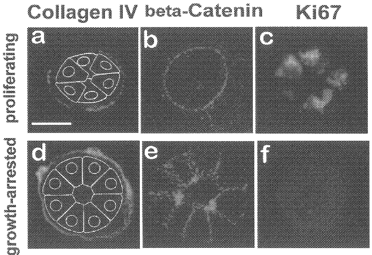
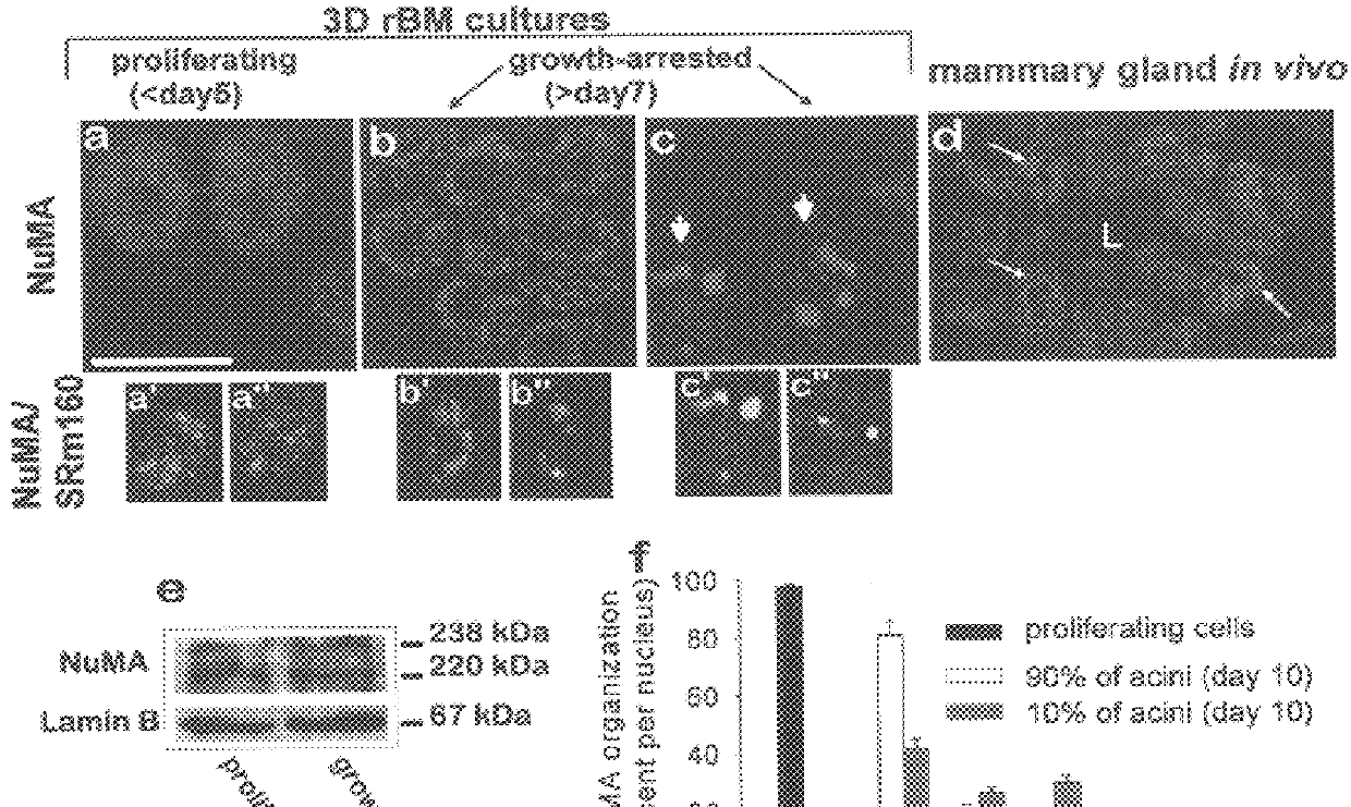
ECM-directed growth-arrest is an early and critical step in mammary epithelial cell morphogenesis. To distinguish between the effect of ECM-directed growth-arrest and changes due to tissue structure and polarity, the localization of NuMA and SRm160 was compared between growth-arrested and proliferating cells cultured as 2D monolayers. Less than five percent of the cells remained in the cell cycle following growth-arrest induced by EGF removal, as indicated by the absence of detectable Ki-67 immunostaining (not shown). NuMA was uniformly distributed in the nuclei of proliferating cells, but coalesced into denser areas upon growth-arrest.

Referring now to FIG. 3, there is shown confocal fluorescence images (0.2.mu. optical sections) of NuMA (Texas red, a-c) and Rb (FITC green, d, e, g, h) in cells proliferating as 2D monolayers (a & d) and within 3D rBMs (g), and cells growth-arrested in monolayer (b & e) and ...

Example

Example 3

Cross-Modulation Between NuMA Distribution, Chromatin Structure, And The Acinar Phenotype

The degree of histone acetylation has been shown to regulate chromatin structure and gene expression. Histone acetylation was altered in the acini using the histone deacetylase inhibitor trichostatin A. After two hours of treatment NuMA foci began to disperse, and several cells entered the cell cycle, as measured by an increase in the Ki-67 labeling index. Referring now to FIG. 4, there is shown confocal fluorescence images (0.2.mu. optical sections) of NuMA (a, e, i), collagen IV (b, f, j), .beta.-catenin (c, g k) and acetylated histone H4 (d, h, l) in control, trichostatin A (TSA)-treated and NuMA monoclonal antibody (mAb)-incubated acini (day 10 of 3D rBM culture). (a-d) Nuclear organization and acinar phenotype in controls: acini exhibit NuMA foci (a), an organized endogenous collagen IV-rich BM (b), cell-cell localized .beta.-catenin (c) and dispersed acetylated H4 histone(d). (e-h...


PUM


Abstract

The localization of nuclear apparatus proteins (NUMA) is used to identify tumor cells and different stages in the tumor progression and differentiation processes. There is a characteristic organization of NuMA in tumor cells and in phenotypically normal cells. NuMA distribution patterns are significantly less diffuse in proliferating non-malignant cells compared to malignant cells. The technique encompasses cell immunostaining using a NuMA specific antibody, and microscopic analysis of NuMA distribution within each nucleus.

Description

The present invention relates to the use of nuclear structural proteins, particularly NuMA (Nuclear Mitotic Apparatus Protein) as: a diagnostic indicator of cell phenotype in histopathology; a transporter protein, to which therapeutic agents of interest may be linked for delivery to the nucleus; and a drug discovery target for agents that block or mimic the interaction between NuMA and its binding partners.Abbreviations: CMS protein, connective membrane skeleton protein; FA, focal adhesion; NLS, nuclear localization signal; NES, nuclear export signal. NM, nuclear matrix; rBM, reconstituted basement membrane; HMEC, human mammary epithelial cells; 2D and 3D, two and three dimensional; Rb, retinoblastoma protein; ECM, extracellular matrix; EGF, epidermal growth factor; mAb, monoclonal antibody;The cell nucleus is organized by a non-chromatin internal structure referred to as the nuclear matrix (NM). Identified NM components include coiled-coil proteins, cell cycle regulators, tissue-sp...

Claims


Application Information

Patent Timeline
IPC IPC(8): G01N33/68G01N33/574
CPCG01N33/6803G01N33/574
Inventor LELIEVRE, SOPHIEBISSELL, MINA
Owner RGT UNIV OF CALIFORNIA
Why Eureka
Try Eureka