Therapeutics of osteoarthritis and inflammatory joint disease

Inactive Publication Date: 2004-09-07
KATO +2
View PDF4 Cites 32 Cited by
  • Summary
  • Abstract
  • Description
  • Claims
  • Application Information

AI Technical Summary

Benefits of technology

As already mentioned, type X collagen and alkaline phosphatase are known to be critical substances that occur during calcification of cartilage or subchondral bone and suppressing the occurrence of type X collagen and the production of alkaline phosphatase is effective for the purpose of ameliorating osteoarthritis. Other findings on osteoarthritis are the lowering of the ability of articular cartilage cells to synthesize the

Problems solved by technology

The currently practised therapeutic regimens against osteoarthritis are no more than nosotropic or indirect as exemplified by preservative therapy, administration of

Method used


Image

  • Therapeutics of osteoarthritis and inflammatory joint disease
  • Therapeutics of osteoarthritis and inflammatory joint disease
  • Therapeutics of osteoarthritis and inflammatory joint disease

Examples


example 1

In the following Examples, grown chondrocytes were separated from the subchondral bone to bone transition area of rabbits 4 weeks old from birth by the method of Shimomura et al. (Shimomura, Y. et al., Calcif. Tissue Res. 19, 179-187, 1975). The separated chondrocytes were cultivated by the method of Iwamoto et al. (Iwamoto, M. et al. Dev. Biol. 136 500-507, 1989) and the method of Kato et al. (Kato, Y. et al. Endocrinology, 127, 114-118, 1990). Stated specifically, the separated chondrocytes were suspended in an Eagle's minimum essential medium (MEM) to give a concentration of 8.times.10.sup.4 cells / mL in the presence of 10% fetal bovine serum (FBS) and 50 .mu.g / mL of ascorbic acid and then seeded in 1-mL portions into 15-mL plastic centrifugal tubes (Corning). After 5-min centrifugation (at 1,500 rpm), the cells were cultivated in a CO.sub.2 gas incubator having a temperature setting of 37.degree. C. Starting on the 6th day of the cultivation, medium changes were made every other ...

example 2

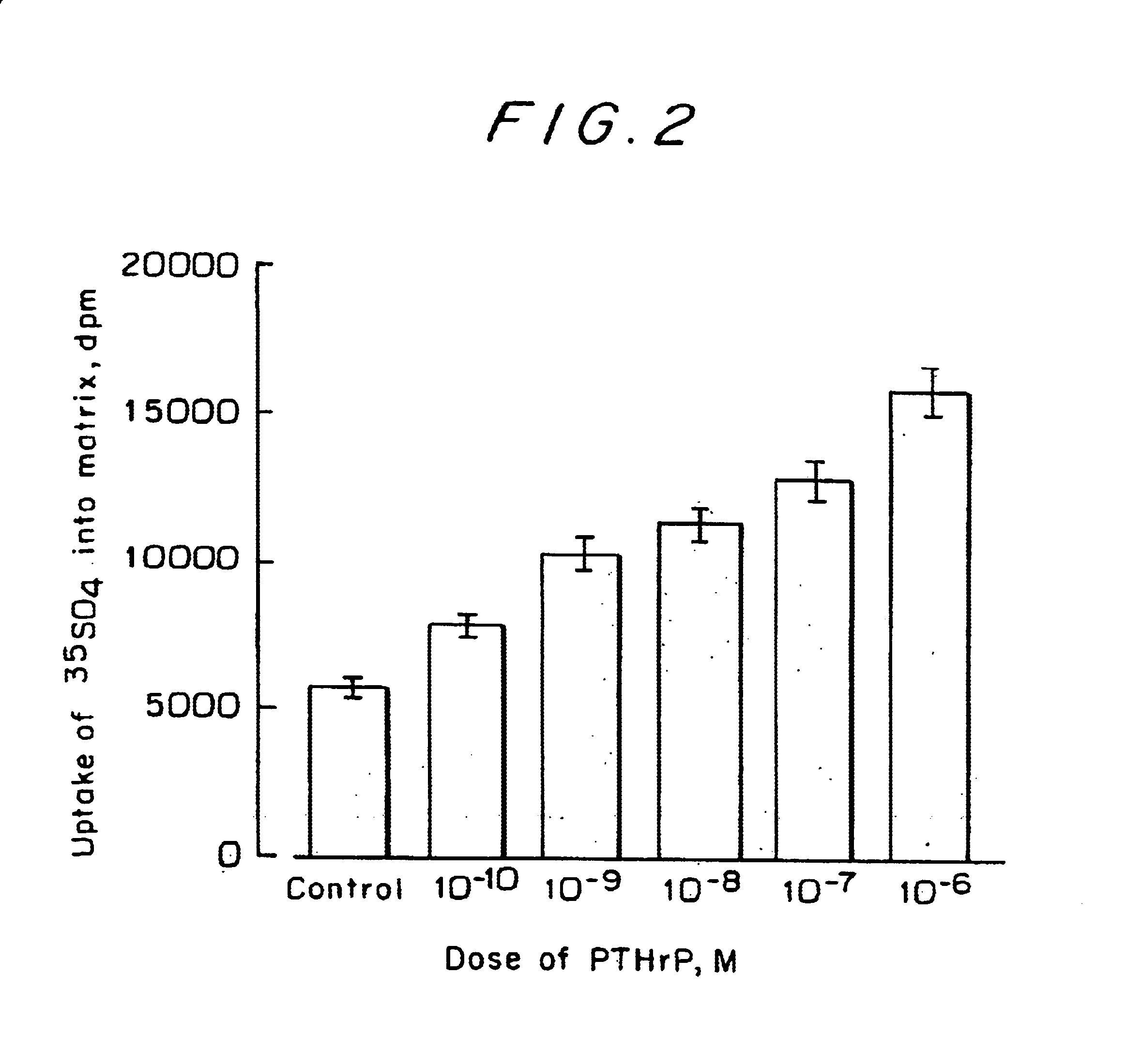
As in Example 1, grown chondrocytes were separated from the subchondral bone to bone transition area of rabbits 4 weeks old from birth by the method of Shimomura et al. (Shimomura, Y. et al., Calcif. Tissue Res. 19, 179-187, 1975). The separated chondrocytes were suspended in an Eagle's minimum essential medium (MEM) containing 10% fetal bovine serum and 50 .mu.g / mL of ascorbic acid and seeded in a 96-well culture plate at a concentration of 5,000 cells in each well. When the cells became confluent, PTHrP was added and, one day later, 1 .mu.Ci of .sup.33 S-sulfuric acid was added per well, followed by 17-h cultivation. The amount of cartilage matrix produced in the supernatant of the culture solution was measured by the method of Kato et al. (Kato, Y. et al., Endocrinology 122, 1991-1997, 1988). Briefly, the quantity of .sup.33 S-sulfuric acid incorporated into proteoglycans was measured.

The results are shown in FIG. 2, which may be regarded as a dose response curve for PTHrP over t...

example 3

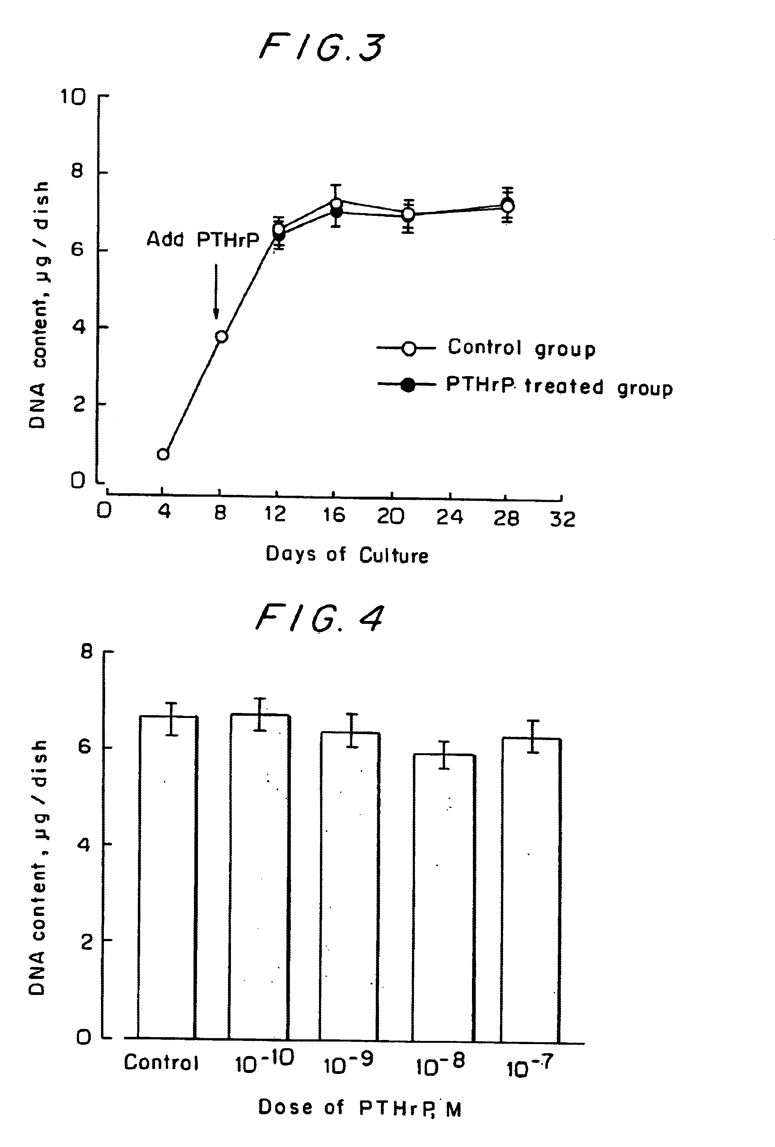
As in Example 1, chondrocytes were cultured in a centrifugal tube. On the 8th day of the cultivation, 10.sup.-7 M of PTHrP was added and the cultivation was continued for 28 days. After the cultivation, the cells were solubilized at appropriate intervals and the time-dependent changes in DNA content were measured. In another experiment, PTHrP was added at varying concentrations from 10.sup.-10 to 10.sup.-7 M and the DNA content on the 21st day of the cultivation was measured.

The time-dependent changes in the DNA content of the chondrocytes are shown in FIG. 3 and the dose-response relationship between PTHrP and the DNA content is shown in FIG. 4. Obviously, the DNA content of the control group increased with time, indicating satisfactory growth of chondrocytes in the centrifugal tube. The DNA content of the chondrocytes was not influenced at all by the addition of PTHrP. This shows that PTHrP is a drug that causes no adverse effects on the growth of chondrocytes.


PUM


Abstract

Drugs for preventing or treating diseases that involve the destruction and degeneration of articular cartilage tissue contain a parathyroid hormone related peptide (PTHrP) or a PTHrP derived substance as an effective ingredient.

Description

TECHNICAL FIELDThis invention relates to drugs having either preventive or therapeutic effect on osteoarthritis and other diseases that involve the destruction and degeneration of the articular cartilage tissue.BACKGROUND ARTOsteoarthritis involves the collapse of the articular cartilage surface and the resulting growth of new cartilage at the articular margins, joint deformity and loss of compliance, which are eventually manifested as the inflammation of the synovial membrane of the joint. Osteoarthritis is divided into two types, primary and secondary. Secondary osteoarthritis has predisposing causes such as trauma and infection that lead to the degeneration of cartilage but no predisposing cause can be identified in primary osteoarthritis. The principal lesion of osteoarthritis is the degeneration of articular cartilage and it may be attributable to the endogenous degeneration of articular cartilage and the mechanical stress on the joint; however, the mechanism of its etiology re...

Claims


Application Information

Patent Timeline
IPC IPC(8): A61K38/29
CPCA61K38/29Y10S514/825A61P19/02
Inventor KATOIWAMOTO, MASAHIROKOIKE, TATSUYA
Owner KATO
Why Eureka
Try Eureka