
Hybrid cells

a hybrid cell and cell technology, applied in the field of hybrid cells, can solve the problems of inability to apply in a clinical treatment setting, inability to achieve the effect of antigen-specific anergy, and inability to achieve the effect of autologous whole tumor cell-based vaccines alone,

Inactive Publication Date: 2005-02-01
ORBIS HEALTH SOLUTIONS
View PDF8 Cites 1 Cited by
  • Summary
  • Abstract
  • Description
  • Claims
  • Application Information

AI Technical Summary

Benefits of technology

A promising approach takes advantage of antigen presentation by cells that lack accessory molecules, like B7. These cells present antigen in the context of MHC, yet, because they lack the accessory interactions required for activation, they induce tolerance specific to the antigen presented. Thus, it is possible to load these cells, which include immature (naïve) B cells, with a specific antigen, and induce antigen-specific anergy. As with the cancer example described above, this antigen-by-antigen approach does not have the general applicability needed for practical clinical use. A methodology is needed which is applicable to any transplant organ, irrespective of the immunogenic antigens the organ displays.

Problems solved by technology

In addition, they are used in a variety instances for research purposes, but their broader application, for example, in a clinical treatment setting has heretofore not been practical.
While these strategies are efficacious against some tumors, their potency is limited because they only enhance the already enfeebled ability of tumor cells to present their “foreign” epitopes to CD8 T-cells, and to generate thereby a tumor-specific cytotoxic T lympocyte (CTL) response.
To date, however, autologous whole tumor cell-based vaccines alone have shown only some isolated or marginal successes.
This approach, however, is limited to application against tumors expressing known tumor antigens.
An additional problem with antigen pulsing techniques is that the antigen presenting system of an APC works more effectively and efficiently when the protein / antigen is synthesized inside the cell rather than outside the cell, a substantial drawback to using antigen-pulsed cells.
However, this gene therapy approach is also fraught with many disadvantages including: 1) the limited ability to identify all of the important specific tumor antigens, 2) the limited ability to map the genes of the specific tumor antigens, 3) only one or a small number of the known tumor antigen genes can be introduced into the dendritic cell and 4) the process is time-consuming and cumbersome.
Since the introduction and selection schemes using markers requires culture and multiple cell division, they cannot be applied to dendritic cells, because DCs are terminally differentiated, non-dividing cells.
Thus, it is no surprise that the previous fusion work relied on well-defined tumor cell lines, bearing such a marker, and DC- and tumor-specific conjugated antibodies, which limits the usefulness of this strategy in cancer treatment.
In summary, the previous cancer-based fusion protocols have the following limitations: 1) they require established tumor cell lines which show specific marker(s); 2) they require both DC and tumor cell specific antibodies to select the fused cells; 3) the selection and expansion of the fused cells takes an impractical amount of time.
Such approaches have the obvious disadvantage of making the patient more susceptible to disorders that otherwise could have been warded off by an intact immune system.
Thus, disruption of either the MHC or the accessory interaction should result in a non-response useful, for example, in preventing transplant rejection.
The problem with such an approach, and the likely reason that it has not be attempted clinically, is that tolerance would pertain to any antigen presented during treatment, not just to transplant antigens.
This would prevent the patient from warding off the pathogen, having perhaps lethal consequences.
As with the cancer example described above, this antigen-by-antigen approach does not have the general applicability needed for practical clinical use.

Method used


Image

  • Hybrid cells
  • Hybrid cells
  • Hybrid cells

Examples


example 1

Animal Studies

This example demonstrates the preparation of certain hybrids between cancer cells and dedritic cells, called dendritomas. These hybrids are used as a cellular vaccine to prevent cancer in a murine metastatic cancer model system.

To prepare dendritic cells from bone marrow, the appropriate number of female C57BL / 6J. mice to support later Dendritoma injections (two mice for every one mouse to be injected) were sacrificed. The femur and tibia of both hind legs were removed from each mouse. Bone marrow was flushed out of the bones using a syringe containing RPMI 1640 with 25 mM Hepes (Gibco BRL). The media containing the bone marrow was filtered through a 40 μm cell strainer into a 50 ml conical centrifuge tube. The bone marrow cells were pelleted by centrifugation at 1500 rpm for five minutes. After removing the supernatant, the tube was gently tapped to loosen the cell pellet. Red blood cell lysis was achieved by adding 5 ml / mouse of ACK Lysing Solution (0.15 M NH4Cl, 1 m...

example 2

Human Studies

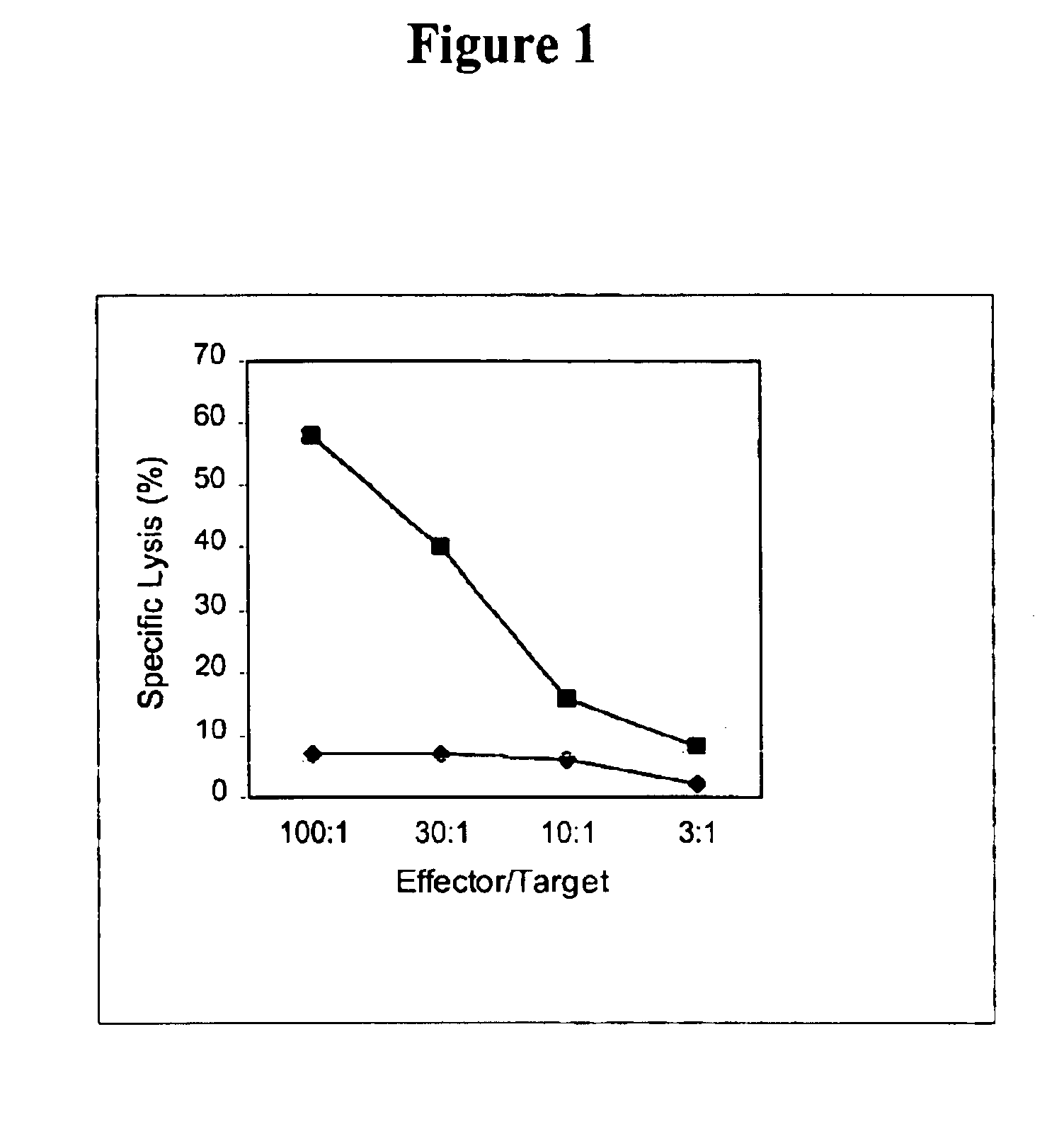
This example demonstrates that the inventive dendritoma hybrid cells induce a cancer cell-specific cytotoxic T cell response. This offers proof of principle that these hybrid cells can induce a therapeutically meaningful immune response against a tumor.

In order to enhance the number of dendritic cells, which were isolated from peripheral blood, a panning technique using anti-CD14 coated plates was utilized. Five hundred micrograms of anti-CD14 antibody was resuspended in 5 ml of 1× PBS with BSA (700 μg of BSA per 100 μl of antibody). A 100 mm tissue culture plate was coated with 5 mls of the antibody. The plate was swirled and incubated for one hour at room temperature or overnight at 4° C. The antibody solution was removed, and the plate was washed five times with 5 mls of 1× PBS.

Peripheral blood mononuclear cells (PBMC's) were isolated from whole blood by obtaining 50 ml of peripheral blood from the patient in preservative-free or sodium heparin tubes. The blood was d...

example 3

Dendritoma Characterization

This example provides further characterization of the reactant cells and the dendritomas described in Examples 1 and 2.

Fluorescent microscopic analysis showed that 100% of the stained cells were successfully labeled. To test whether the dye can interstain between the two different type of cells, green DCs and red tumor cells were mixed together and incubated overnight. Fluorescent microscopic examination showed there was no interstaining. Immediate examination of the fusion product demonstrated that the green DCs and the red tumor cells were fused together and after an overnight recovery, the fused cells showed both colors. The double colored cells (approximately 10% of the total cells), instant dendritomas, were then purified by FACS sorting. More than 95% of the sorted cells were double colored fused cells.

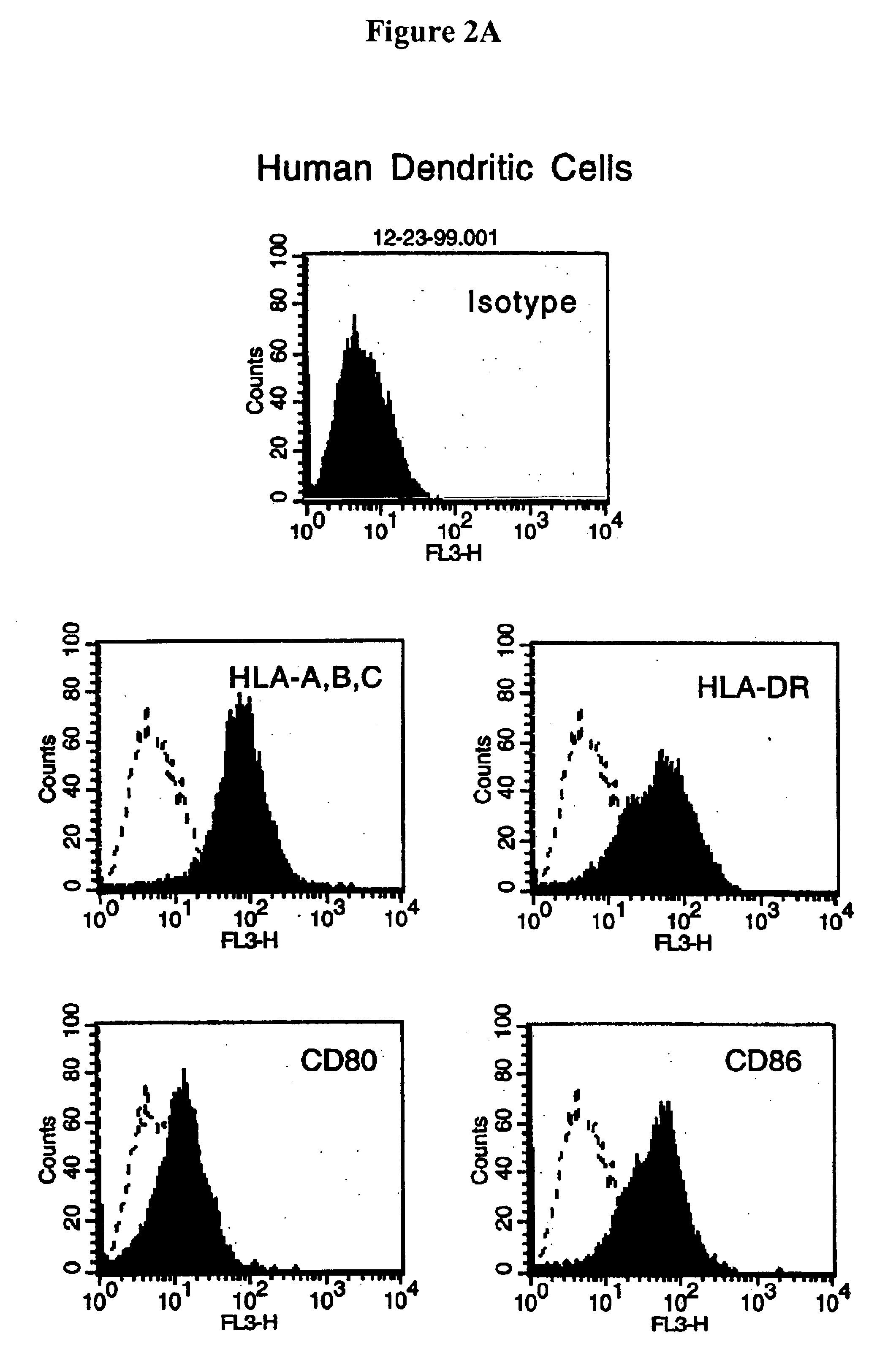
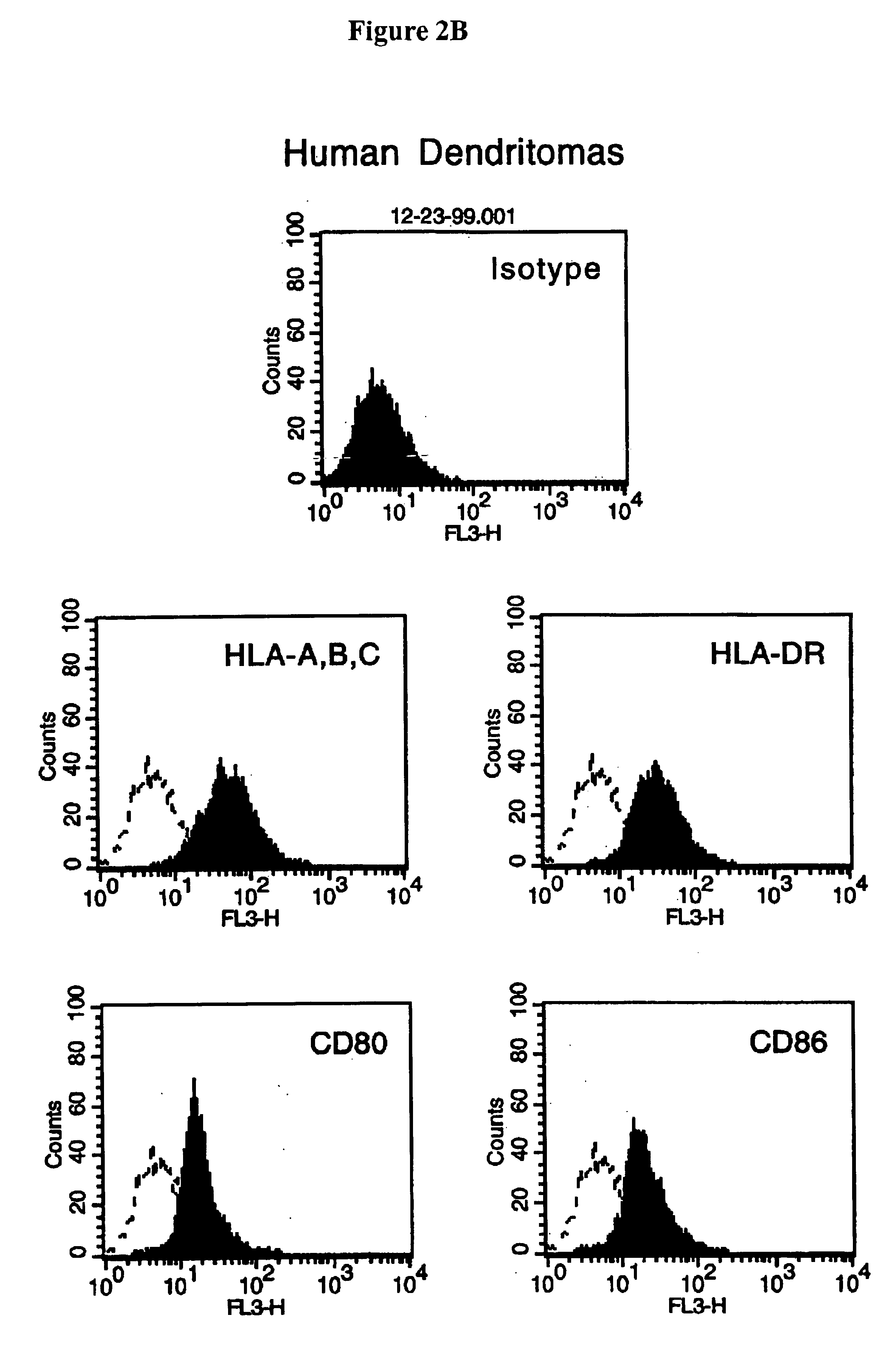
Instant dendritomas express all the molecules necessary for antigen presentation. FACS analysis showed that instant dendritomas express the molecules ...


PUM

PropertyMeasurementUnit
timeaaaaaaaaaa
temperatureaaaaaaaaaa
fluorescence activated cell sortingaaaaaaaaaa

Abstract

A rapid, simple-to-use method for preparing hybrid cells, applicable to fully differentiate, non-dividing cells, entails bringing at least two different cells into contact under conditions that promote cell fusion and then purifying the resultant hybrid without antibiotic or metabolic selection. This approach yields hybrid cells useful in a variety of applications, including clinical treatment regimens, as cellular modulators of the immune system.

Description

BACKGROUND OF THE INVENTIONThe present invention relates to hybrid cells and methods of making and using hybrid cells. Perhaps the most substantial practical application of hybrid cells is the production of hybridomas, which are used to produce monoclonal antibodies. In addition, they are used in a variety instances for research purposes, but their broader application, for example, in a clinical treatment setting has heretofore not been practical. These clinical applications include the cellular vaccines for treating or preventing cancer and other disorders, as well as preventing transplant rejection. The present invention responds to such deficiencies by providing methods and reagents that make the broad applicability of hybrid cells a reality.Although a cancer treatment goal for more than half a century, recent advances in molecular immunology now make immunotherapy a truly viable option for the treatment of patients with cancer and metastatic disease. The past decade has seen the...

Claims


Application Information

Patent Timeline
Patent Type & Authority Patents(United States)
IPC IPC(8): A61K35/14A61K39/00C12N5/16A61K35/12C12N15/02A61K35/13A61K35/15A61K38/00A61P3/10A61P31/00A61P31/04A61P31/10A61P33/00A61P33/02A61P33/04A61P33/10A61P35/00A61P37/02A61P37/04A61P37/06A61P43/00C12N5/02C12N5/07C12N5/0784C12N5/09C12N5/10
CPCA61K35/13A61K35/15A61K38/2013A61K39/0011C12N5/16C12N5/163A61K2039/5152A61K2039/5154A61K2300/00A61P31/00A61P31/04A61P31/10A61P33/00A61P33/02A61P33/04A61P33/10A61P35/00A61P37/00A61P37/02A61P37/04A61P37/06A61P43/00A61P3/10A61K39/4622A61K39/4615A61K2239/31A61K39/464838A61K39/464499A61K2239/57A61K2239/38
Inventor WAGNER, THOMAS E.WEI, YANZHANG
Owner ORBIS HEALTH SOLUTIONS
Learn More