Nanostructured separation and analysis devices for biological membranes

a biological membrane and nanostructure technology, applied in the field of nanostructured matrices fabrication, can solve the problems of lack of reusability, set of separation strategies that rely on this technique, and difficulty in incorporation of these techniques

Inactive Publication Date: 2005-07-05
STC UNM
View PDF50 Cites 200 Cited by
  • Summary
  • Abstract
  • Description
  • Claims
  • Application Information

AI Technical Summary

Benefits of technology

[0014]It is therefore an object of the present invention to provide an e

Problems solved by technology

However, the set of separation strategies that rely on this technique are hampered by: (1) inconvenience of preparation of the variety of gels needed for the separations, (2) inherent inconsistencies in production conditions; and therefore, irreproducibility between different batches of gels, (3) susceptibility of the polymer to degradation under high electric fields, (4) lack of reusability, (5) difficulty in incorporation of these techniques into strategies for development of multi-dimensional (multi-technique) integrated separation systems, and (6) limited resolution and dynamic range of biomolecular separations.
However, their utility is greatly hampered by the need for cumbersome gel preparation protocols and lack of reproducibility.
These currently available systems, however, suffer from a number of drawbacks: (1) the matrices formed are generally composed of non-uniform structures, (2) even where a gradation in size of structures is required, they may be random or at best have to be serially and sequentially arrayed through a cumbersome process of lithography, (3) fabrications of separation devices pose problems in terms of batch-to-batch variations; and consequently, poor reproducibility of results therefrom, (4) lack of efficiency of separation, (5) loss of sample volume, and (6) biomolecules may not be amenable to separation by many of the available systems.
Although such prior work demonstrated that relatively simple 3-dimensional architectures could lead to effective separation, the developments have not gained ground with the biotechnological community.

Method used


Image

  • Nanostructured separation and analysis devices for biological membranes
  • Nanostructured separation and analysis devices for biological membranes
  • Nanostructured separation and analysis devices for biological membranes

Examples


example 1

[0131]Design and construction of microscale electrophoresis cells incorporated much of the characteristics of the present invention into a compact system. The cell preferably has the following characteristics: (1) electrochemical current and fluid flow is restricted to occur only through the separation matrix; (2) loading and stacking functions are included; (3) monitoring of mobility and biomolecular detection is possible (e.g., through fluorescence imaging); and (4) for certain applications, separated compounds are recoverable. Simple methods have been used for incorporating nanostructured silicon / silica chips into electrophoresis cells that satisfy criteria (1-3) above. For example, simple methods of rapid prototyping of elastomeric gasket materials have been used.

example 2

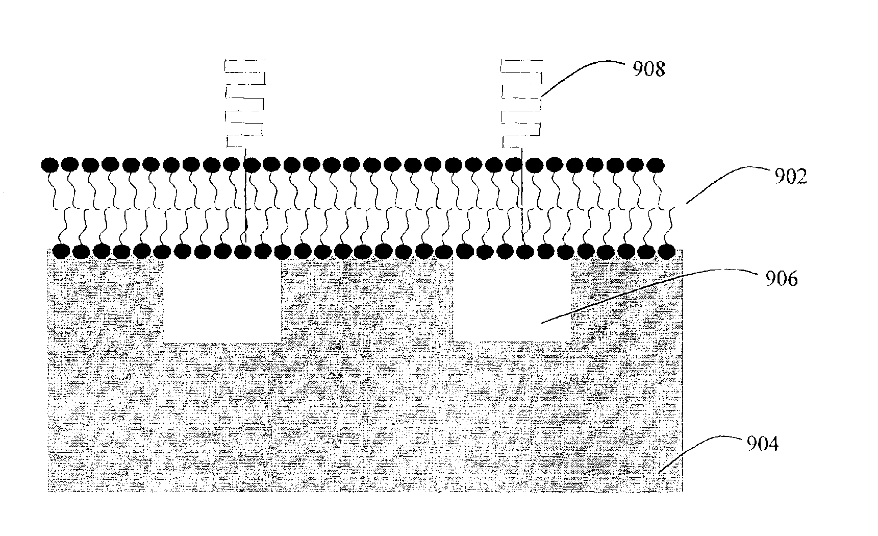
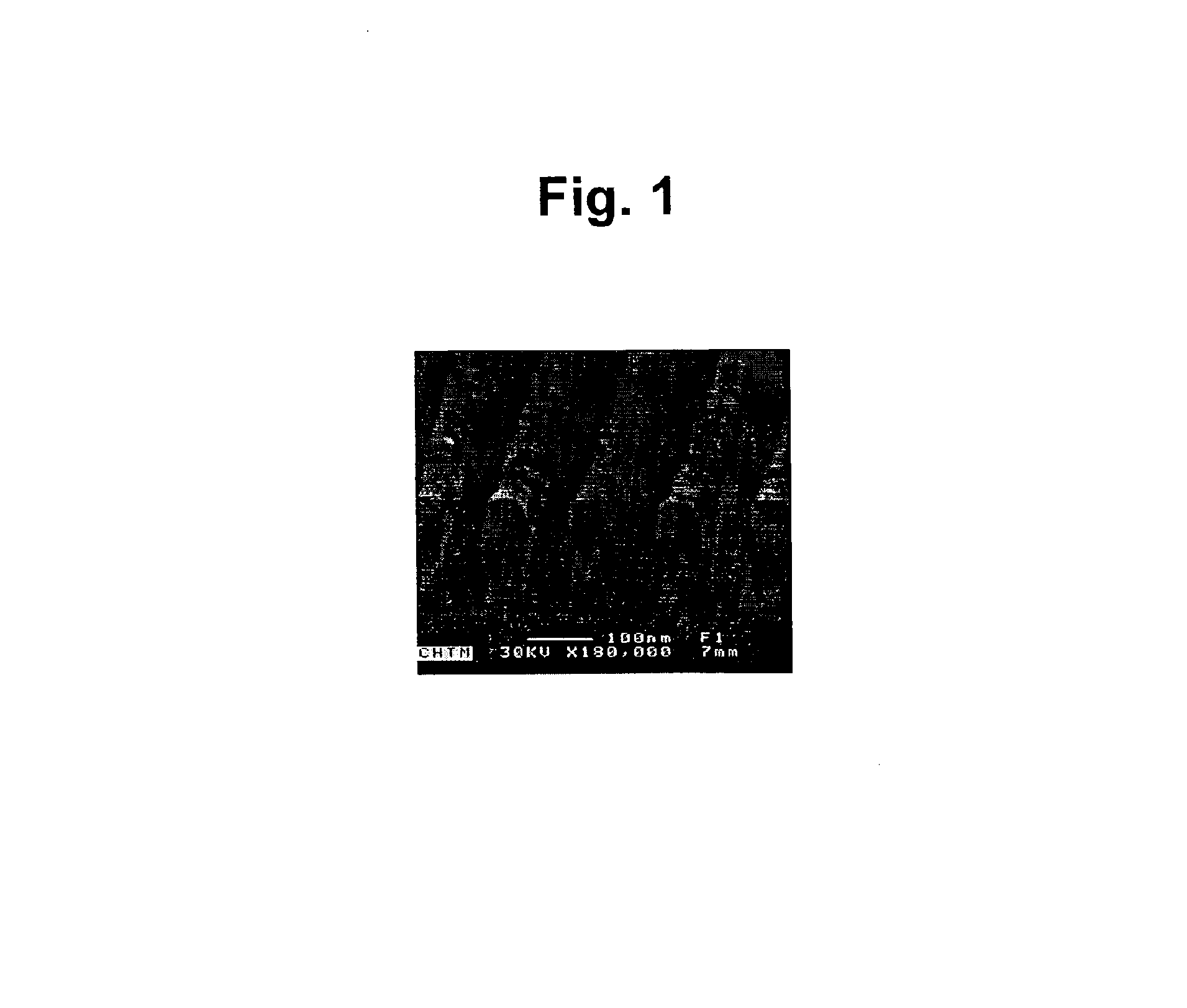
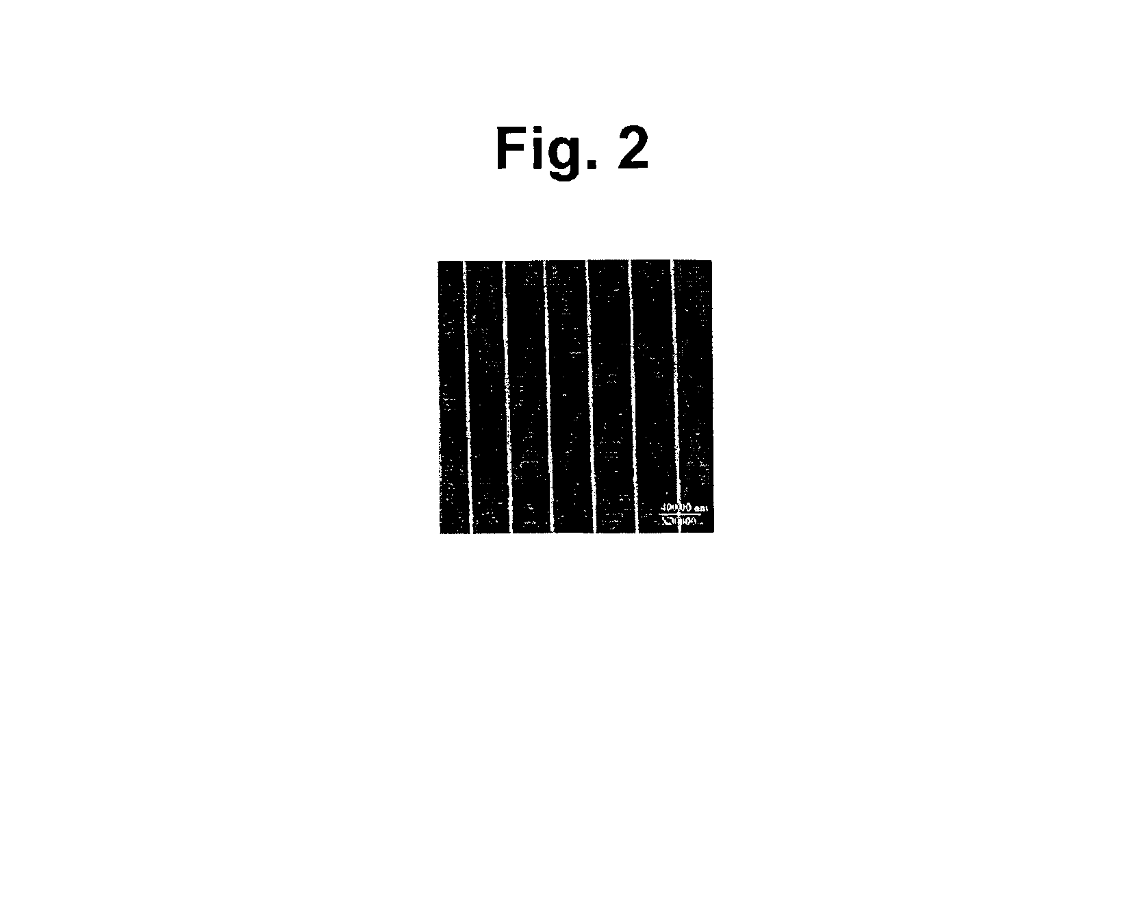
[0132]Supported phospholipid bilayers (SPBs) of egg phosphatidyl choline (Egg PC) were formed by vesicle fusion on nanostructured silicon wafers containing troughs ˜180 nm in width on a 360 nm pitch. An intercalating dye was introduced, and the membranes were imaged by scanning laser microscopy. The resultant fluorescence micrographs indicated that the SPBs formed uniformly over the surface and simple FRAP measurements indicated that the bilayers were fluid and that recovery of fluorescence was preferentially in the direction parallel to the nanotroughs.

example 3

[0133]Transmembrane or membrane associated proteins may be incorporated into an SPB, from incorporation in the vesicle stage, insertion on the membrane or through incorporation of cell ghosts (i.e., intact membranes isolated from cells or organelles). The architecture and / or chemistry of the underlying nanotextured support would be then used to guide the movement of membrane proteins through the supported or suspended bilayer, either by size exclusion of the trough over which the membrane is supported, or by chemical interactions with modifications on the nanostructured support.


PUM

PropertyMeasurementUnit
Forceaaaaaaaaaa
Pressureaaaaaaaaaa
Lipophilicityaaaaaaaaaa

Abstract

The present invention provides a nanostructured device comprising a substrate including nanotroughs therein; and a lipid bilayer suspended on or supported in the substrate. A separation method is also provided comprising the steps of supporting or suspending a lipid bilayer on a substrate; wherein the substrate comprises nanostructures and wherein the lipid bilayer comprises at least one membrane associated biomolecule; and applying a driving force to the lipid bilayer to separate the membrane associated biomolecule from the lipid bilayer and to drive the membrane associated biomolecule into the nanostructures.

Description

CROSS-REFERENCE TO RELATED APPLICATIONS[0001]This application is a Continuation-in-Part of and claims priority to U.S. patent application No. 10 / 073,935, entitled “Nanostructured Devices for Separation and Analysis,” filed on Feb. 14, 2002, now U.S Pat. No. 6,685,841 B2 issued on Feb. 3, 2004, which claims priority to U.S. Provisional Patent Application No. 60 / 268,365, entitled “Nanostructured Devices for Separation and Analysis,” filed Feb. 14, 2001. This application also claims priority to U.S. Provisional Patent Application No. 60 / 347,002, entitled “Nanostructured Devices,” filed on Jan. 11, 2002. The entire contents and disclosures of the above applications are hereby incorporated by reference.GOVERNMENT INTEREST STATEMENT[0002]This invention is made with government support under grant number DAAD19-99-1-0196 awarded by the United States Army Research Office. The government may have certain rights in this invention.BACKGROUND OF THE INVENTION[0003]1. Field of the Invention[0004]...

Claims


Application Information

Patent Timeline
IPC IPC(8): B01D11/00B01D61/14
CPCG01N27/44773B82Y30/00G01N30/00Y10S977/717Y10S977/845Y10T436/255G01N30/88G01N2030/8813Y10T436/25375
Inventor LOPEZ, GABRIEL P.BRUECK, STEVEN R.ISTA, LINNEA K.
Owner STC UNM
Why Eureka
Try Eureka