Non-invasive approach for assessing tumors in living animals

Inactive Publication Date: 2005-08-09
VOGELSTEIN BERT +2
View PDF1 Cites 7 Cited by
  • Summary
  • Abstract
  • Description
  • Claims
  • Application Information

AI Technical Summary

Benefits of technology

[0012]Another embodiment of the invention provides a device for collecting urine from a small mammal. The device has a floor comprising a plurality of liquid-impermeable wells for deposition of urine by a small mammal. The device also has one or more walls enclosing at least a portion of the plurality of wells. The walls of the device are of sufficient height to inhibit or prevent escape of the small mammal. The floor of the device comprises sufficient wells within the walls so that at least 50% of the wells which are occupied by urine are not also occupied by feces after a period of time sufficient for 90% of a population of healthy said small mammals to have urinated.
[0013]Yet another embodiment of the invention provides another device for collecting urine from a small mammal. The device has a floor co

Problems solved by technology

Moreover, the size of subcutaneous tumors often does not reflect the true tumor load, as the volume composed of stroma, necrosis, and scar tissue cannot be distinguished from that occupied by neoplastic cells.
Surgical approaches to detect smaller internal tumors can be useful, but anesthetics and surgical manipulation can di

Method used


Image

  • Non-invasive approach for assessing tumors in living animals
  • Non-invasive approach for assessing tumors in living animals
  • Non-invasive approach for assessing tumors in living animals

Examples


example 1

Materials and Methods

[0050]B 16-F10 (mouse melanoma cell line), EMT6 (mouse mammary carcinoma cell line), and SW480 (human colon carcinoma cell line) cells were obtained from the American Type Culture Collection. B16 and EMT6 cells were maintained in DMEM growth medium (Life Technologies, Gaithersburg, Md.), and SW480 cells were cultivated in McCoy's 5A medium (Life Technologies). Media was supplemented with 10% fetal bovine serum (HyClone, Logan, Utah), 100 U / ml of penicillin, and 100 μg / ml of streptomycin.

[0051]To generate a β-hCG expression vector, a hCG cDNA fragment (534 bp) was amplified via PCR from human placental cDNA (Clontech, Palo Alto, Calif.), using the primers 5′-TGTGCTCTAGATCATGACCAAGGATGGAGATGTTCCAG-3′ (SEQ ID NO:1) and 5′-GCACAGTCTAGATTATTGTGGGAGGATCGGG-3′ (SEQ ID NO: 2). The PCR product was sequenced to confirm that it was mutation-free and cloned through several steps into the pCIneo expression vector (Clontech, Palo Alto, Calif.), generating pCI-hCG. The tgCMV / H...

example 2

Generation of Recombinant Tumor Cells

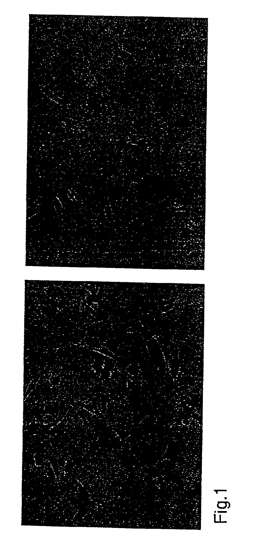
[0052]Cells of three different cancer cell lines were transfected with the β-hCG expression vector as described below. A total of 2×10⁶ cells were transfected with 9 μg of linearized plasmid DNA using the FuGene 6™ reagent (Boehringer Mannheim, Indianapolis, Ind.) according to the manufacturer's instructions. Approximately 70%, 60%, and 25% of tested clones derived from EMT-6, B16, and SW480 cells, respectively, stained uniformly and intensely with an antibody to β-hCG (FIG. 1).

[0053]Transfectants which constitutively expressed β-hCG were maintained in medium supplemented with 10% fetal bovine serum and 2 mg / ml geneticin. EMT6-CG-TK cells, which expressed herpes simplex virus thymidine kinase (TK) as well as β-hCG, were maintained in DMEM with 10% fetal bovine serum, 2 mg / ml geneticin, and 1.5 mg / ml Hygromycin B (Calbiochem, La Jolla, Calif.).

[0054]After ten days of selection in geneticin, clones of the recombinant tumor cells were evaluated for ...

example 3

Urine Collection and Measurement of Urinary β-hCG and Creatinine

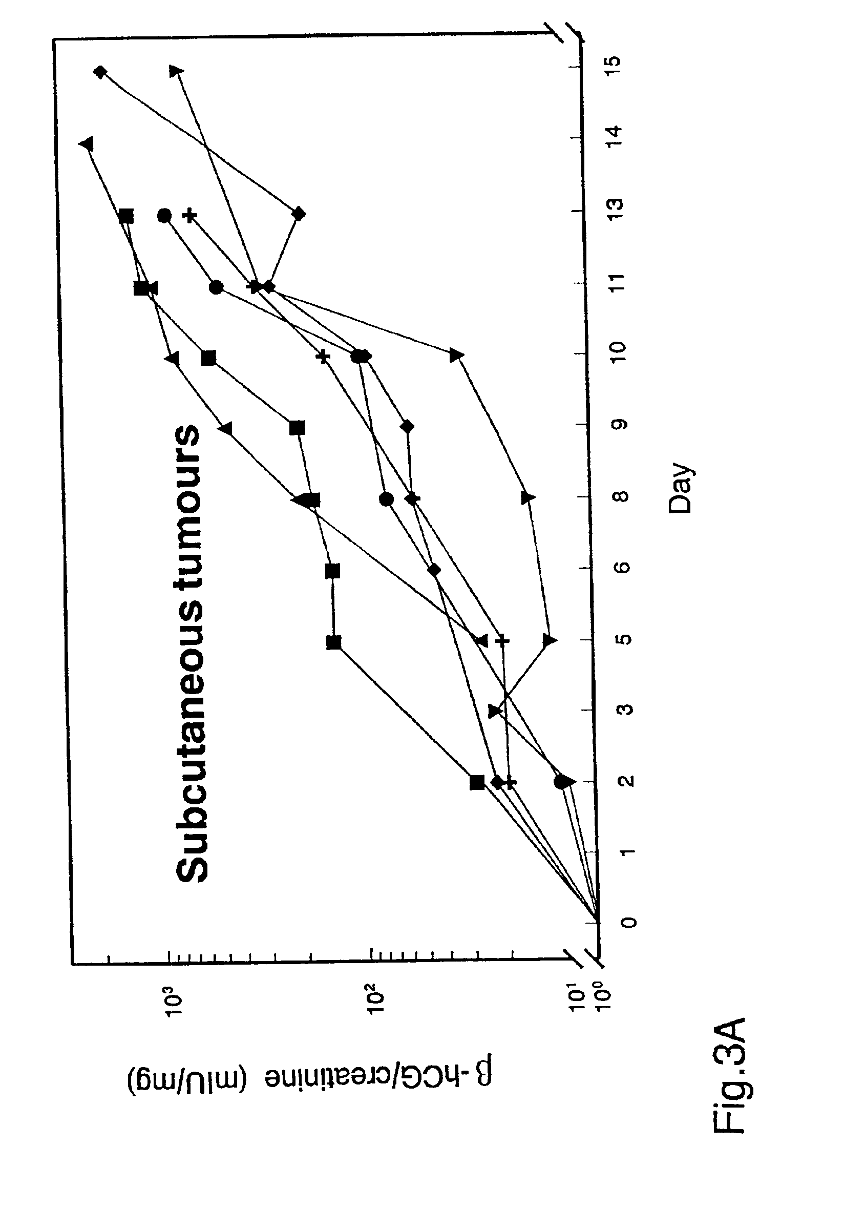
[0056]Female mice, 10-12 weeks old, were obtained from Harlan (Indianapolis, Ind.). For tail vein injections, 105 B 16-CG cells in 0.1 ml PBS were injected into C57BL / 6 mice. For intraperitoneal administration, 2×10⁶ EMT6-CG cells in 0.3 ml PBS were injected intraperitoneally into Balb / c mice. For intrasplenic injection, 106 B16-CG cells in 0.1 ml PBS were injected into the spleens of athymic (nu / nu) mice. For subcutaneous injection, 3×10⁶ B16-CG cells or 8×10⁶ SW480-CG cells in 0.1 ml PBS were injected into subcutaneous tissues adjacent to the lower spine of athymic (nu / nu) mice.

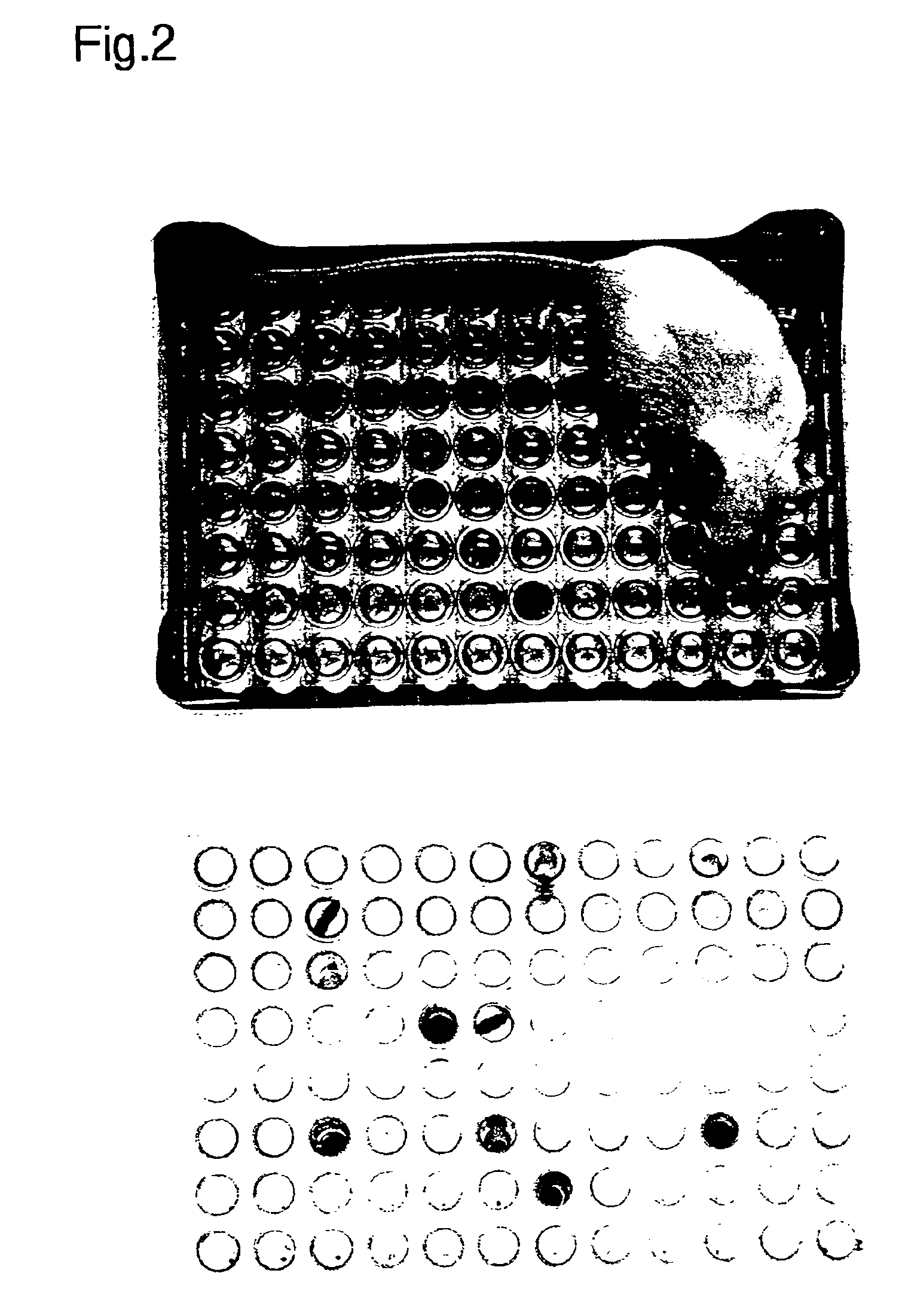
[0057]Standard laboratory plasticware was reconfigured to form a urine collection device. A “house” was made by placing a 96-well 0.2 ml PCR plate (Research Products International Corp., Mount Prospect, Ill.) on an inverted pipette tip rack inside the box (see FIG. 2). Mice were placed in the “house” and the wells were checked for urine deposit...


PUM

PropertyMeasurementUnit
Ratioaaaaaaaaaa
Therapeuticaaaaaaaaaa
Timeaaaaaaaaaa

Abstract

A means for following the growth of experimental neoplasms involves administering recombinant tumor cells containing an expression construct encoding a secretable marker to an experimental animal and measuring secreted marker in the urine of animals bearing tumors formed by such recombinant tumor cells. Urinary marker levels are quantitatively related to tumor loads. Urinary marker can be detected before tumors are grossly visible or clinically apparent. Marker levels decrease following surgical excision or chemotherapeutic treatment, with an estimated half-life of 11 hours. This approach is applicable to the study of many experimental tumor systems.

Description

[0001]This is a Continuation application of parent application Ser. No. 09 / 517,740, filed Mar. 3, 2000, U.S. Pat. No. 6,419,896.[0002]The U.S. Government has a paid-up license in this invention and the right in limited circumstances to require the patent owner to license others on reasonable terms as provided for by the terms of Grant Nos. CA 57345, CA 43460, and CA 62924 awarded by the National Institutes of Health.BACKGROUND OF THE INVENTION[0003]There is no tool more essential to cancer research than the experimental tumor. Every therapeutic and preventative strategy employs such tumors, and tumors growing in animals are instrumental to basic studies of cancer biology. Many experimental tumors are injected subcutaneously because subsequent growth can be followed visually. However, it is often preferable to have tumors grow internally. Such internal growths more closely mimic the environment of naturally occurring tumors, because host factors (blood supply, immune recognition and ...

Claims


Application Information

Patent Timeline
IPC IPC(8): G01N33/574G01N33/74G01N33/76
CPCG01N33/57484G01N33/76G01N2800/52
Inventor VOGELSTEIN, BERTKINZLER, KENNETH W.SHIH, LE-MING
Owner VOGELSTEIN BERT
Why Eureka
Try Eureka