Gene therapy for cerebrovascular disorders

a cerebrovascular disorder and gene therapy technology, applied in cardiovascular disorders, drug compositions, peptide/protein ingredients, etc., can solve the problems of limiting the effect of vegf on endothelial cells, no effective methods for improving the reduced and no effective strategies for improving the reduction of blood flow in these cerebrovascular disorders have been established, so as to achieve the effect of reducing blood flow and delayed neuronal death

Inactive Publication Date: 2005-08-30
ANGES MG INC +1
View PDF16 Cites 14 Cited by
  • Summary
  • Abstract
  • Description
  • Claims
  • Application Information

AI Technical Summary

Benefits of technology

[0012]The present inventors investigated in vivo whether the introduction of HGF gene and VEGF gene can induce angiogenesis on the surface of an ischemic brain. As a result, we have revealed that: (a) after the transfection of HGF gene or VEGF gene, these proteins are detected in the brain over a prolonged period of time, (b) therapy with HGF gene or VEGF gene transfection can induce angiogenesis on the surface of an ischemic brain, (c) the transfection of HGF gene or VEGF gene is effective In treating reduced blood flow in the brain caused by vascular obstruction, and (d) the therapy is also useful when performed before obstruction. Furthermore, we have also demonstrated that the introduction of these genes can be more effectively attained by a novel method of administration i.e., introduction into subarachnoid space.
[0013]In addition, the present inventors have found that delayed neuronal death due to ischemia in the hippocampus CA-1 region can be suppressed by the introduction of HGF gene.

Problems solved by technology

However, no effective methods for improving the reduced blood flow in these cerebrovascular disorders have been established yet.
However, the effectiveness of the strategy in cerebral ischemia has not been known so far.
The binding sites of VEGF including tyrosine kinase receptor Flt, Flk-1, and KDR occur on the endothelial cell but not on other cell types, thereby limiting the effect of VEGF to the endothelial cell.
However, it is not known whether therapeutic angiogenesis using recombinant HGF or VEGF is actually feasible for cerebral ischemia etc.
Furthermore, recombinant angiogenic growth factors rapidly disappear from the brain and thus require continuous injection into the brain, which procedure is rather dangerous and impractical in the clinical settings.

Method used


Image

  • Gene therapy for cerebrovascular disorders
  • Gene therapy for cerebrovascular disorders
  • Gene therapy for cerebrovascular disorders

Examples


experiment i

[0086]A Study on Angiogenesis and Effect of Improving Blood Flow in the Brain with HGF Gene and VEGF Gene

Materials and Experimental Methods

1) Ligation of the Bilateral Carotid Arteries

[0087]Male Sprague Dawley rats (350-400 g; Charles River Japan, Atsugi city, Japan) were anesthetized with pentobarbital sodium (50 mg / kg, intraperitoneal), and were allowed to breathe spontaneously during surgery. By midline neck incision, the bilateral carotid arteries were exposed, and were tightly ligated by 2-0 silk.

2) Preparation of HVJ-liposome Complex

[0088]The method used to prepare HVJ-liposome is as previously described (J. Clin. Invest. 93:1458-1464 (1994); Am. J. Physiol. 271:R1212-1220 (1996)). Briefly, phosphatidyl serine, phosphatidyl choline, and cholesterol were mixed at a weight ratio of 1:4.8:2. Tetrahydrofuran was removed by rotary evaporator to allow the lipid mixture (10 mg) to deposit on the side wall of the flask. The dried lipid was hydrated in 200 μl of a balanced salt solutio...

example 1

Effect of the HVJ-liposome Delivery System on In vivo Transfection of β-galactosidase Gene

[0102]As a gene to be introduced, β-galactosidase gene (manufactured by Invitrogen, concentration in HVJ-liposome: 20 μg / ml) was used to prepare HVJ-liposome as described in the above materials and experimental method.

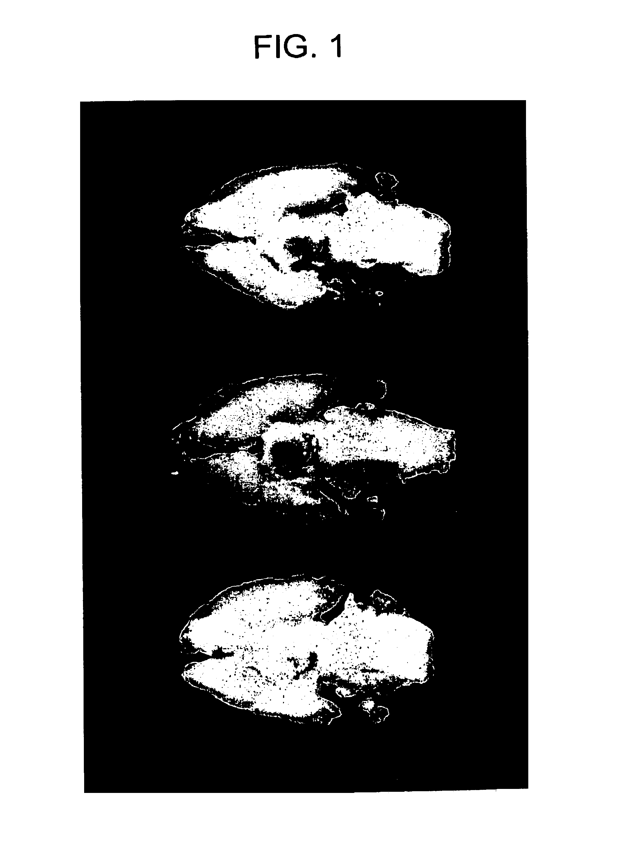
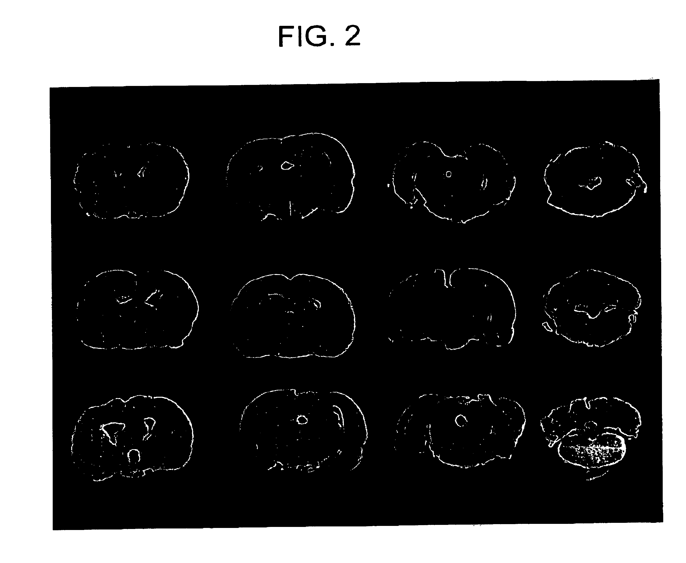
[0103]First, the HVJ-liposome complex was injected directly into the internal carotid artery and was allowed to reach the brain. However, in the intraarterial injection in the above carotid artery, little expression in the brain or microvascular endothelial cells was generated on day 3 and 7 after the injection (data not shown). Therefore, HVJ-liposome was injected into the lateral ventricle and the subarachnoid space. The injection of β-galactosidase gene by the HVJ-liposome method gave rise to marked expression of β-gal on day 3 and 7 after the injection (FIG. 1 and FIG. 2). When injected into the lateral brain, β-gal expression was mainly observed in the lateral ventricle and t...

example 2

In vivo Transfection of HGF Gene and VEGF Gene

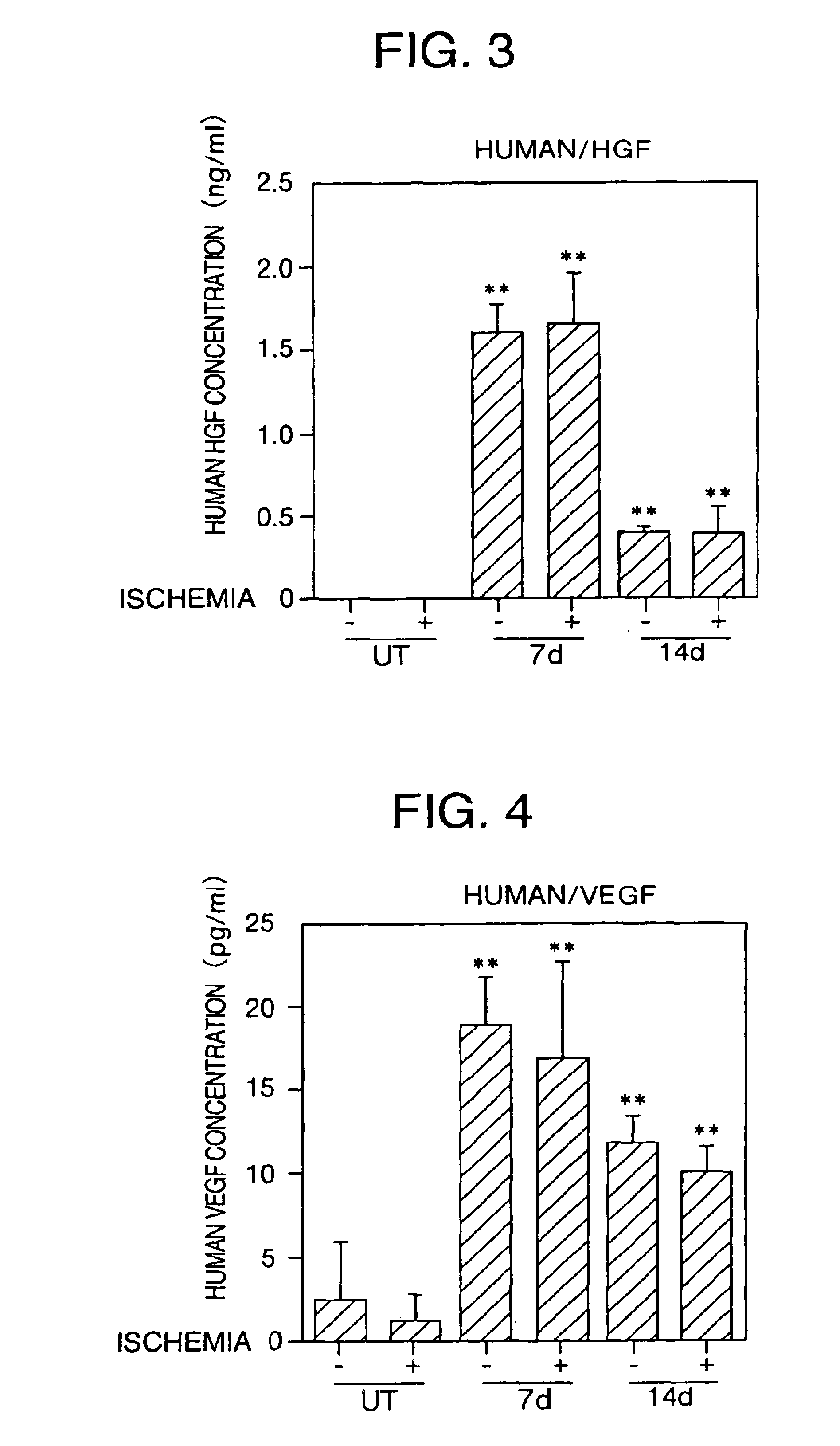
[0104]In order to understand the effect of introduction of HGF gene and VEGF gene, the protein expression of these molecules in the cerebrospinal fluid (CSF) was determined by an ELISA method (n=4, each group). First, human HGF and VEGF were determined in the CSF of the control rats (treated with an expression vector in which no HGF gene or VEGF gene were introduced) before, 7 and 14 days after the obstruction of the bilateral carotid arteries, and no concentration of these proteins was detected (FIG. 3 and FIG. 4).

[0105]Next, the concentration of human HGF protein was determined in the CSF of the rats in which HGF gene (concentration in HVJ-liposome: 20 μg / ml) was introduced into the subarachnoid space immediately after the carotid artery obstruction. On day 7 after transfection, human HGF was detected but not rat HGF (FIG. 3). There were no marked differences observe between the rats (1.63±0.16 ng / ml) in which the carotid artery was ob...


PUM

PropertyMeasurementUnit
weightaaaaaaaaaa
pHaaaaaaaaaa
total volumeaaaaaaaaaa

Abstract

By introducing hepatocyte growth factor (HGF) gene and/or vascular endothelial growth factor (VEGF) gene into the subarachnoid space in humans, cerebrovascular disorders such as cerebrovascular obstruction, cerebral infarction, cerebral thrombosis, cerebral embolism, stroke, cerebral bleeding, moyamoya disease, cerebrovascular dementia, and Alzheimer's dementia can be effectively treated or prevented.

Description

TECHNICAL FIELD[0001]The present invention relates to novel gene therapy agents for treating or preventing cerebrovascular disorders, and novel methods for administration of said gene therapy agents. More preferably, the present invention relates to therapeutic or preventive agents for cerebrovascular disorders comprising hepatocyte growth factor (HGF) gene and / or vascular endothelial growth factor (VEGF) gene as an active ingredient, or novel administration methods comprising administering said therapeutic or preventive agents to the subarachnoid space.BACKGROUND ART[0002]Cerebral obstructive diseases, moyamoya disease and the like caused by atherosclerosis in the cerebral artery often result in chronic reduction in cerebral blood flow. This state may lead not only to the subsequent cerebral ischemic events but also to neuropathological changes including dementia (Stroke 25:1022-1027, Stroke 29:1058-1062 (1998)); Stroke 24:259-264 (1993); Ann. N. Y. Acad. Sci. 695:190-193 (1993)). ...

Claims


Application Information

Patent Timeline
Patent Type & Authority Patents(United States)
IPC IPC(8): A61K38/18A61K38/19A61K48/00A61K35/76A61P9/00A61P9/10A61P25/00A61P25/28A61P43/00
CPCA61K38/1833A61K38/1866A61K48/00A61P25/00A61P25/28A61P43/00A61P9/00A61P9/10
Inventor MORISHITA, RYUICHIOGIHARA, TOSHIO
Owner ANGES MG INC
Why Eureka
Try Eureka