Plastic microfluidics enabling two-dimensional protein separations in proteome analysis

a microfluidic and proteome technology, applied in the field of microfluidic equipment, can solve the problems of inability to detect protein separation in microchannels, time-consuming tasks prone to irreproducibility and poor quantitative accuracy, and lack of methods to introduce different separation media into different dimensions in the same unit, etc., to achieve high resolution detection of proteins, low florescence, and simple procedures

Inactive Publication Date: 2005-12-13
CALIBRANT BIOSYST +1
View PDF23 Cites 38 Cited by
  • Summary
  • Abstract
  • Description
  • Claims
  • Application Information

AI Technical Summary

Benefits of technology

[0010]One advantage of the invention is that it overcomes these and other drawbacks in existing systems by providing a microfluidic apparatus for performing 2-D biomolecular separations. The microfluidic 2-D device may comprise first and second planar substrates which include at least a first dimension microchannel extending in a first direction and an array of second dimension microchannels extending in a second direction, preferably, orthogonal to the first dimension. The ends of at least some of the microchannels are in fluid communication with a plurality of reservoirs. The substrates may further comprise a number of microchannels and reservoirs. The reservoirs are in electrical communication with a plurality of electrodes and voltage power sources. The device enables two dimensional separations of proteins and other biomolecules. According to another aspect of the invention, an isoelectric point based separation is enabled in a first dimension, and a size based separation in a second dimension.
[0011]Another advantage of the invention is that it enables introduction of two different media in different microchannels of the same 2-D microfluidic device (e.g., a media for isoelectric point based separation, and a media for size based separation). In one embodiment, a pressure filling technique may be used to introduce the two different media. Electroosmotic or other electrokinetic pumping may also be used to introduce the two different media. In some embodiments, a polymeric membrane sandwiched between the upper and the lower microchannels may serve as a hydrodynamic barrier, enabling the introduction of two different separation media in the upper and the lower microchannels. Other filling approaches may be used.
[0012]Another advantage of the invention is that it enables simultaneous transfer of proteins from first dimension microchannels to second dimension microchannels (e.g., by changing the electric potentials at the reservoirs connected to the microchannels). Any separation accomplished in the first dimension may be completely retained upon transfer to the second dimension. In some embodiments, the transfer of material (e.g. proteins) from the first to the second dimension may be achieved by hydrodynamic pressure at the reservoirs connecting first dimension microchannels. In other embodiments, isoelectric focused proteins in the first dimension may be electrokinetically injected into the second dimension, by altering the electric potentials at the reservoirs connecting microchannels. This simultaneous transfer approach also significantly simplifies the procedures compared to those involved in continuous sampling and separation of the eluants from the first dimension.
[0013]Another advantage of the invention is that it enables high resolution detection of proteins in microchannels. In one embodiment, proteins may be covalently labeled with a florescent dye. During first and second dimension separations, the labeled proteins may be monitored using a florescent detector attached to the microfluidic system. In some embodiments, microchannels fabricated by polydimethylsiloxane (PDMS) substrates may be used which provide low florescence background during detection and enable better signal to background resolution. According to another embodiment of the invention, laser induced florescent detection (LIFD) may be employed for the detection of SDS-protein complexes using non-covalent, environment-sensitive, fluorescent probes.
[0014]Another advantage of the invention is that it enables integration of 2-D microfluidic networks formed in plastic substrates (e.g., using template imprinting, injection molding, laser machining, or a combination of these technologies) with LIFD and mass spectrometry detection for automation, high throughput, reproducibility, robustness, and ultrahigh resolution. These capabilities are advantageous for large-scale proteome analysis and for “differential display” of protein expressions under various physiological conditions.
[0015]The microfluidic 2-D PAGE of the present invention may be advantageous for the study of organisms having fully sequenced genomes, and may identify proteins (and their modifications in many cases) as well as provide quantitative measurements of expression levels. Other uses will be apparent.

Problems solved by technology

Despite the selectivity of 2-D PAGE, existing techniques are a collection of manually intensive procedures and time-consuming tasks prone to irreproducibility and poor quantitative accuracy.
One drawback of existing systems is a lack of methodology to detect protein separations in microchannels.
Another drawback of the application of existing microfluidic techniques to 2-D PAGE devices is a lack of methods to introduce different separation media into different dimensions in the same unit.
Another drawback of the application of existing microfluidic techniques to 2-D PAGE devices is a lack of methods to transfer proteins simultaneously from first to second dimensions without significant loss in resolution.
To date, sufficient resolution has not been achieved using existing methods.

Method used


Image

  • Plastic microfluidics enabling two-dimensional protein separations in proteome analysis
  • Plastic microfluidics enabling two-dimensional protein separations in proteome analysis
  • Plastic microfluidics enabling two-dimensional protein separations in proteome analysis

Examples


Embodiment Construction

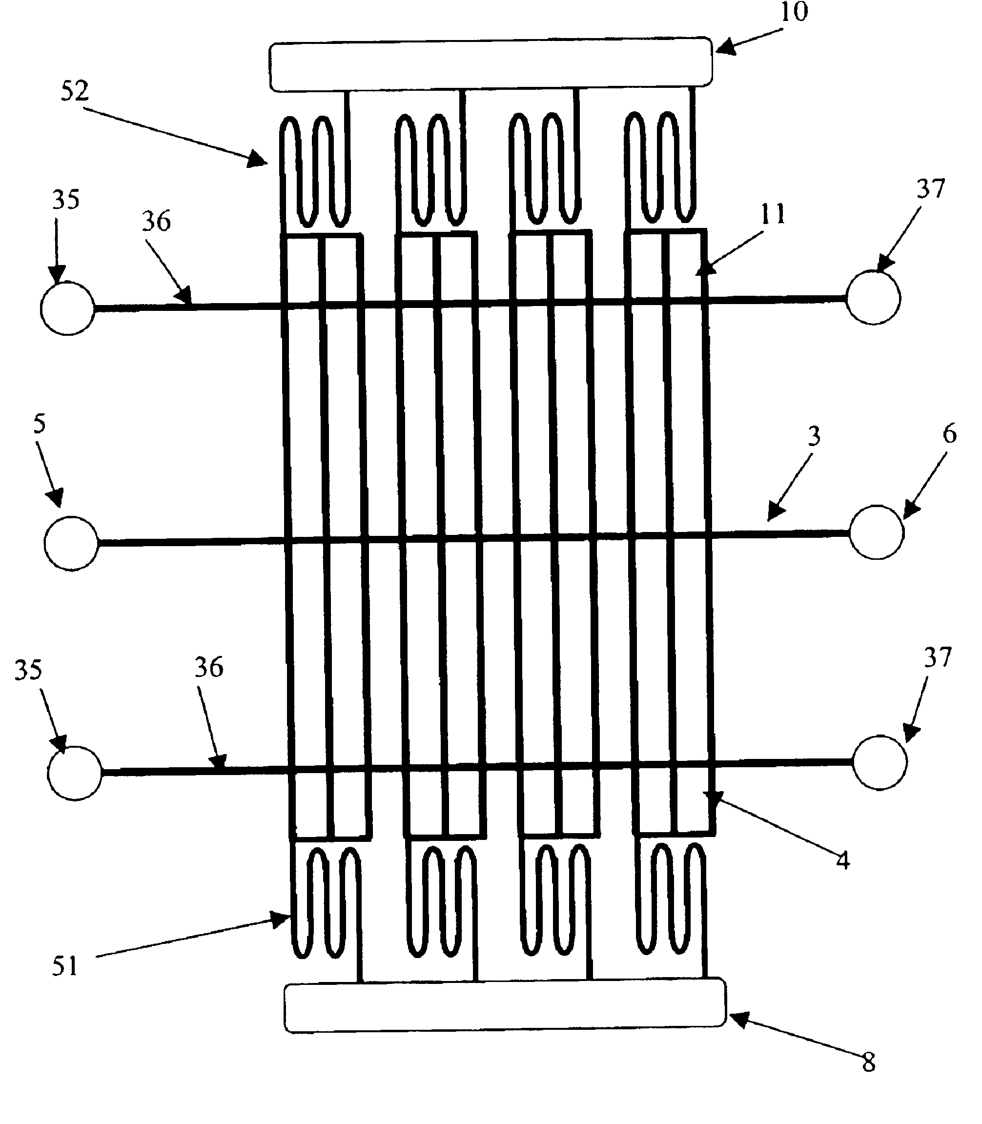
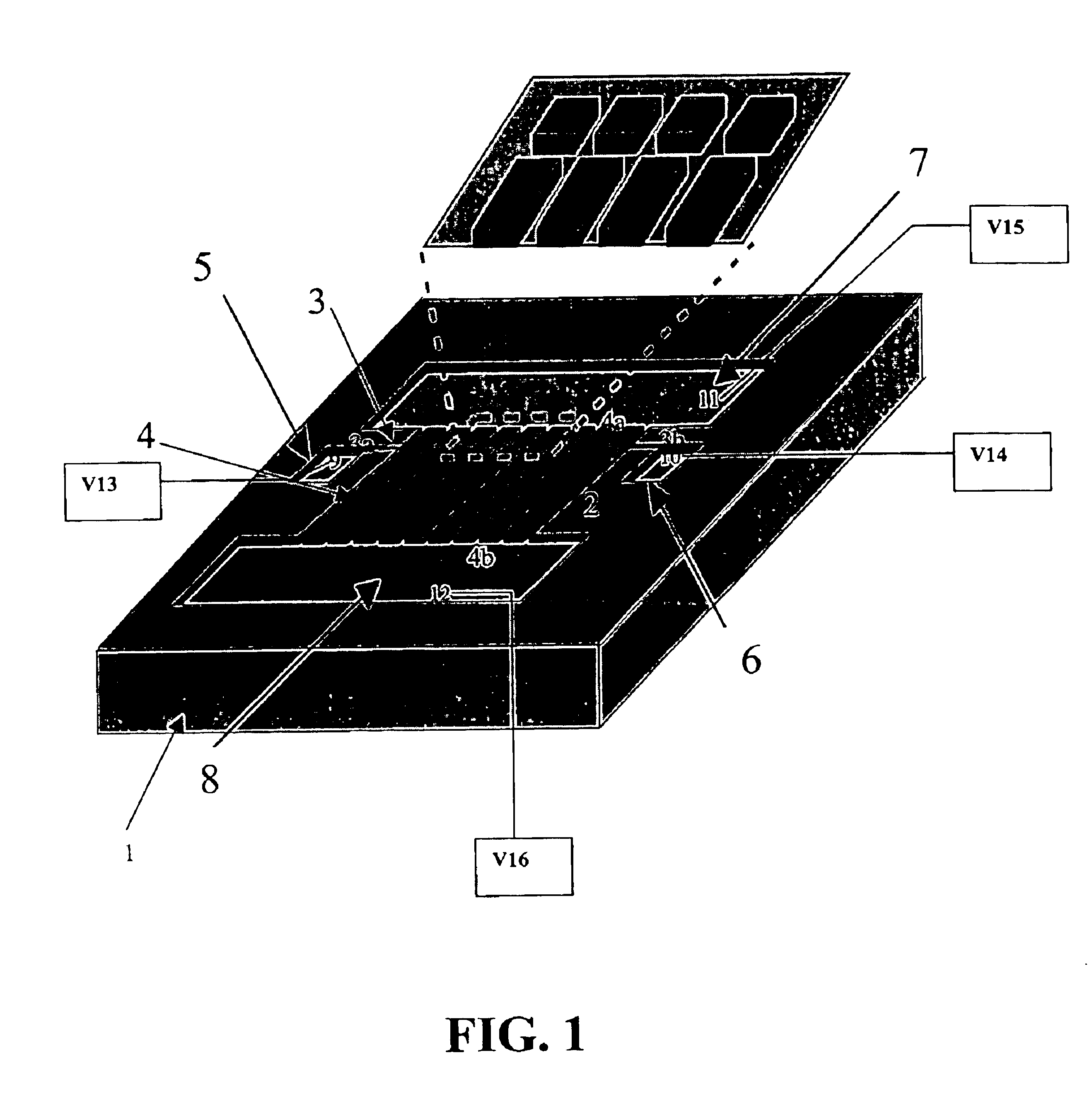
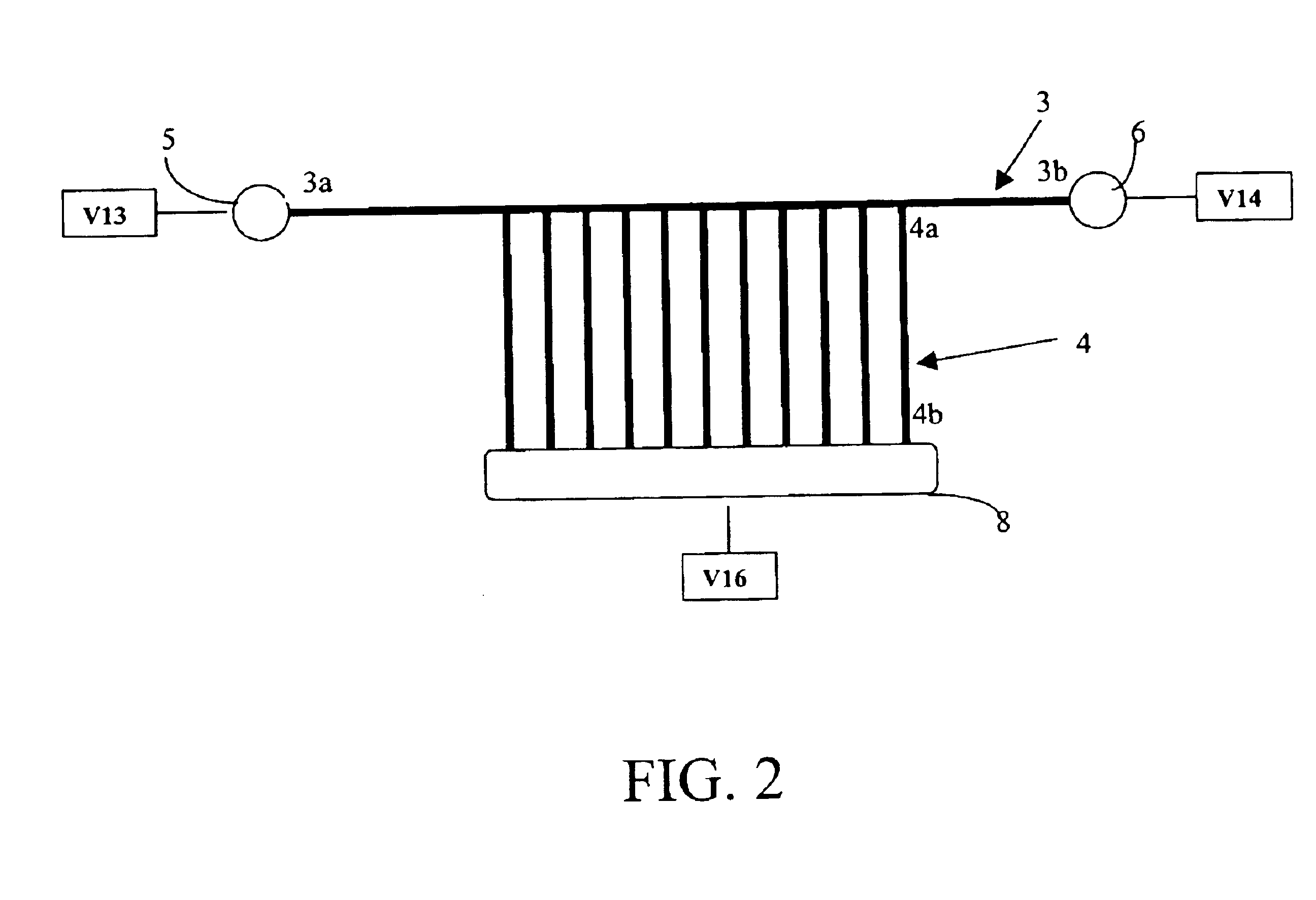
[0028]According to one embodiment of the invention as illustrated in FIG. 1, for example, a microfluidic 2-D gel electrophoresis apparatus is provided. Microfluidic 2-D gel electrophoresis apparatus may comprise a first planar substrate 1 containing one or more first-dimension microchannels 3 for first dimension separation, as well as a second planar substrate 2 (bonded to first planar substrate 1) to provide enclosure for one or more second-dimension microchannels 4 for second dimension separation.

[0029]According to one embodiment, the first-dimension microchannel 3 may extend in a first direction, while an array of one or more second-dimension microchannels 4 may extend from, or intersect with, the first-dimension microchannel 3 in a second direction. Preferably the second direction is orthogonal to the first direction. The first-dimension microchannel 3 may have a first end 3a and a second end 3b. Similarly, an array of one or more second-dimension microchannels 4 may each have a...


PUM

PropertyMeasurementUnit
inner widthaaaaaaaaaa

Abstract

The invention provides a microfluidic apparatus for performing 2-D biomolecular separations. The microfluidic 2-D device may include first and second planar substrates which include at least a first dimension microchannel extending in a first direction and an array of second dimension microchannels extending in a second direction, preferably, orthogonal to the first dimension. The ends of at least some of the microchannels are in fluid communication with a plurality of reservoirs. The substrates may further include a number of microchannels and reservoirs. The reservoirs are in electrical communication with a plurality of electrodes and voltage power sources. The device enables two dimensional separations of proteins and other biomolecules. According to another aspect of the invention, an isoelectric point based separation is enabled in a first dimension, and a size based separation in a second dimension.

Description

CROSS-REFERENCE TO RELATED APPLICATIONS[0001]This application claims priority from U.S. provisional patent application Ser. No. 60 / 287,754 filed on May 1, 2001, which is incorporated by reference in its entirety.GOVERNMENT LICENSE RIGHTS[0002]The U.S. Government has a paid-up license in this invention and the right in limited circumstances to require the patent owner to license others on reasonable terms as provided for by the terms of Grant Numbers: R43CA092819, and R43GM062738, awarded by the National Institutes of Health, and Grant Number: DAAH01-02-C-R136, awarded by the Defense Advanced Research Projects Agency.FIELD OF THE INVENTION[0003]The invention relates to a microfluidic apparatus and methods for using the apparatus to perform two-dimensional (2-D) protein separations.BACKGROUND OF THE INVENTION[0004]Existing protein analysis technology is largely based upon two-dimensional polyacrylamide gel electrophoresis (2-D PAGE), which has undeniably assumed a major role and is ce...

Claims


Application Information

Patent Timeline
Patent Type & Authority Patents(United States)
IPC IPC(8): B81B1/00G01N27/447
CPCG01N27/44773G01N27/44791G01N27/44795
Inventor LEE, CHENG SHENGDEVOE, DON
Owner CALIBRANT BIOSYST
Why Eureka
Try Eureka