
Computer system and a method for segmentation of a digital image

a computer system and digital image technology, applied in image enhancement, image analysis, instruments, etc., can solve the problems of reducing objectivity, unable to fully automate the thresholding and morphology system in many cases, and many specific segmentation problems that have not been satisfactorily solved, etc., to achieve fast and with computational costs, increase image resolution, and accurate measurement of volume

Active Publication Date: 2006-01-10
FRAUNHOFER GESELLSCHAFT ZUR FOERDERUNG DER ANGEWANDTEN FORSCHUNG EV
View PDF26 Cites 90 Cited by
  • Summary
  • Abstract
  • Description
  • Claims
  • Application Information

AI Technical Summary

Benefits of technology

[0031]It is another object of the present invention to provide for a segmentation procedure that is robust even against considerable image non-uniformity as well as image noise.
[0078]In order to reduce noise and artifacts, or to increase or decrease the amount of information in an image, resampling and interpolation of the original image data can be applied prior to the WT.

Problems solved by technology

Still many specific segmentation problems persist that have not been solved satisfactorily.
There are several problems with manual segmentation preventing it from being routinely used in many cases: (i) Interaction and time costs (manual tracing takes several minutes per image), (ii) requirement for sufficient training (the segmentation result is highly dependent on the level of training), (iii) requirement for care and concentration during segmentation (quality and precision of human interaction is not constant), and (iv) reduction of objectivity (different operators will generate different results).
In most cases, however, the structure representing the brain in the binary image is connected to neighboring structures (caused by nerves, blood vessels, image non-uniformity, or limitations of image resolution).
A fully automated system of thresholding and morphology tends to fail in many cases, e.g., when the image acquisition parameters vary.
Furthermore, the results often need final manual editing.
The algorithm will fail in presence of moderate contrast non-uniformity, since no threshold can be found that is appropriate for all parts of the image.
In general, this weakness against image inhomogeneities is characteristic for all segmentation methods comprising global thresholding.
Statistical segmentation methods are useful, but target another class of segmentation problems.
In general, statistical methods only converge to an appropriate solution after the original image has been segmented, so that a small set of known tissue types remains plus additional noise and non-uniformity.
Conversely, statistical methods such as described in U.S. Pat. No. 5,425,368 are not applicable to the original MR images directly.
Compared with manual image analysis, the semi-automated method resulted in smaller systematic and random differences for volume, ejection fraction, and wall thickness of the left cardiac ventricle.
A problem with template driven segmentation is caused by geometrical or anatomical variations.
For pathological cases, e.g., templates often are not applicable.
Interaction techniques integrated in template based segmentation algorithms can only serve to constrain parameters, but not to deal with variations beyond the model.
However, there is a class of problems that can be solved by the direct application of the WT.
In these cases the main problem consists of separating an object of interest from neighboring structures that exhibit a similar image intensity, but are separated by a thin layer of slightly darker or brighter tissue.
However, the gradient information of an image, to which the WT is applied in accordance with the prior art, is only suited to yield appropriate results if the object borders exhibit image gradients that are higher than the gradients inside the object.

Method used


Image

  • Computer system and a method for segmentation of a digital image
  • Computer system and a method for segmentation of a digital image
  • Computer system and a method for segmentation of a digital image

Examples


Embodiment Construction

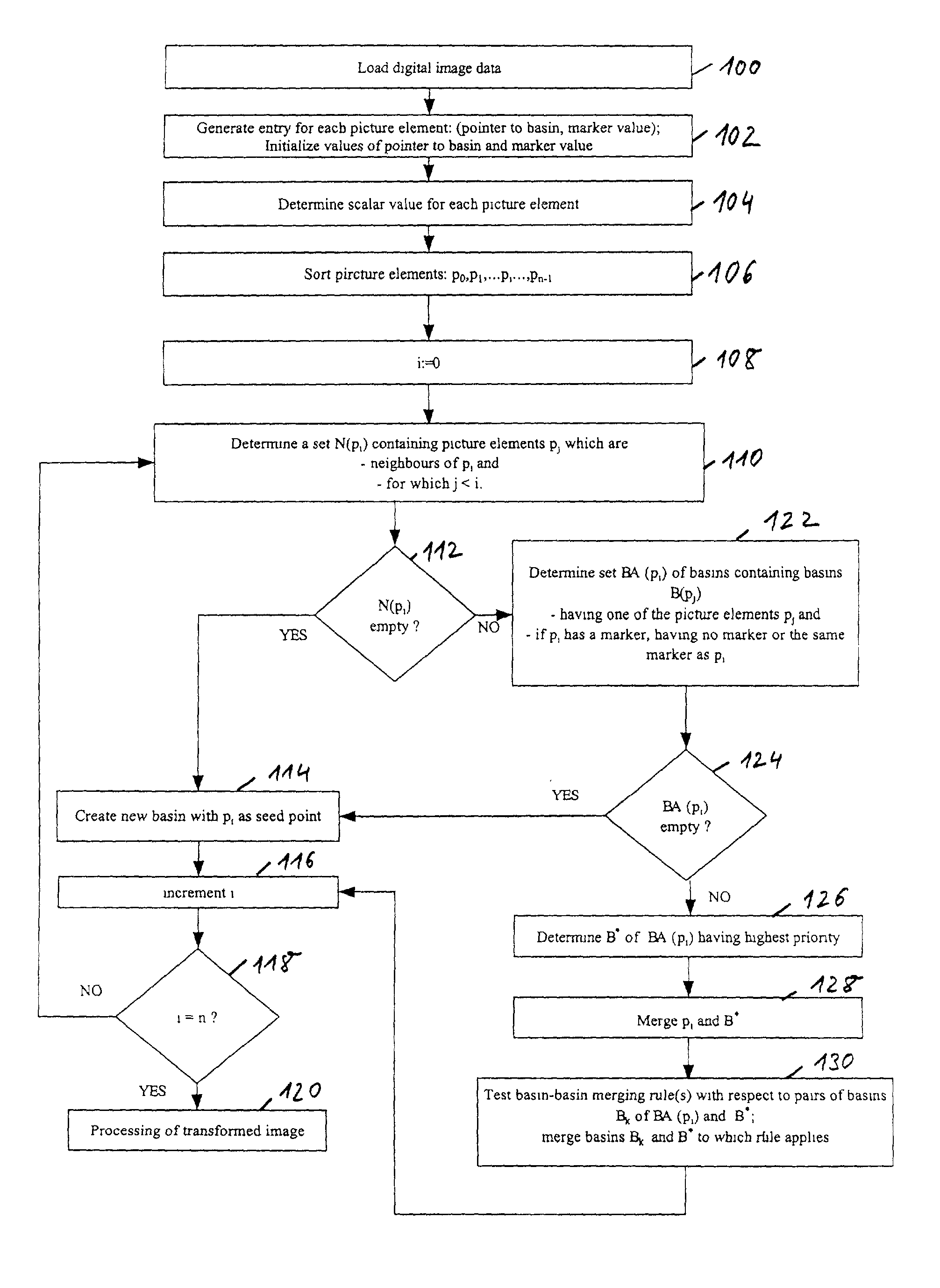
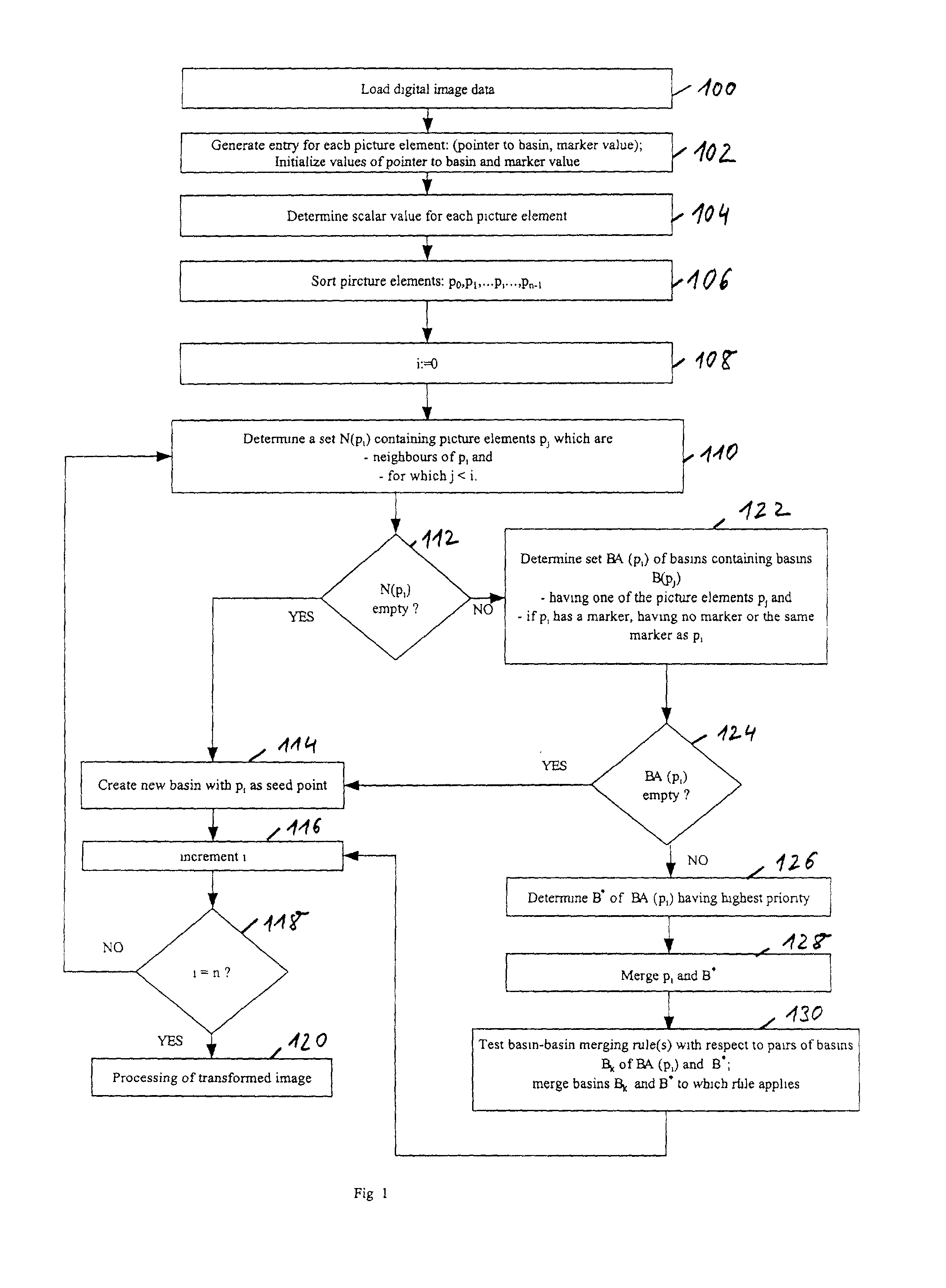
[0098]FIG. 1 shows a preferred embodiment of a watershed transformation method in accordance with a first embodiment of the invention.

[0099]In step 100 digital image data is loaded. The present invention is not restricted with respect to the kind of image data and is broadly applicable to a variety of classes of image data. The present invention is particularly useful for medical image data, in particular digital image data acquired from computer tomographic images, magnetic resonance images and T1-weighted magnetic resonance images. Further it can be advantageously employed to other radiologic images.

[0100]In step 102 a data entry is generated for each picture element of the digital image data. Typically digital image data comprises a large number of picture elements, i.e. pixels for two-dimensional images and voxels for three-dimensional images. In addition there are also four-dimensional images where the fourth dimension is time. Such four-dimensional images are acquired for obse...


PUM


Abstract

The invention relates to a method for segmentation of a digital image being represented by picture element data for the purposes of visualization, segmentation and quantification, such as determining the volume of a segment. The method comprises the steps of performing a watershed transformation of the digital image by applying a watershed transformation method on the picture element data to provide one or more basins and post-processing by processing the picture element data belonging to at least one of the basins, where post-processing is one or a combination of the following steps: (i) gray-scale based segmentation, (ii) visualization using a transfer function, or (iii) volumetry using histogram analysis.

Description

FIELD OF THE INVENTION[0001]The present invention relates to the field of segmentation of digital images, in particular medical digital images, and more particularly to the field of visualization of segments corresponding to anatomical structures and to the measurement of the volumes of such anatomical structures.BACKGROUND AND PRIOR ART[0002]Segmentation is a crucial task in visualization and quantification based on digital medical image data. The number of digitally stored medical images and the amount of information stored in each image both are steadily increasing, thus increasing the importance of robust and fast segmentation methods at the same time. Robustness refers to the ability of the method to: (i) work in presence of image disturbance, (ii) with a low failure rate, and (iii) to work for a wide range of different images and patients.[0003]Still many specific segmentation problems persist that have not been solved satisfactorily. Each problem is defined by: (i) the imagin...

Claims


Application Information

Patent Timeline
IPC IPC(8): G06K9/00G06T5/00G06T7/60
CPCG06K9/342G06T7/0081G06T7/0091G06T7/602G06K2209/05G06T2207/30048G06T2207/10088G06T2207/20152G06T2207/30016G06T7/11G06T7/155G06T7/62G06V10/267G06V2201/03
Inventor HAHN, HORST
Owner FRAUNHOFER GESELLSCHAFT ZUR FOERDERUNG DER ANGEWANDTEN FORSCHUNG EV
Learn More