Method of treating edematous retinal disorders

a retinal disorder and retinal edema technology, applied in the field of retinal edema, can solve the problems of no pharmacological approach employed in the treatment of rhegmatogenous retinal, loss of vision, irreversible blindness,

Inactive Publication Date: 2006-07-18
MERCK SHARP & DOHME LLC
View PDF20 Cites 13 Cited by
  • Summary
  • Abstract
  • Description
  • Claims
  • Application Information

AI Technical Summary

Benefits of technology

[0015]The pharmaceutical formulations useful in this invention comprise adenine-, uridine-, and cytidine-containing dinucleoside polyphosphates, and derivatives thereof, and hydrolysis-resistant monucleoside triphosphates, which are selective agonists of the P2Y receptor on epithelial cells of the retinal pigment epithelium.

Problems solved by technology

Retinal detachment or edema in the central part of the retina (macula) produces significant loss of vision, and can ultimately lead to irreversible blindness.
There are no pharmacological approaches employed in the treatment of rhegmatogenous retinal detachment (RRD).
Intra-retina accumulation of fluid in the macula results in decreased visual acuity and is the most common cause of vision loss in patients with diabetic retinopathy, AMD and other ischemic retinopathies such as branch and central retinal vein occlusion.
Macular edema is also a major cause of decreased vision following intraocular surgery (called cystoid macular edema).
Breakdown of the blood-retinal barrier can result in focal leakage of fluid from the vasculature and fluid accumulation within retinal layers or in the subretinal space.
These therapies show limited utility in patients.
A number of pharmacological and surgical approaches are employed to treat cystoid and diabetic macular edema, but they are generally considered empirical and often ineffective.
Acetazolamide also alleviates certain types of macular edema and is postulated to work via activation of the RPE pump, but systemic tolerance to acetazolamide is poor.
However, both ATP and UTP are rapidly degradeable by ubiquitous extracellular nucleotidases.
Drug delivery into the subretinal space is widely regarded to be unacceptably risky for patients because it involves the insertion of a needle between the retina and RPE, which can result in complications and blindness.

Method used


Image

  • Method of treating edematous retinal disorders
  • Method of treating edematous retinal disorders
  • Method of treating edematous retinal disorders

Examples


example 1

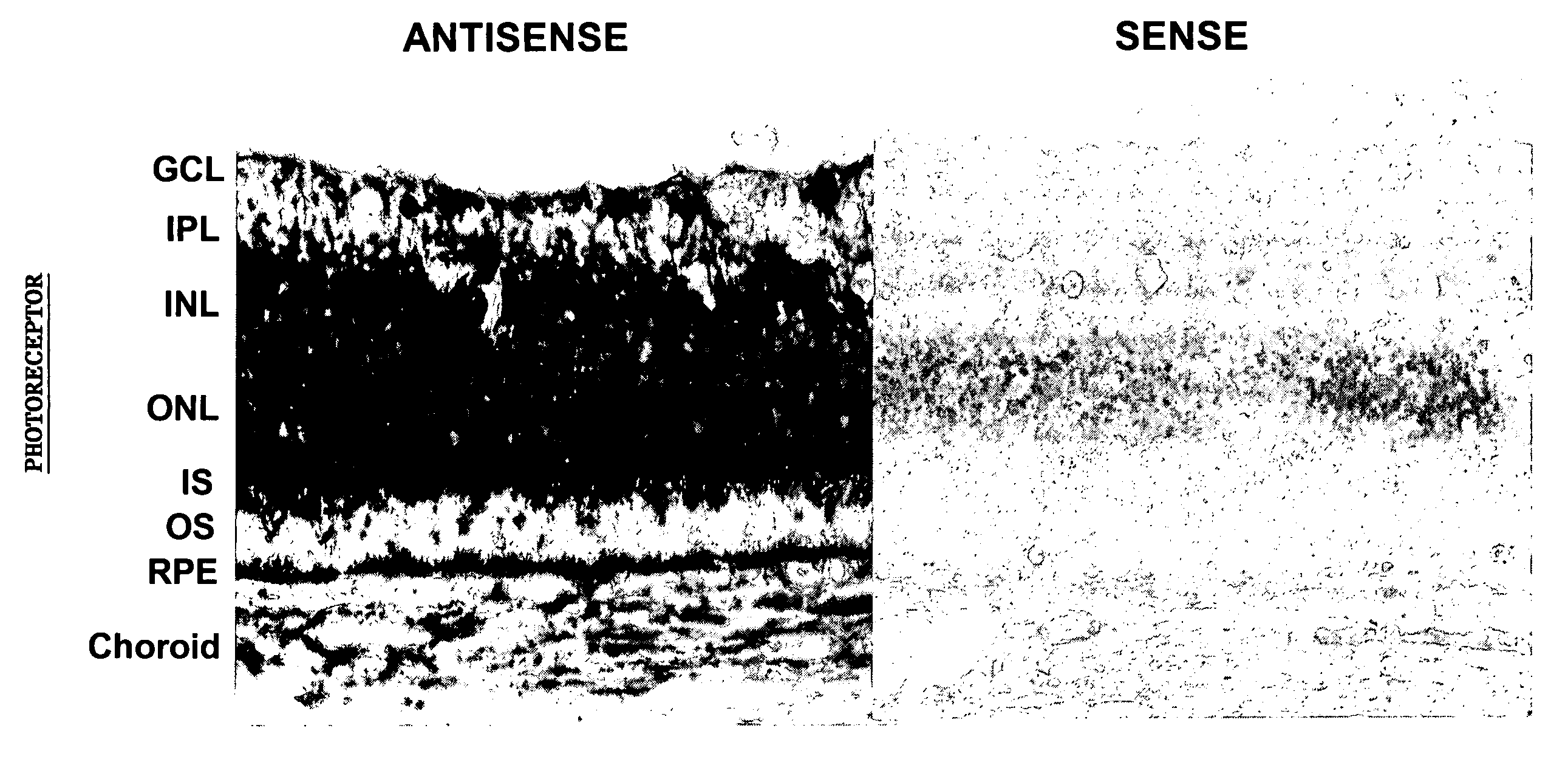
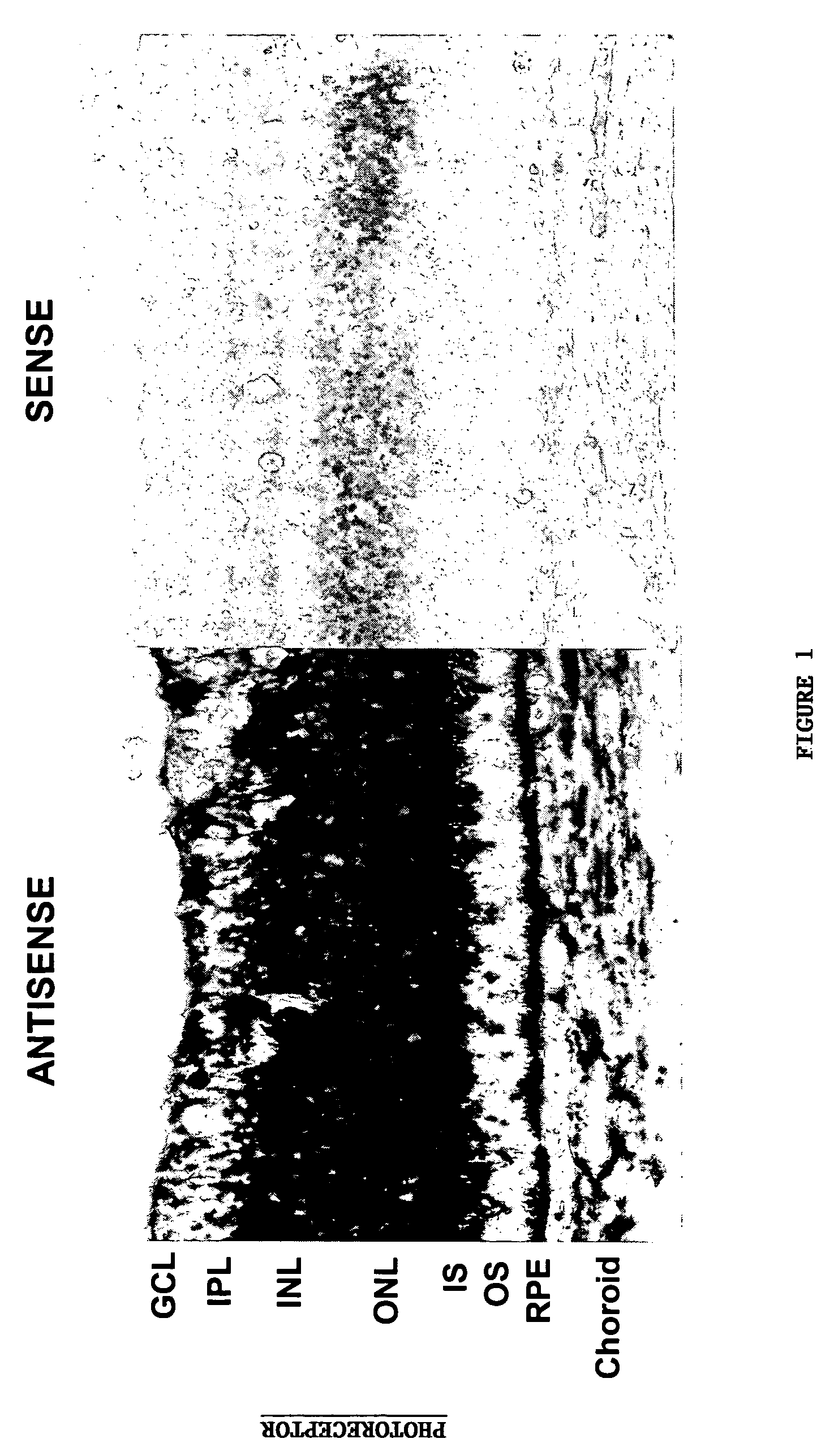
Localization of P2Y2-Receptor mRNA in Retina and RPE

[0096]Cellular localization of P2Y2-receptor mRNA in fresh frozen cross-sections of albino rabbit retina / RPE / choroid tissue was investigated by using nonisotopic in situ hybridization techniques. FIG. 1 shows a representative in situ hybridization result from antisense and sense digoxigenin (DIG)-labeled riboprobes engineered based on the P2Y2 receptor mRNA sequence. Hybridization of antisense and sense riboprobes was visualized by immunohistochemistry using alkaline phosphatase-conjugated anti DIG antibody, and DIG-specific signal was detected using a chromophore reaction against the alkaline phosphatase, yielding purple / black staining. The tissues were also counterstained with nuclear fast red. The control sense probe (right) shows no specific labeling. Labeling with the anti-sense probe showed P2Y2 receptor mRNA localization in scattered nuclei in the ganglion cell and inner nuclear layers and through the inner segment layer of ...

example 2

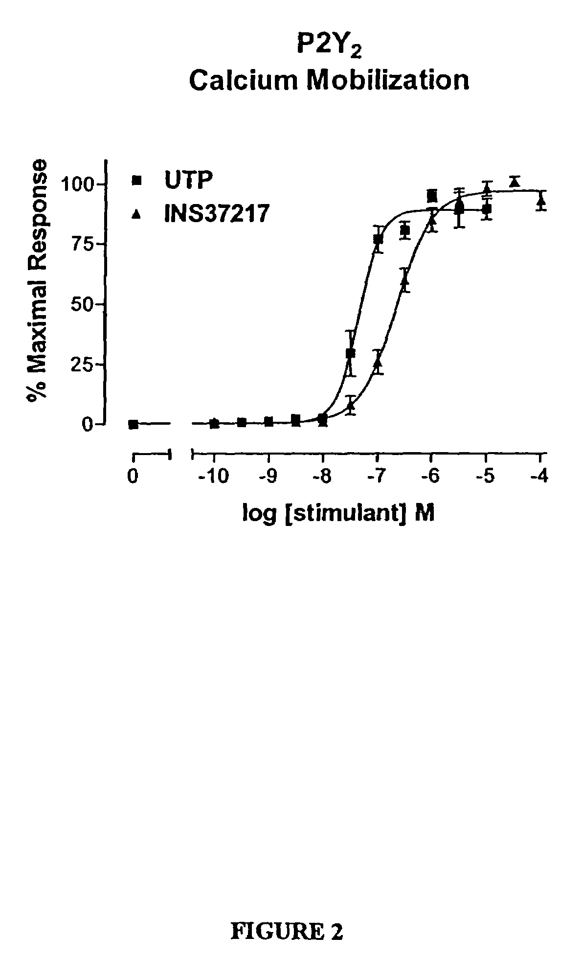
Effects of Synthetic P2Y2 Agonist UP4dC on Cloned Human P2Y2 Receptors

[0097]The dinucleotide, [P1-(uridine 5-)-P4-(2′-deoxycytidine 5′-)tetraphosphate tetrasodium salt](UP4dC) INS37217, was tested for its activity (potency, efficacy, and selectivity) at cloned human P2Y receptor subtypes, which were stably expressed in 1321N1 astrocytoma cells. Activity was assessed using two in vitro indices of cell activation: 1) mobilization of intracellular calcium stores, and 2) accumulation of [3H]-inositol phosphates ([3H]-IP). UP4dC was evaluated for activity in both assays against cells expressing the P2Y1, P2Y2, P2Y4, or P2Y6 receptors.

[0098]UTP and UP4dC induced mobilization of cytosolic calcium in 1321N1 astrocytoma cells expressing human P2Y2 (FIG. 2) receptors with EC50 values of 0.22 μM and 0.8 μM, respectively. The calcium response to 100 μM UP4dC was 100% of the maximal response to UTP at P2Y2 receptors. In conclusion, UP4dC is a full agonist for calcium mobilization at P2Y2 recepto...

example 3

UP4dC Stimulates Fluid Absorption in Freshly Isolated RPE Monolayers

[0100]Fluid transport across freshly isolated, intact bovine and human fetal RPE monolayers was studied using a modified capacitance probe technique (Frambach, et al., Biophys. J 47(4):547–52 (1985); Hughes, et al., J. Gen. Physiol. 83(6):875–99 (1984)). The RPE was mounted vertically in a modified Ussing chamber such that apical and basolateral membranes were separately exposed to Ringer's solutions held in bathing reservoirs. Stainless steel capacitive probes were lowered into the apical and basolateral bathing reservoirs to sense the capacitance of the air gap between the probe and fluid meniscus. Fluid transport rate Jv([2L cm−2 hr−1) was determined by monitoring fluid movement-induced changes in the air gap capacitance at the apical and basolateral baths.

[0101]Representative effects of agonist on J, in human fetal RPE are shown in FIG. 4. Positive Jv values reflect fluid absorption (apical-to-basolateral) and n...


PUM

PropertyMeasurementUnit
osmolarityaaaaaaaaaa

Abstract

The present invention provides a method of treating edematous retinal disorders. The method comprises administration of a pharmaceutical formulation comprising a hydrolysis-resistant P2Y receptor agonist to stimulate the removal of pathological extraneous fluid from the subretinal and retinal spaces and thereby reduce the accumulation of said fluid associated with retinal detachment and retinal edema. The P2Y receptor agonist can be administered with therapeutic and adjuvant agents commonly used to treat edematous retinal disorders. The pharmaceutical formulation useful in this invention comprises a P2Y receptor agonist with enhanced resistance to extracellular hydrolysis, such as dinucleoside polyphosphate compounds, or hydrolysis-resistant mononucleoside triphosphates.

Description

[0001]This application is divisional of U.S. application Ser. No. 10 / 397,795, filed Mar. 25, 2003 now Pat. No. 6,818,629, which is a continuation-in-part of U.S. application Ser. No. 09 / 774,752, filed Jan. 30, 2001, now U.S. Pat. No. 6,596,725; which is a continuation-in-part of U.S. application Ser. No. 09 / 101,395, filed Jul. 10, 1998, which issued as U.S. Pat. No. 6,348,589 on Feb. 19, 2002; which was the National Stage of International Application No. PCT / US98 / 02702, filed Feb. 6, 1998, published Aug. 13, 1998 under PCT Article 2 1(2) in English; and was a continuation-in-part of U.S. application Ser. No. 08 / 798,508, filed Feb. 10, 1997, which issued as U.S. Pat. No. 5,837,861 on Nov. 17, 1998. All U.S. applications and patents cited herein are specifically incorporated herein by reference in their entirety.TECHNICAL FIELD[0002]This invention relates to a method of treating edematous retinal disorders. Specifically, this invention relates to a method of removing pathological flui...

Claims


Application Information

Patent Timeline
Patent Type & Authority Patents(United States)
IPC IPC(8): A61K31/70C07H21/00A61K31/7084C07H19/04C07H19/10C07H19/20C10M101/02C10M105/38C10M107/10C10M111/02C10M111/04C10M169/04
CPCA61K31/7084C07H19/10C07H19/20C07H21/00C10M101/02C10M105/38C10M107/10C10M111/02C10M111/04C10M169/041C07H19/04Y10S514/851C10M2203/10C10M2203/1006C10M2203/102C10M2203/1025C10M2203/1045C10M2203/1065C10M2203/1085C10M2205/00C10M2205/02C10M2205/028C10M2205/0285C10M2207/2825C10M2207/2835C10M2207/2855C10M2209/084C10N2240/06C10N2040/06
Inventor PETERSON, WARD M.YERXA, BENJAMIN R.
Owner MERCK SHARP & DOHME LLC
Why Eureka
Try Eureka