Ultra-miniature in-vivo electrode used for measuring bioelectrical neural activity

a bioelectrical neural activity and ultra-miniature technology, applied in the field of ultra-miniature in-vivo electrodes, can solve the problems of mechanical damage, difficult to maintain good insulation performance in the living body for longer than, and difficulty in achieving reliable signal processing, etc., to achieve stable setting of thin peripheral nerves, better contact, and stable operation

Inactive Publication Date: 2006-09-26
NIPPON CABLE SYSTEM INC +1
View PDF15 Cites 13 Cited by
  • Summary
  • Abstract
  • Description
  • Claims
  • Application Information

AI Technical Summary

Benefits of technology

[0010]In this in-vivo electrode, the periphery of the separated portion of the measuring object can be enclosed with a pair of electric conductors by leading a measuring object into the through-hole of the electric conductors. Because wires are connected to each electric conductor, the electrical potential difference between the separated portions of the measuring object can be measured easily by observing the electric potential difference between the other ends of the wires. Further, since the electric conductor encloses the whole periphery of the measuring object, better contact to the measuring object can be stably maintained compared with the conventional crank like, furcated, and needle like electrode, thereby enabling stable setting to thin peripheral nerves etc. The use of polymeric material enables molding of thin, short electrodes, which enables a number of settings and the continuation of multidimensional measurements without any damage against a thin nerve bund. Since the electric conductor is insulated by the surrounded insulator and the wires exposed to the outside area are also insulated, the measurement can be performed by embedding the electrode into a living body, thereby enabling a long term stable continuous measurement.
[0011]In the in-vivo electrode in which the conductors and the insulator are made of an elastic body like rubber, respectively, and a slit which reaches the through-hole from the outside surface, is formed in order to guide the measuring object into the through-hole wherein the slit is always closed by elastic force, the through-hole is exposed outside when the slit is opened with the use of elasticity. Therefore, the measuring objects such as neural fascicles can easily be led into the through-hole. When the force expanding the slit is loosened, the slit closes naturally to enclose the measuring object by its elasity. Accordingly, setting and removing of measuring objects such as a thin nerve is easily performed. That is to say, this in-vivo electrode not only enables a continuously stable setting to the measuring object but also allows for ease in setting and removal of the measuring object.
[0012]An in-vivo electrode, which is provided with a thread binding the periphery of the insulator so as to keep the slit closed, binds the periphery of the insulator using a textile thread, which makes the contact of the slit tight thereby reducing the possibility of insulation breakdown due to infiltration of body fluid etc. through the slit.
[0013]The in-vivo electrode provided with an earth electrode laid on the surface of the insulator and with the earth wire having an insulated surface and connected to the earth electrode, so as to embed the whole body into a living body, allows the end of the earth wire to be electrically and stably connected to the living body. It saves the labor of having to connect an earth wire separately to the living body for a reference of the electrical potential and also saves the labor of having to remove the wire. Further, it allows the earth wire to be lead out of the living body together with the wires connected to the conductors.
[0014]In the in-vivo electrode which is provided with the insulator being composed of an intervening member in between the conductor, an edge member covering each edge of the conductors, and a cylindrical member enclosing whole periphery of the conductors, the intervening member, and the edge members, easy assembling is accomplished by shoving in one piece of the conductor, the intervening member, and the other piece of the conductor from one end of the cylindrical member and then fitting the edge members.

Problems solved by technology

However, the sympathetic nerves to the other visceral organs such as the adrenal, splenic, and digestive organs etc. or the nerves distributing to skeletal muscles, skin, and brain / spinal chord have extremely smaller diameters on the order of less than 100 μm, and they are vulnerable to mechanical damages.
Furthermore, it is difficult to maintain good insulation performance in the living body for longer than about one week, for which conventional measurements can be achieved.
Finally, low signal levels of some microvolts measured in the small nerve bundle cause difficulties in achieving reliable signal processing.
Further, the measuring object is a microscopic-level nerve fascicle in the living body; which can easily be mechanically damaged or killed.
Furthermore, it is difficult to maintain good insulation performance of an implantable electrode for a long period of time and to keep it stably fitted to the thin nerve fascicle.

Method used


Image

  • Ultra-miniature in-vivo electrode used for measuring bioelectrical neural activity
  • Ultra-miniature in-vivo electrode used for measuring bioelectrical neural activity
  • Ultra-miniature in-vivo electrode used for measuring bioelectrical neural activity

Examples


Embodiment Construction

[0015]The best mode for carrying out of the invention is described with reference to accompanying drawings in which:

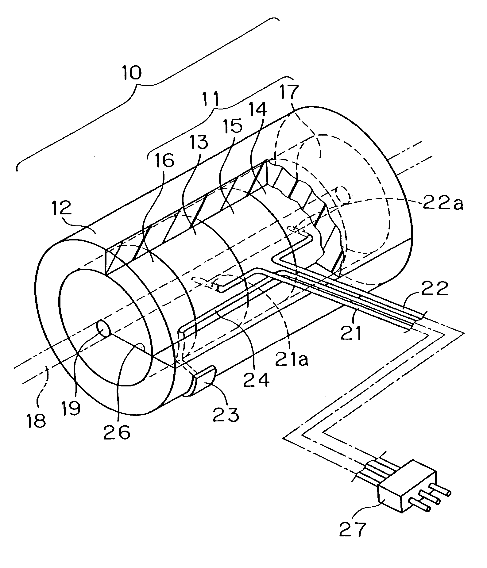
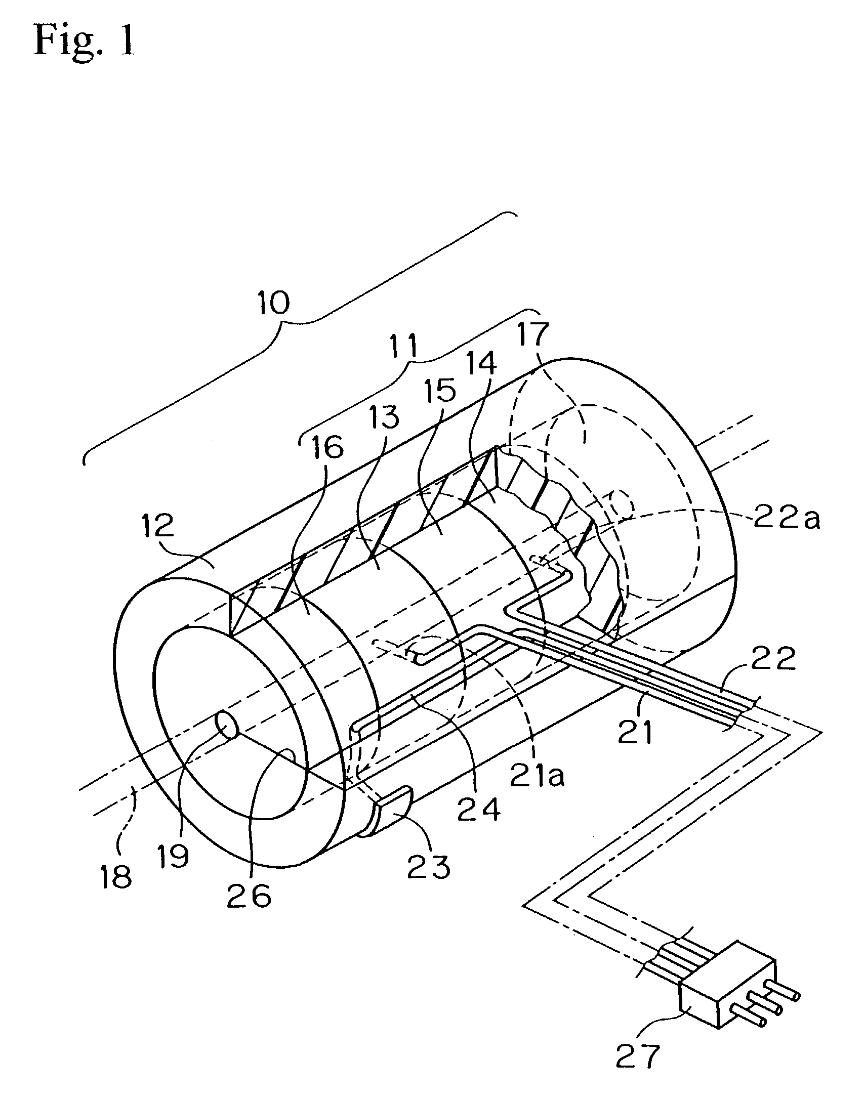
[0016]FIG. 1 is a perspective view with a fragmentary sectional view showing an embodiment of the in-vivo electrode of this invention;

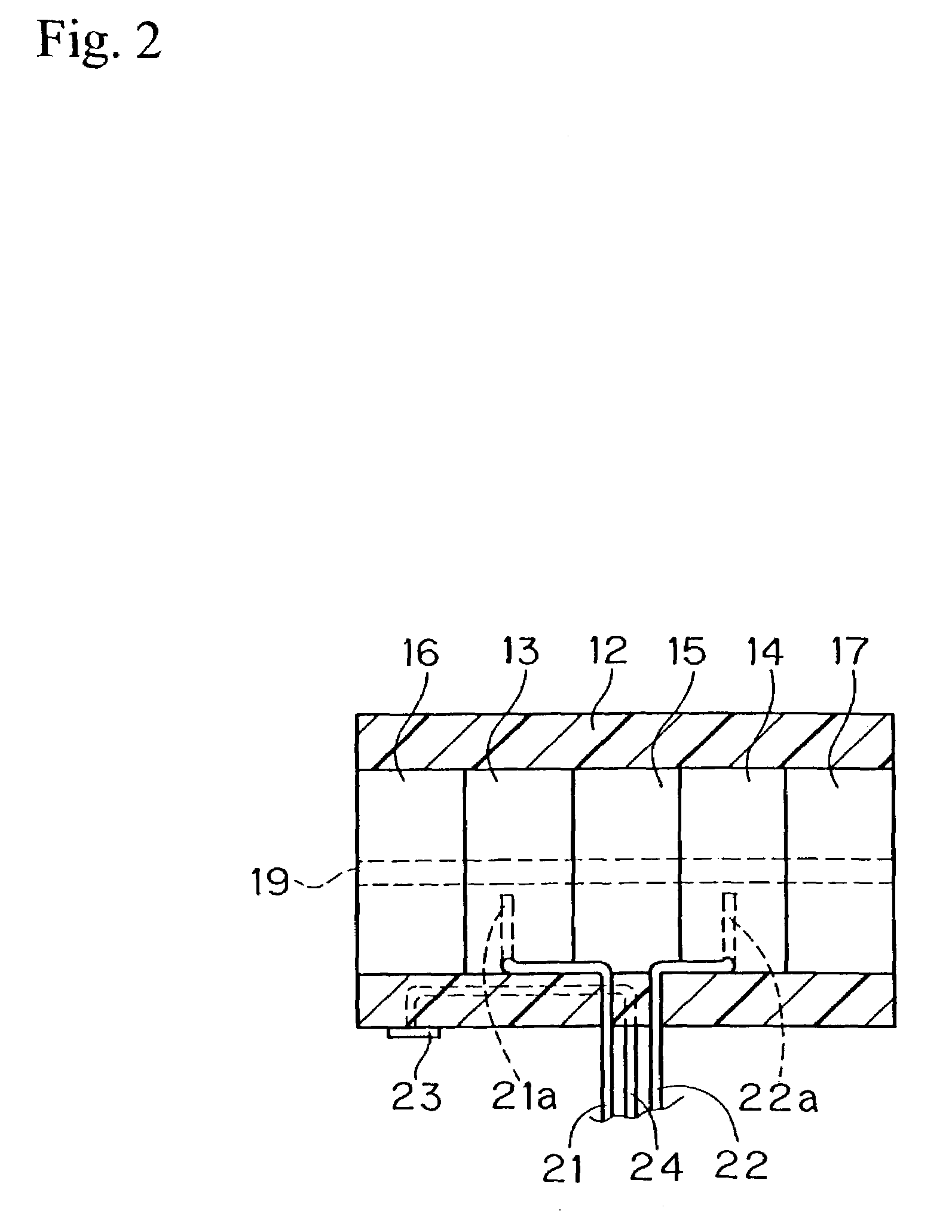
[0017]FIG. 2 is a partial sectional view of the in-vivo electrode;

[0018]FIG. 3a and FIG. 3b are side views of the conductor and the edge member respectively;

[0019]FIG. 4 is a perspective view of the other embodiment of the in-vivo electrode;

[0020]FIG. 5 is a process drawing showing the process of use of the in-vivo electrode of FIG. 2;

[0021]FIG. 6 is a partial sectional view of the other embodiment of the in-vivo electrode of this invention;

[0022]FIGS. 7a, 7b, and 7c are side views of the other embodiment of the in-vivo electrode of this invention respectively;

[0023]FIG. 8a is a side view of the other embodiment of the in-vivo electrode of this invention;

[0024]FIG. 8b is a cross sectional view taken along the line VIII—VIII of FIG. 8a;

[...


PUM


Abstract

An ultra-miniature in-vivo electrode used for measuring the bioelectrical neural activity etc. in a living body with small cubic volume, slight burden to the living body, and high insulation performance, thereby facilitating long term stable measurements. Structurally the in-vivo electrode comprises conductors 13, 14 made of polymeric material having a through-hole 19 in each center so as to lead a measuring object 18; a intervening member 15 made of non-conductive polymeric material provided in between the conductors so as to insulate the periphery of the conductors; edge members 16, 17 provided in the outer edge of the conductors; a clad body 12 made of cylindrical insulant enclosing the periphery of them; measuring wires 21,22 to lead electricity to outside from each conductor 21, 22; a slit 26 which reaches the through-hole 19 from the outside surface being formed in all of the conductors, the intervening member, the edge members, and the clad body.

Description

FIELD OF THE ART[0001]This invention generally relates to an ultra-miniature in-vivo electrode or a microelectrode for measuring the minimal bioelectric potential difference or minimal current of nerve fascicles, and particularly to an ultra-miniature in-vivo electrode usable under the embedded condition in a living body with only a slight burden to the living body, thereby facilitating long-term continuous measurements of bioelectrical neutral activity.BACKGROUND ART[0002]In conventional measurements of the neural activity of a living body, electrodes using such materials as biomolecular collagen and metallic silver wires are contacted to a nerve fascicle, and the measurement of a minimal current flow in the nerve fascicle is performed through insulated wires affixed to the electrode. These electrodes are used for the measurements of neural activities including peripheral nerves having the diameter of the order of millimeters, and sympathetic nerves to the heart and kidney having t...

Claims


Application Information

Patent Timeline
Patent Type & Authority Patents(United States)
IPC IPC(8): A61B5/04
CPCA61B5/04001A61B5/4041A61B5/24A61B5/294
Inventor MATSUKAWA, KANJIITO, TAKAKOKONDOH, KAZUHIKOSEKI, YASUO
Owner NIPPON CABLE SYSTEM INC
Why Eureka
Try Eureka