
Optical imaging of blood circulation velocities

a technology of optical imaging and blood circulation, applied in the field of sensing blood velocity, can solve the problem of difficult direct measurement of rbc velocity, and achieve the effects of reducing the effect of local tissue irregularities, eliminating registration errors, and improving the accuracy of velocity computation

Inactive Publication Date: 2006-09-26
WINTEC LLC
View PDF15 Cites 174 Cited by
  • Summary
  • Abstract
  • Description
  • Claims
  • Application Information

AI Technical Summary

Benefits of technology

[0017]The invention combines digital and single-exposure photography to obtain velocity estimates over large sensing areas. Since it utilizes spatial averaging, it loses spatial resolution as compared with the LDV method. However, the spatial resolution can be improved by increasing optical magnification of the imaging system and / or by using a detector array such as a charged-couple device with small pixel size. This invention has the advantage of being a fill-field method that provides a global picture of the velocity pattern without the need for scanning or contact with the sampled tissue.Mathematical Relations
[0036]The invention reduces the effect of local tissue irregularities, by incorporating the analysis of phase randomization, thus improving the accuracy of the velocity computation. A device based on the invention is developed using a laser, an imager to record and process the speckle information, a display unit, and a data storage unit. Since the image of the sampled area and the speckle data can be collected using a common aperture system, the magnification factor and the image size will be the same, eliminating registration errors due to these factors. The computed blood velocity map can be superimposed onto a digitized image of the sampled area for easy visualization. Motion artifacts due to slight motions of the sampled tissue and the device reduce the signal-to-noise ratio of both LDV and LSCA methods. The LDV method reduces the effects of motion artifacts by filtering out low frequency (<30 Hz) signals, whereas this invention applies fast exposures to reduce these effects.

Problems solved by technology

The convoluted nature of the capillaries makes the direct measurement of RBC velocity difficult.

Method used


Image

  • Optical imaging of blood circulation velocities
  • Optical imaging of blood circulation velocities
  • Optical imaging of blood circulation velocities

Examples


Embodiment Construction

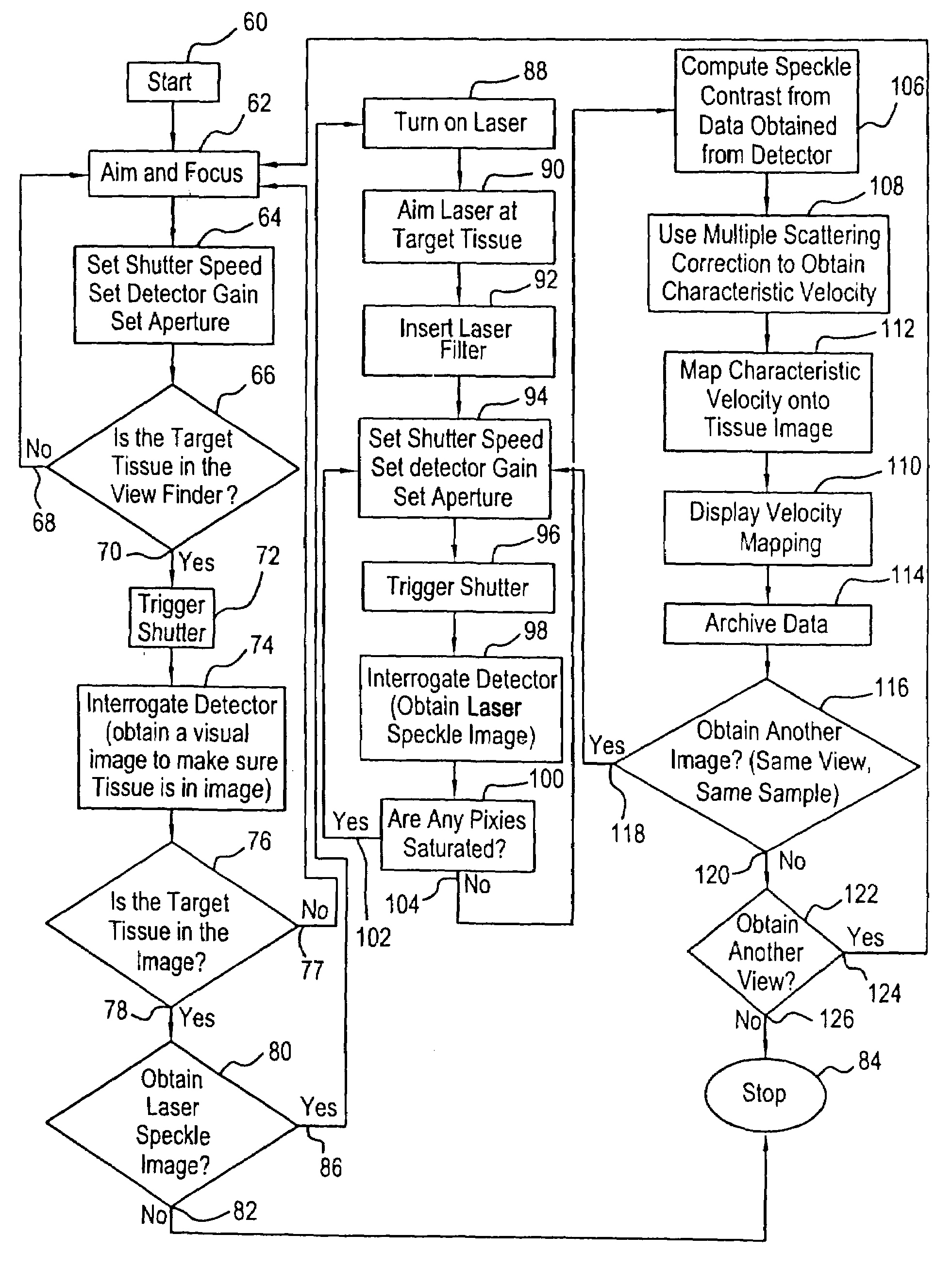
[0049]The LSCA / MS device consists of a laser system for illuminating the sampled tissue, a collection system for collecting tissue images and laser speckle data from tissue, a data processing system, and a display system.

[0050]The laser system consists of a laser and a beam expanding telescope that consists of a focusing lens and a pinhole to illuminate the sampled area with a uniform beam.

[0051]The collection system consists of a laser filter, a multi-element collection lens, a shutter, and a detector unit. The laser filter is used to eliminate the ambient light from laser speckle images. A visual tissue image can be obtained by removing the laser filter. The multi-element lens is used to collect the scattered light and to focus an image onto a detector array. The shutter is used to control the exposure (integration) time.

[0052]The data processing unit initializes the device by setting the exposure time, coordinates the timing for collecting speckle images with the blood pulse of t...


PUM


Abstract

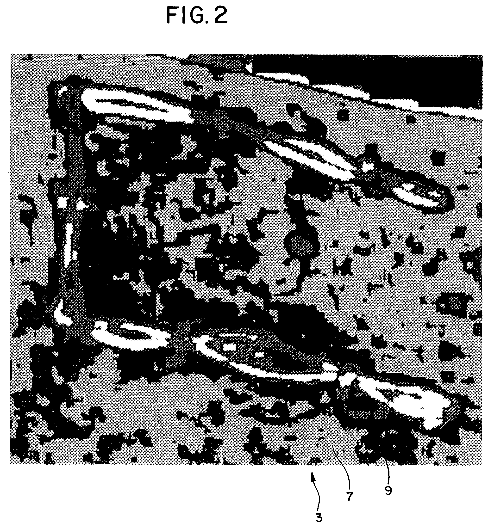
New devices and methods are provided for noninvasive and noncontact real-time measurements of tissue blood velocity. The invention uses a digital imaging device such as a detector array that allows independent intensity measurements at each pixel to capture images of laser speckle patterns on any surfaces, such as tissue surfaces. The laser speckle is generated by illuminating the surface of interest with an expanded beam from a laser source such as a laser diode or a HeNe laser as long as the detector can detect that particular laser radiation. Digitized speckle images are analyzed using new algorithms for tissue optics and blood optics employing multiple scattering analysis and laser Doppler velocimetry analysis. The resultant two-dimensional images can be displayed on a color monitor and superimposed on images of the tissues.

Description

BACKGROUND OF THE INVENTION[0001]The invention relates to sensing blood velocity in tissues such as skin, skin flaps, transplants, breasts, retinas, and internal organs and tissue.[0002]The evaluation of hemodynamics is an important diagnostic subject and has been one of the most difficult challenges in medicine. In skin studies it is important to assess blood velocities over wide areas to determine blood perfusion and predict tissue viability. For surgical procedures involving skin flaps, a reliable method of quantitatively monitoring tissue blood velocity can provide predictive value in assessing tissue conditions during partial detachments and before, during, and after reattachment to avoid tissue necrosis. The same is true in the transplant of tissues and organs and before, during, and following surgery.[0003]The invention is also useful in diagnosing tissue damage due to complications of diabetes, addressing practicality and viability of tissue repairs and vascular densities, a...

Claims


Application Information

Patent Timeline
IPC IPC(8): A61B6/00
CPCA61B3/1233A61B5/0261A61B5/0059A61B2562/0242A61B5/7425
Inventor WINCHESTER, JR., LEONARD W.CHOU, NEE-YIN
Owner WINTEC LLC
Learn More