Osteoimplant and method for making same

a technology of osteoimplant and bone graft, which is applied in the field of preparing a partially demineralized bone graft, can solve the problems of difficulty in repairing itself, difficulty in integrating the host's blood supply into the cortical bone, and inability to fully integrate the remodeling process, etc., and achieves the effect of convenient placemen

Inactive Publication Date: 2007-02-20
WARSAW ORTHOPEDIC INC
View PDF14 Cites 97 Cited by
  • Summary
  • Abstract
  • Description
  • Claims
  • Application Information

AI Technical Summary

Benefits of technology

[0021]Also provided is a osteoimplant implantation kit containing an osteoimplant and a delivery tool useful in performing the placement of the osteoimplant during a surgical procedure. The osteoimplant may be provided in a presterillzed, prepackaged form held by the delivery tool, which can be used without repositioning the osteoimplant in the tool. The implantation tool carries the osteoimplant so as to avoid damage to the osteoimplant prior to and during surgical implantation. The tool also permits the surgeon to securely hold the osteoimplant during implantation and to easily place it at the proper location.

Problems solved by technology

One problem associated with many structural osteoimplants is that they are never fully incorporated by remodeling and replacement with host tissue.
This is due, in part, to the difficulty with which the host's blood supply may penetrate cortical bone.
Since repair is a cellular-mediated process, dead (non-cellular, allograft or xenograft) bone is unable to repair itself.
It has been observed that fatigue damage is usually the result of a buildup of unrepaired damage in the tissue.
In applications where the mechanical load-bearing requirements of the osteoimplant are challenging, e.g., intervertebral spinal implants for spinal fusion, lack of substantially complete replacement by host bone tissue may compromise the osteoimplant by subjecting it to repeated loading and cumulative unrepaired damage in the tissue (mechanical fatigue) within the implant material.
However, the prior art has not addressed the need to provide a demineralized osteoimplant with sufficiently mineralized regions for engagement of insertion instrumentation.
However, removal of the mineral components of bone significantly reduces mechanical strength of bone tissue.
Thus, demineralization sacrifices some of the load-bearing capacity of mineralized cortical bone and as such is not suitable for all osteoimplant designs.
Another disadvantage to the demineralization process is the likelihood of creating dimensional changes in the osteoimplant.
Such changes of dimension can make it difficult for a configured piece to mechanically engage with surgical instruments, other implants, or the prepared surgical site.

Method used


Image

  • Osteoimplant and method for making same
  • Osteoimplant and method for making same
  • Osteoimplant and method for making same

Examples


example 1

Surface Demineraliztion

[0072]A study was undertaken to determine the effect of varying penetration of surface demineralization on the osteoinductive response to human cortical bone implants using an athymic rat intramuscular assay. Osteoinductivity is often defined as the ability of the agent to recruit cells to the site, and to convert them to the osteoblastic lineage. Osteoinductivity has been observed to follow an “endochondral pathway” similar to that of embryonic bone when it is first formed. This bone goes though stages, including the penetration and infiltration of cells induction of chondrogenic (cartilage-forming) cells at the site, laying down of cartilage tissue, and the transformation of that cartilage tissue to bone tissue. All of these changes can be tracked over time in histological samples.

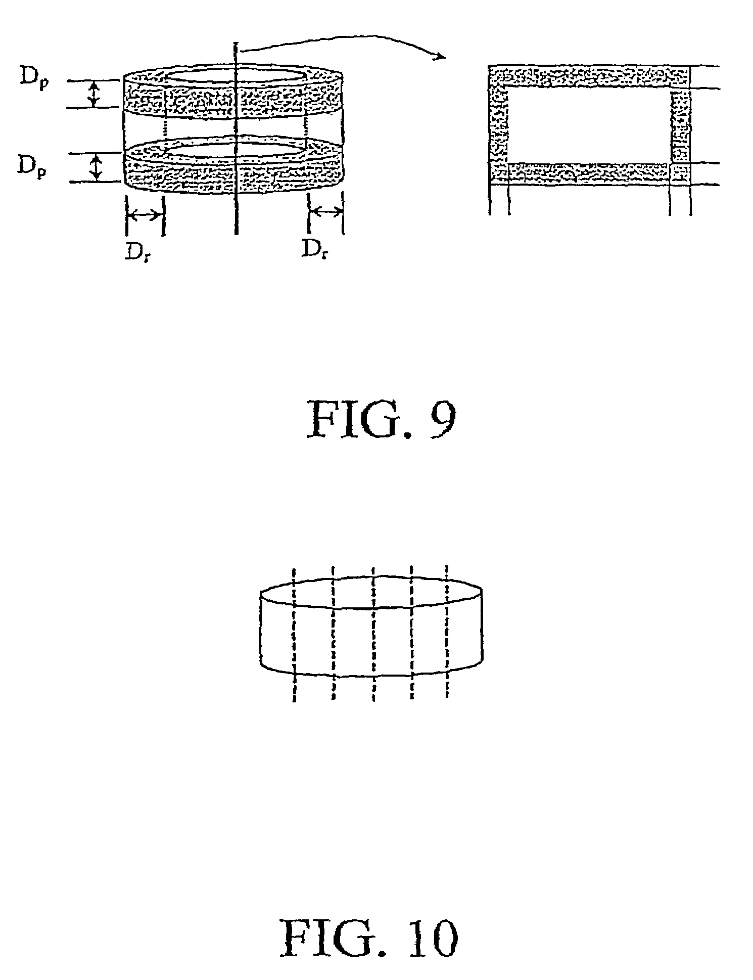
[0073]Implants were constructed aseptically from human cortical bone. Cylindrical discs (7±1 mm in diameter and 3±0.5 mm thick) were cut from cortical bone of the same donor. Befor...

example 2

Spinal Fusion Procedure

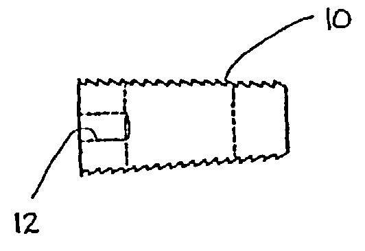
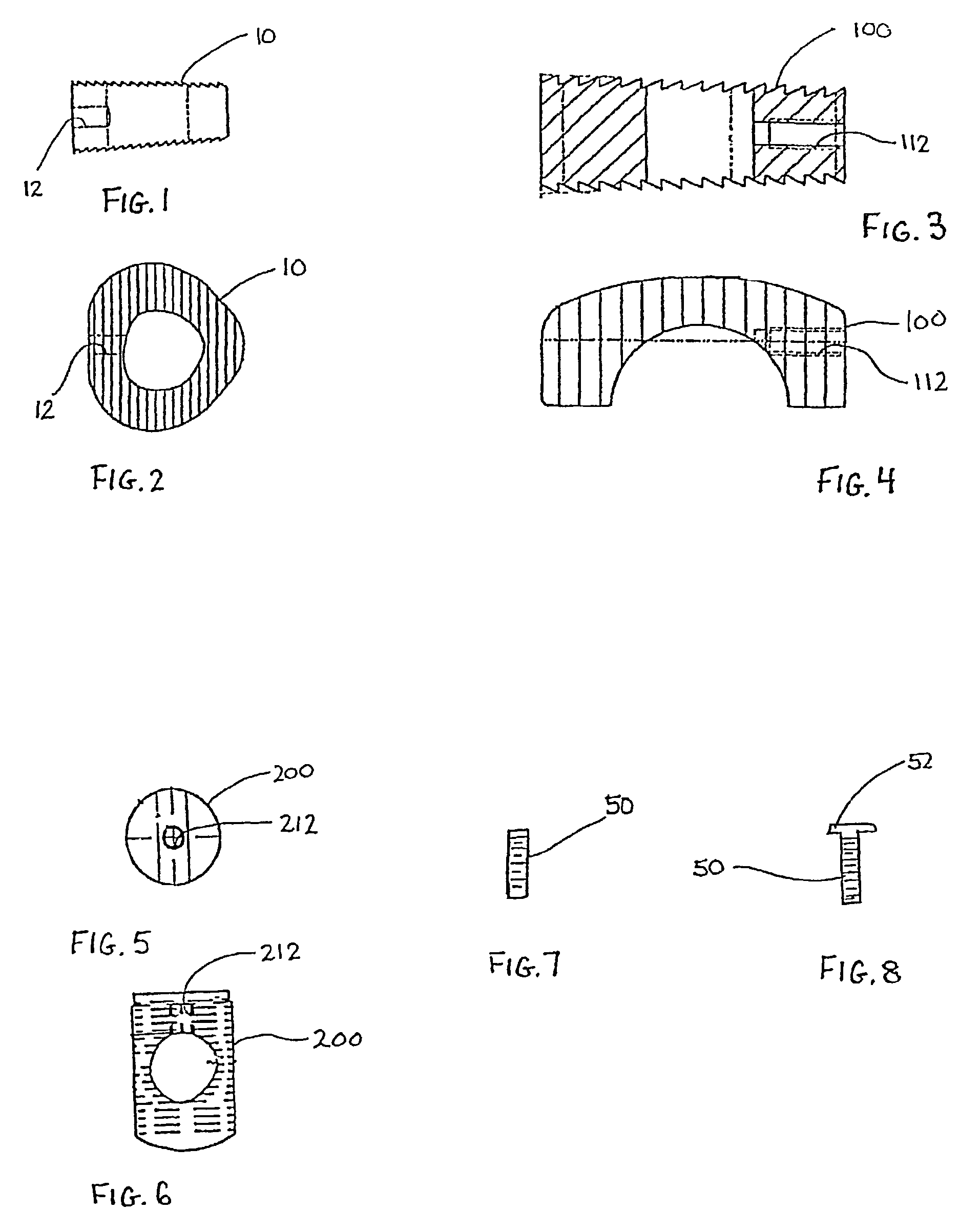
[0084]In this example, the insertion of osteoimplants having an engagement structure (12, 112, 212) in conjunction with a posterior approach for lumbar discectomy and spinal fusion will be discussed. It is to be appreciated that other surgical approaches, e.g., anterior, postero-lateral, etc., may be utilized to perform the discectomy and insert engagement structure as well.

[0085]Initially, the vertebral column is accessed via a posterior approach with the use of appropriate retractors to retract neighboring muscle tissue, blood vessels and / or nerve tissue. Thereafter, at least a portion of the disc is removed with an appropriate rongeur or cutting implements. A retractor is mounted to the posterior faces of the vertebrae. One retractor suitable for this purpose is the Cloward Lumbar Lamina Spreader manufactured by Codman. The retractor includes a pair of retractor arms which are mountable to the posterior vertebral faces via screws. With the retractor appropr...


PUM

PropertyMeasurementUnit
thickaaaaaaaaaa
thickaaaaaaaaaa
thickaaaaaaaaaa

Abstract

A method of manufacturing an osteoinductive osteoimplant is provided which comprises the steps of: demineralizing part or all of at least one surface of a monolithic section of cortical bone to a depth of at least about 100 microns; and, configuring the monolithic section of cortical bone to provide an osteoimplant possessing an outer surface possessing at least one demineralized zone and a non-demineralized zone. An implant produced according to the above method demonstrates improved osteoinduction without producing any clinically significant reduction of strength in critical regions of the osteoimplant.

Description

CROSS-REFERENCE TO RELATED APPLICATIONS[0001]This is a continuation of co-pending application Ser. No. PCT / US01 / 15599 filed in English on May 14, 2001 and which claims benefit under 35 U.S.C. §119(e) of Provisional Appln. Ser. Nos. 60 / 204,069 and 60 / 221,056 respectively filed May 12, 2000 and Jul. 27, 2000, and a continuation-in-part of U.S. application. Ser. No. 09 / 328,242 filed Jun. 8, 1999 and now U.S. Pat. No. 6,277,149 issued Aug. 21, 2001, the entire contents of all of which are incorporated herein by this reference.BACKGROUND OF THE INVENTION[0002]1. Field of the Invention[0003]The present disclosure is directed to a method for preparing a partially demineralized bone graft. More specifically, this invention relates to a load-bearing osteogenic osteoimplant fabricated from a monolithic section of cortical bone and to a method for making the osteoimplant as well as a method of using same.[0004]2. Description of the Related Art[0005]Shaped or cut bone segments have been used ex...

Claims


Application Information

Patent Timeline
Patent Type & Authority Patents(United States)
IPC IPC(8): A61F2/28A61L27/36
CPCA61L27/3608A61L27/3683A61L27/365A61L2430/02A61P19/00
Inventor EDWARDS, JEAN T.SHIMP, LAWRENCE A.DIEGMANN, MICHELEBOYCE, TODD M.SCARBOROUGH, NELSON L.RUSSELL, JAMES L.
Owner WARSAW ORTHOPEDIC INC
Why Eureka
Try Eureka