Saliva immunoassay for detection of antibodies for cardiovascular disease

a technology of antibody detection and saliva, which is applied in the direction of material analysis, biological material analysis, instruments, etc., can solve the problems of tissue damage, fibrous lesions or plaque formation, and release of tissue antigens

Inactive Publication Date: 2007-08-21
IMMUNOSCI LAB
View PDF2 Cites 7 Cited by
  • Summary
  • Abstract
  • Description
  • Claims
  • Application Information

AI Technical Summary

Problems solved by technology

These toxins may directly or indirectly induce tissue damage and cause release of tissue antigens.
If this vicious cycle of inflammation continues, it can result in fibrous lesions or plaque formation.
When pieces of the plaque break loose, they can start blood clots and cause heart attack.
However, most autoantibodies are not complement fixing, and removal of the immunoglobulin-coated platelets occurs in the spleen and other sites of reticuloendothelial tissue.
However, this concept may be an oversimplification, since although immunization in the lung may lead to antibodies in distant secretory sites, such as salivary glands and immunization in the lacrimal glands has also been shown to lead to the production of antibodies in saliva.
While this concept of oral tolerance to high doses of soluble antigen may be correct, certain conditions—such as overloading of the GI tract with bacterial toxins—may not lead to oral tolerance.
This is due to the fact that bacterial toxins will cause the opening of tight junctions, which will in turn lead to the absorption of ingested proteins and bacterial antigens from the gut in significant amounts.

Method used


Image

  • Saliva immunoassay for detection of antibodies for cardiovascular disease
  • Saliva immunoassay for detection of antibodies for cardiovascular disease
  • Saliva immunoassay for detection of antibodies for cardiovascular disease

Examples


example 1

General Procedures for Immunity

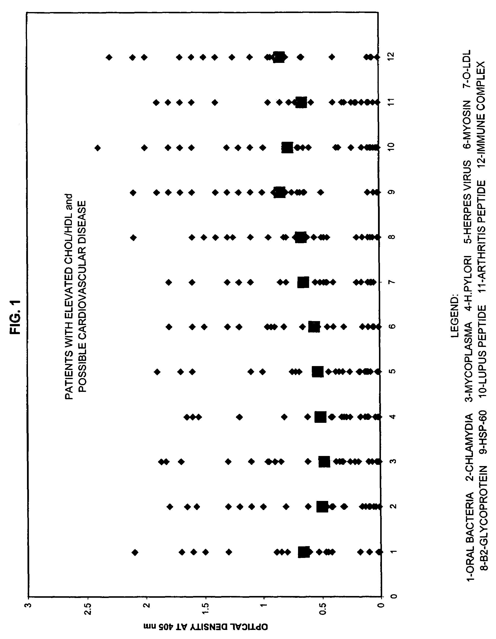
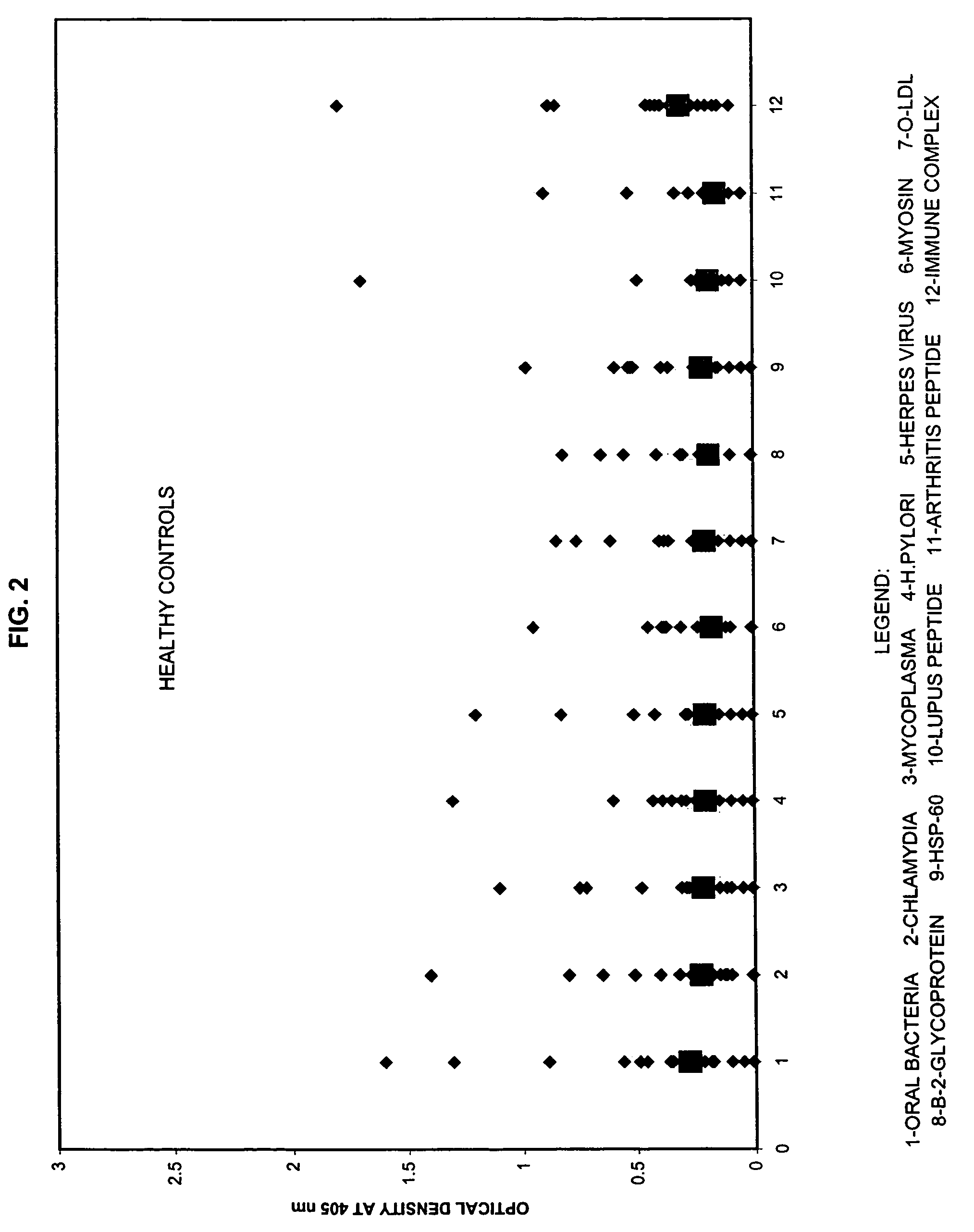
Panel for Cardiovascular Disease

[0060]For the test, about 2 ml of patient saliva was collected. Saliva specimen was kept at −20° C. until the performance of the assays.

[0061]The purified antigens were immobilized by attachment to a solid surface, such as a microtiter plate. The saliva sample was added to the plate followed by incubation and washing. Antibody bound to antigen was revealed by adding enzyme labeled monoclonal antibody directed against the first immunoglobulin. After addition of substrate, color development was measured by microtiter reader at 405 nm. The intensity of the color was directly related to the concentration of antibodies to these antigens present in patient's specimen.

[0062]Saliva samples were collected in the morning, before brushing teeth, smoking, or drinking. 2 ml of saliva was collected. Saliva was collected after a gentle chewing action in a test tube containing 0.1 ml of preservative. Saliva specimen was kept at −20° C. ...

example 2

Test for Myosin Antibody

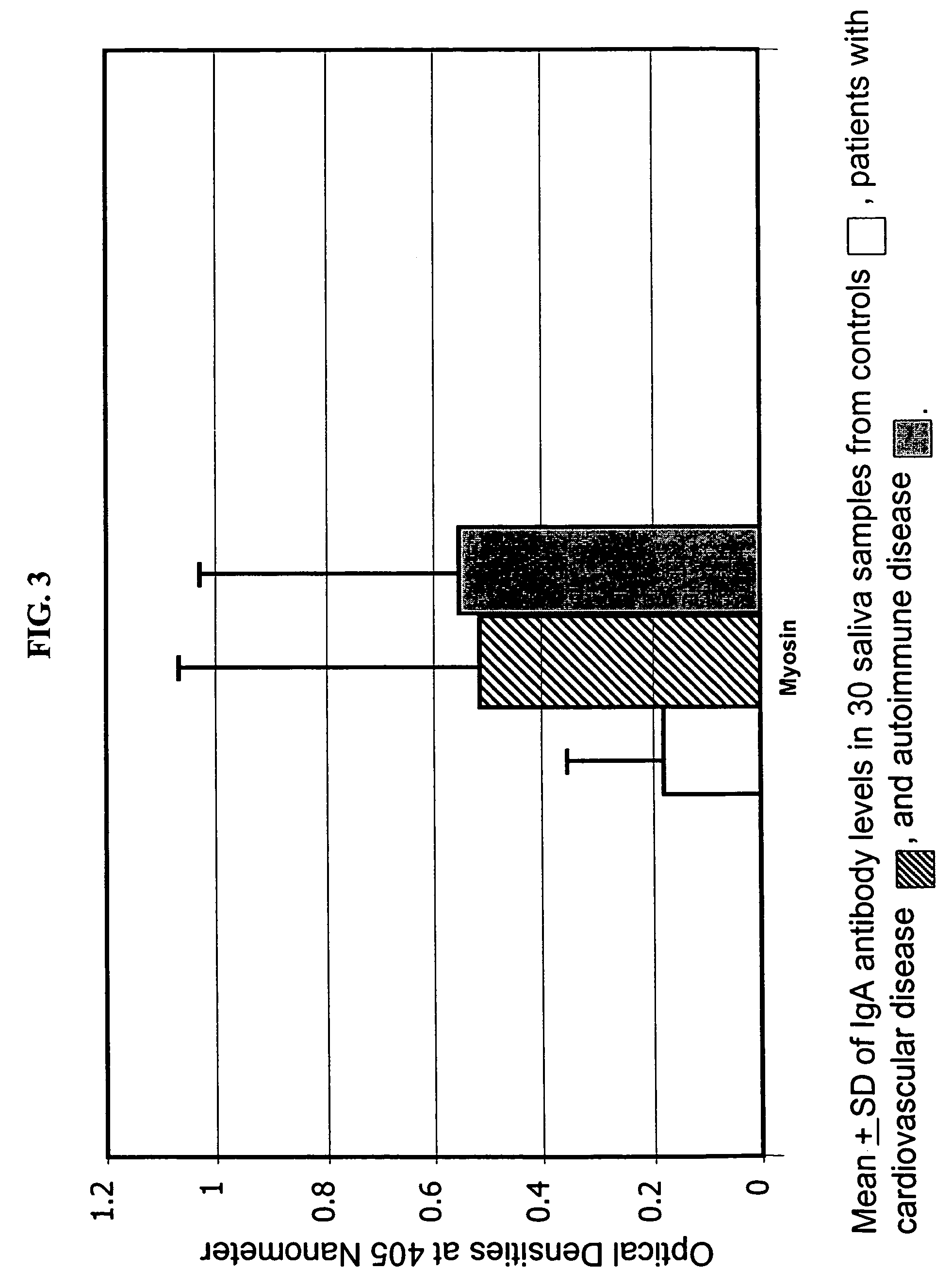
[0073]Myosin pathogenic peptide “SLKLMATLFSTYASA” was synthesized by a robotic multiple peptide synthesizer and resin was used as solid support. Peptide was characterized by reversed-phase HPLC and electrospray mass-spectrometry with purity greater than 80%. This peptide was bound to bovine serum albumin and used for coating microtiter plates.

[0074]Each well of microtiter plate was coated with 3 μg peptide in 0.1 M carbonate buffer pH 9.5. After 24 hours incubation and washing, 200 ml of 2% BSA was added and incubated for an additional 2 hours. Plates were washed, dried, and used for measurement of myosin antibodies. The test specimen was added to the plate followed by incubation and washing. The procedure in Example 1 was followed to measure for the myosin antibodies.

[0075]The ELISA values for the calibrators used in this test system were as follows: Calibrator I=7.5, Calibrator II=15, and Calibrator III=30.

[0076]The ELISA values for each test specimen were ...

example 3

Test for Oxidized LDL Antibody

[0077]Wells of microtiter polystryrene plate were coated with 3 μg of oLDL in 100 μl of 0.1 M carbonate buffer pH 9.6 and were kept overnight at 4° C. The plates were then washed with PBS and blocked with 2% BSA for 2 hours at room temperature. Plates were washed, dried, and used for detection of antibodies against oLDL. The test specimen was added to the plate followed by incubation and washing. The procedure in Example 1 was followed to measure for oLDL antibodies.

[0078]The ELISA values for the calibrators used in this test system were as follows: Calibrator I=37, Calibrator II=75, Calibrator III=300, and Calibrator IV=1200.

[0079]The ELISA values for each test specimen were determined using the formula in Example 1.


PUM

PropertyMeasurementUnit
molecular massaaaaaaaaaa
temperatureaaaaaaaaaa
soak timeaaaaaaaaaa

Abstract

A method for diagnosing the likelihood and severity of cardiovascular disease in a patient is disclosed. The method determines the levels of antibodies against autoantigens, including myosin, oxidized LDL, β-2-glycoprotein, heat shock protein-60, platelet glycoprotein, and immune complexes. It then compares the results to normal levels to determine the likelihood and severity of cardiovascular disease.

Description

RELATED APPLICATION DATA[0001]This application is a divisional application of U.S. patent application Ser. No. 10 / 005,710 filed Nov. 8, 2001 now abandoned, the entirety of which is hereby incorporated by reference.BACKGROUND OF THE INVENTION[0002]1. Field of the Invention[0003]The invention relates to a saliva immunoassay for detection of antibodies for cardiovascular disease.[0004]2. Description of the Related Art[0005]Cardiovascular disease is predicted to be the most common cause of death worldwide by the year 2020. Half of heart disease patients lack established risk factors such as elevated lipids, hypertension, tobacco abuse, and positive family history. Additionally, these risk factors are generally associated with the disease, and the exact mechanism by which they may contribute to the development of atherosclerosis is not clear. However, previous and recent studies point to a linkage between infection with different bacteria and heart disease in the other 50% of observed in...

Claims


Application Information

Patent Timeline
Patent Type & Authority Patents(United States)
IPC IPC(8): G01N33/53G01N33/564G01N33/68
CPCG01N33/564G01N33/6893G01N2800/32Y10S436/811
Inventor VOJDANI, ARISTO
Owner IMMUNOSCI LAB
Why Eureka
Try Eureka