Method for extracting quantitative information relating to interactions between cellular components

a technology of cellular components and quantitative information, applied in the field of measuring interactions, can solve the problems of poor specificity and damaging side effects, damage to the membrane, and difficulty in controlling the release of cellular contents by permeabilization

Inactive Publication Date: 2007-10-16
FISHER BIOIMAGE
View PDF8 Cites 20 Cited by
  • Summary
  • Abstract
  • Description
  • Claims
  • Application Information

AI Technical Summary

Benefits of technology

[0078]One major advantage of the present invention is that changes in mobility can be measured as a change in light intensity. As will be illustrated below, and as described in the examples, this technique allows Redistribution™ to be detected as a fluorescence intensity change.
[0079]A variety of instruments e

Problems solved by technology

Since such compounds will be inherently very selective and have their action without the need for gross inhibition of catalytic activity, it can be expected that therapies based on their use will not carry with them the problems of poor specificity and damaging side effects commonly associated with more traditional active-site inhibitors.
It is difficult to control the release of cellular contents by permeabilization since detergents have the tendency to not only disrupt the membrane but also over time disrupt intracellular components.
Beside this, damaging the membrane will start some uncontrolled protease activity with the same unintentional result.
The mobility of the previously soluble component is greatl

Method used


Image

  • Method for extracting quantitative information relating to interactions between cellular components
  • Method for extracting quantitative information relating to interactions between cellular components
  • Method for extracting quantitative information relating to interactions between cellular components

Examples


example 1

Construction of the Probes and Fusions

[0109]PS462 contains a fusion of PDE4A4 and EGFP under the control of a CMV promoter and has resistance to G418 as selectable marker. To construct the PDE4A4-EGFP fusion, the ca. 1.9 kb C-terminal part of HSPDE4A4 (GenBank Acc. no. L20965), which is common to all PDE4A isoforms, is amplified using PCR with primers 4A-Ct-top and 4A-bottom described below. The sequence of the top primer contains a silent mutation, which introduces a Dra1 site exactly at the beginning of the shared 4A region. The bottom primer includes the common C-terminal sequence minus the stop codon, a BamH1 cloning site, and two extra nucleotides to preserve the reading frame when cloned into pEGFP-N1. The unique ca. 0.8 kb N-terminal part of HSPDE4A4 is amplified using PCR in the presence of 5% DMSO with primers 4A4-top and 4A4N-bottom described below. The top primer includes specific HSPDE4A4 sequences following the ATG, a Kozak sequence, and a Hind3 cloning site. The bottom...

example 2

Protocols and Methods

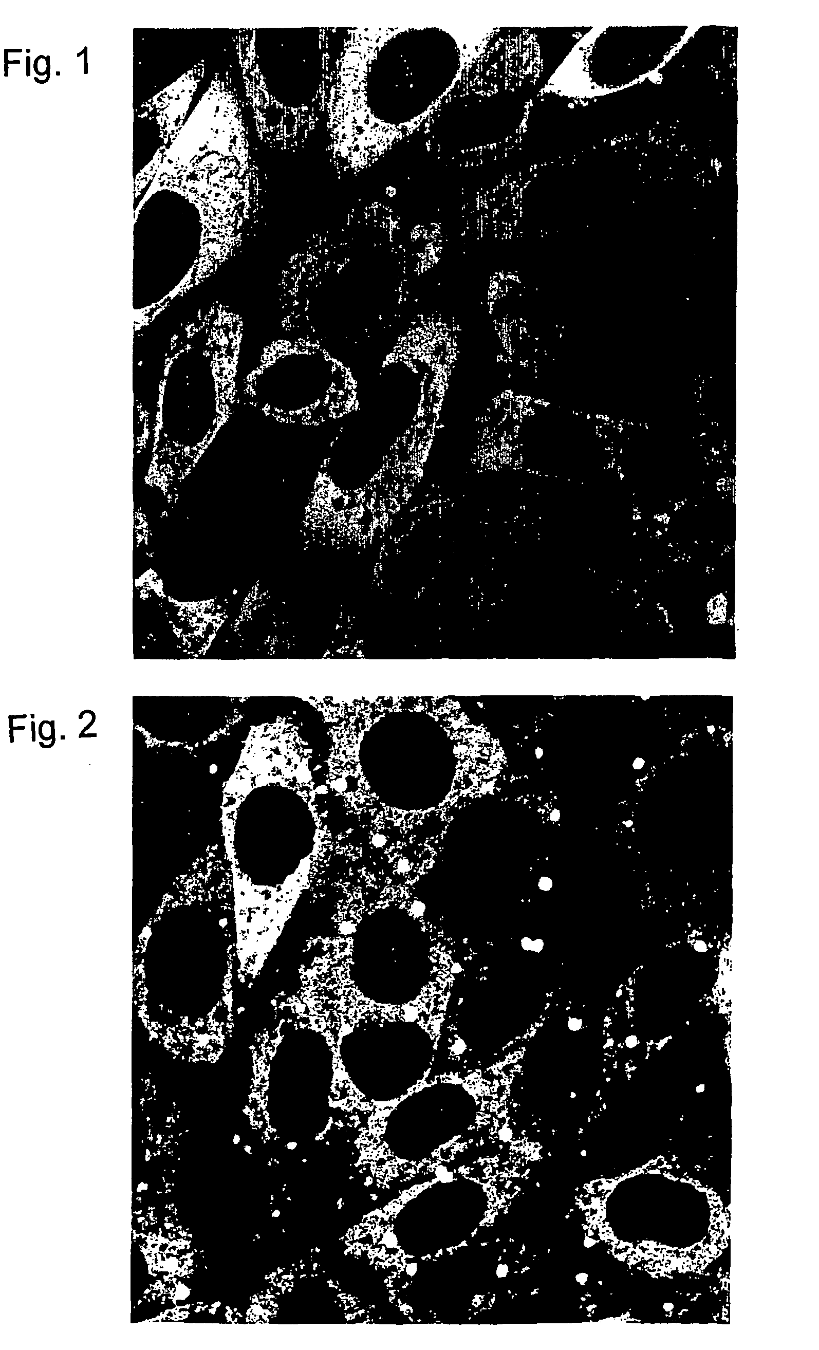
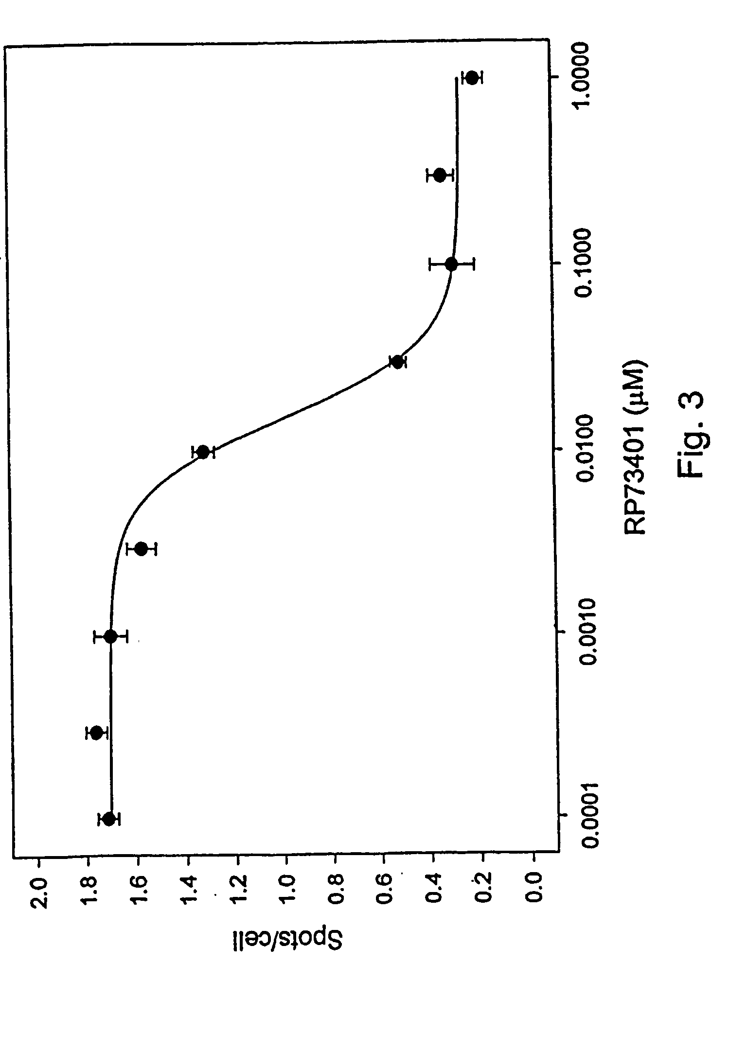
[0150]This example describes protocols and methods used for in vivo expression of the probes described in Example 1, and the visualisation and measurement of changes undergone by EGFP fusion probes, either transfected singly or as co-transfections with anchor probes in CHO cells.

Transfection and Cell Culture:

[0151]Chinese hamster ovary cells (CHO), are transfected with the plasmids described in Example 1 above, either using a single species of plasmid, or pairs of plasmids co-transfected simultaneously, using the transfection agent FuGENE™ 6 (Boehringer mannheim Corp, USA) according to the method recommended by the suppliers. Stable transfectants of single GFP probes are selected using the appropriate selection agent, usually 0.5 mg / ml G418 sulphate (Calbiochem) in the growth medium (HAM's F12 nutrient mix with Glutamax-1, 10% foetal bovine serum (FBS), 100 μg penicillin-streptomycin mixture ml−1 (GibcoBRL, supplied by Life Technologies, Denmark). Co-transfected...

example 3

Probes for Proteins X and Y with PDE4A4 Anchor Protein

[0163]The present example and Example 4, Example 5 and Example 6, describe generic ways to produce a cell line suitable for screening compounds targeting against a specific interaction between two partner components X and Y. In these examples the cells are derived from CHO cells co-transfected with two plasmids, one coding for fusion probes with X or Y attached to either the C or N terminal of the anchor moiety (the anchor probe), and the second with the other partner, Y or X, attached to either the C or N terminal of GFP (the detectable probe). There are therefore 4 possible anchor probes and 4 possible detectable probes that can be made for the interacting pair X-Y, and 8 useful combinations to co-transfect. Anchor and detectable probes use different selection markers to ensure that cells under selection maintain both plasmids; for example, the anchor may confer resistance to zeocin, the detectable to neomycin. Cells that maint...


PUM

PropertyMeasurementUnit
Digital informationaaaaaaaaaa
Compositionaaaaaaaaaa
Fluorescenceaaaaaaaaaa

Abstract

A method is described to assay for protein interactions in living cells, e.g. by the introduction of two heterologous conjugates into the cell. The method uses the measurement of cellular distribution of a detectable component (e.g. a GFP-labeled˜fluorescent probe) to indicate the presence or absence of an interaction between that component and a second component of interest. The method uses the knowledge that certain components can be stimulated to redistribute within the cell to defined locations. Inducible redistribution systems make it possible to determine if specific interactions occur between components. Inducible systems are described where it is demonstrated that the redistribution stimuli are essentially “null”, in that they affect no other system in the cell during the assay period, other than the component whose redistribution can be induced. Also described is an extraction buffer which is useful in high throughput screening for drugs which affect the intracellular distribution of intracellular components. The extraction buffer comprises a cellular fixation agent and cellular permeabilisation agent. Optimizing the composition of the extraction buffer and its application to various cell types is described.

Description

[0001]This application is the national phase under 35 U.S.C. § 371 of PCT International Application No. PCT / DK01 / 00466 which has an International filing date of Jul. 3, 2001, which designated the United States of America.FIELD[0002]The present invention relates to measurement of interactions between two components wherein the two components are present in a cell, and where both components are most usually wholly or mainly proteinaceous in composition (i.e. the interaction is a protein-protein interaction, or protein-protein binding event). This is presently referred to as “GFP assisted Readout of Interacting Proteins (GRIP)”[0003]The present invention also relates to an extraction buffer used in high throughput screening for drugs that affect the intracellular distribution of intracellular components. The extraction buffer comprises a cellular fixation agent and a cellular permeabilization agent.BACKGROUND[0004]The interaction of proteins with each other and with other cellular comp...

Claims


Application Information

Patent Timeline
IPC IPC(8): C07H21/04C12N5/06C12N9/12C12Q1/68G01N33/50C12N15/09C12Q1/02G01N21/77G01N21/78G01N33/15G01N33/53G01N33/566G01N33/68
CPCG01N33/5008G01N33/502G01N33/6845G01N33/68G01N33/5032
Inventor BJORN, SARA PETERSENTHASTRUP, OLETERRY, BERNARD ROBERTHAGEL, GRITHNIELSEN, SOREN JENSBY
Owner FISHER BIOIMAGE
Why Eureka
Try Eureka