Medium for growing human embryonic stem cells

a technology of human embryonic stem cells and medium, which is applied in the field of cell biology of embryonic cells, can solve the problems of considerably less advanced technology for culturing and differentiating human pluripotent stem cells, and achieve the effects of rapid production of high-quality pps cells, easy growth and manipulation of undifferentiated, and expanding primate pluripotent stem cells

Inactive Publication Date: 2007-11-20
ASTERIAS BIOTHERAPEUTICS INC
View PDF44 Cites 142 Cited by
  • Summary
  • Abstract
  • Description
  • Claims
  • Application Information

AI Technical Summary

Benefits of technology

[0011]This disclosure provides an improved system for expanding primate pluripotent stem (pPS) cells. The technology allows the user to rapidly produce high-quality pPS cells for use in therapy are drug discovery, free of undesired contamination by cells of other species and other tissue types.

Problems solved by technology

As a result, technology for culturing and differentiating human pluripotent stem cells is considerably less advanced.
A significant challenge to the use of pluripotent stem cells for therapy is that they are traditionally cultured on a layer of feeder cells to prevent differentiation (U.S. Pat. No. 5,843,780; U.S. Pat. No. 6,090,622).

Method used


Image

  • Medium for growing human embryonic stem cells
  • Medium for growing human embryonic stem cells
  • Medium for growing human embryonic stem cells

Examples


example 1

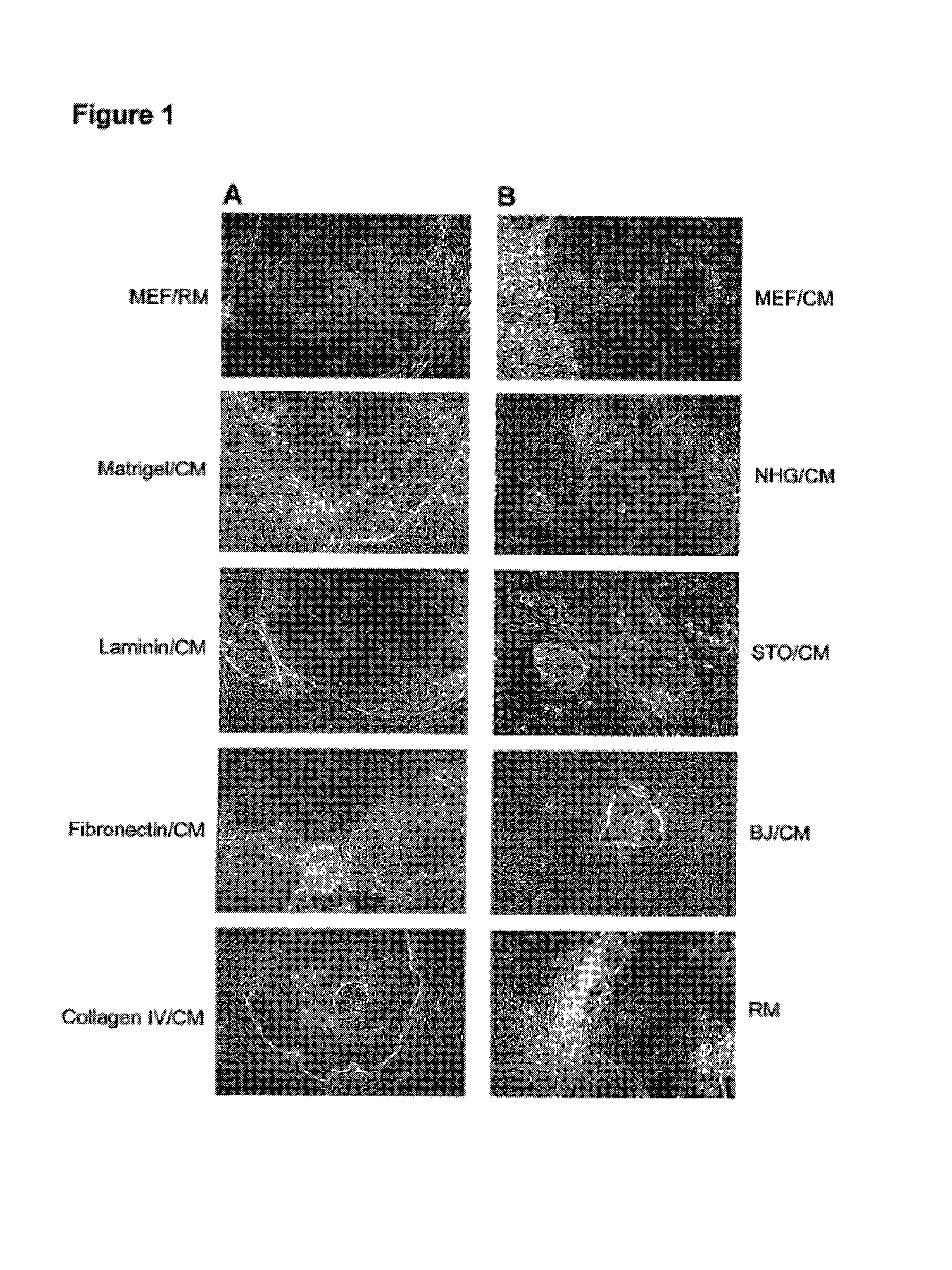
Growing hES Cells Without Feeder Cells in Conditioned Medium

[0104]In this example, undifferentiated hES cells that had been maintained on primary mouse embryonic feeder cells were maintained in the absence of feeders. The culture wells were coated with Matrigel®, and the cells were cultured in the presence of conditioned nutrient medium obtained from a culture of irradiated primary fibroblasts.

[0105]Conditioned medium (CM) was prepared as follows. The fibroblasts were harvested from T150 flasks by washing once with Ca++ / Mg++ free PBS and incubating in trypsin / EDTA (Gibco). After the fibroblasts detached from the flask, they were collected in mEF medium (DMEM+10% FBS). The cells were irradiated at 4000 rad, counted and seeded at about 55,000 cells cm−2 in mEF medium. After at least 4 hours, the medium was exchanged with SR containing ES medium. Conditioned medium was collected daily for feeding of hES cultures. Alternatively, medium was prepared using mEF plated in culture flasks, ex...

example 2

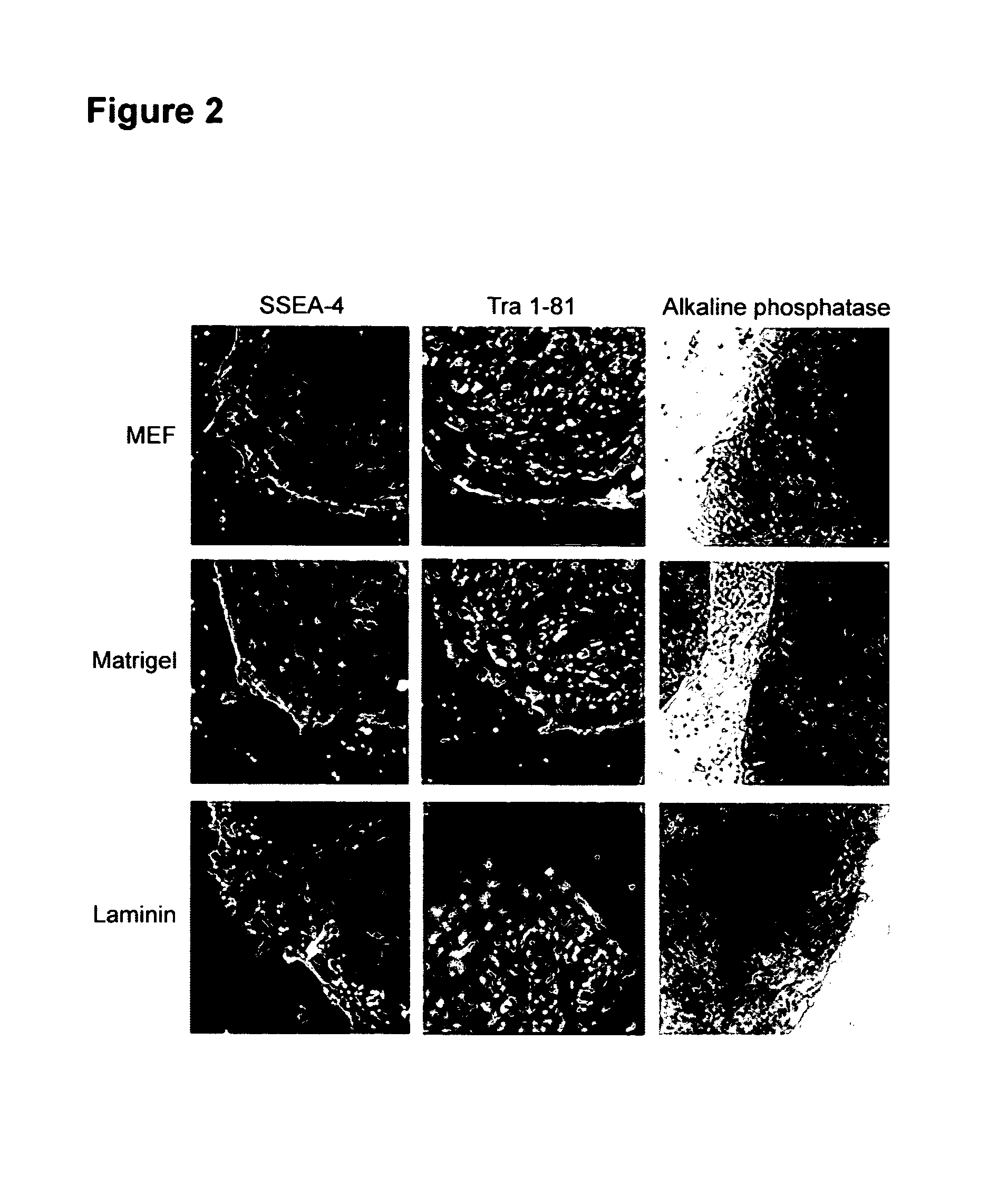
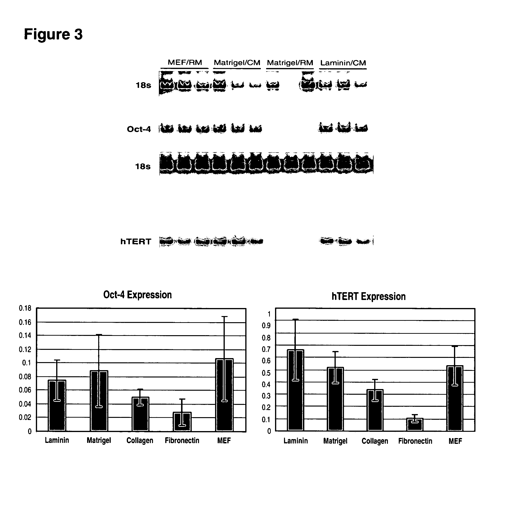
Phenotypic Markers of hES Cells in Feeder-free Culture

[0112]Undifferentiated hES cells express SSEA-4, Tra-1-60, Tra-1-81, OCT-4, and hTERT. In order to assess whether the cells maintained in feeder-free conditions retained these markers, cells were evaluated by immunostaining, reverse transcriptase PCR amplification, and assay for telomerase activity.

[0113]For analysis by fluorescence-activated cell sorting (FACS), the hES cells were dissociated in 0.5 mM EDTA in PBS and resuspended to about 5×10⁵ cells in 50 μL diluent containing 0.1% BSA in PBS. They were labeled with specific primary antibody and then fluorescent second antibody, and analyzed on a Flow Cytometer.

[0114]Similar to the hES cells on feeders, cells on Matrigel®, laminin, fibronectin or collagen IV expressed SSEA-4, Tra-1-60 and Tra-1-81. There was very little expression of SSEA-1, a glycolipid that is not expressed by undifferentiated hES cells.

[0115]FIG. 2 shows marker expression detected by immunocytochemistry. Cel...

example 3

Pluripotency of hES Cells in Feeder-free Culture

[0121]In vitro differentiation was induced in H1 hES cells maintained in conditioned medium on Matrigel®, laminin, fibronectin or collagen IV for 26 days. The hES cells were dissociated into small clumps by incubating in ˜200 U / mL collagenase IV at 37° C. for 10 min, and cultured in suspension to form embryoid bodies (EBs) in medium containing DMEM, 20% FBS (Hyclone), 1 mM glutamine, 0.1 mM β-mercaptoethanol, and 1% non-essential amino acids (Gibco). After 4 days in suspension, the aggregates were transferred onto poly-ornithine-coated plates, and cultured for additional 7 days. The cultures were then examined for the presence of beating cells, and processed for immunocytochemistry.

[0122]The staining patterns were consistent with cells of the neuron and cardiomyocyte lineages (β-tubulin III and cardiac troponin 1, respectively). About 8 days after differentiation, beating regions were identified in all cultures. There were also cells s...


PUM


Abstract

This disclosure provides an improved system for culturing human pluripotent stem cells. Traditionally, pluripotent stem cells are cultured on a layer of feeder cells (such as mouse embryonic fibroblasts) to prevent them from differentiating. In the system described here, the role of feeder cells is replaced by components added to the culture environment that support rapid proliferation without differentiation. Effective features are a suitable support structure for the cells, and an effective medium that can be added fresh to the culture without being preconditioned by another cell type. Culturing human embryonic stem cells in fresh medium according to this invention causes the cells to expand surprisingly rapidly, while retaining the ability to differentiate into cells representing all three embryonic germ layers. This new culture system allows for bulk proliferation of pPS cells for commercial production of important products for use in drug screening and human therapy.

Description

REFERENCE TO RELATED APPLICATIONS[0001]This application is a divisional of U.S. patent application Ser. No. 10 / 235,094, filed Sep. 4, 2002 (pending), through which it claims the priority benefit of U.S. provisional application U.S. Ser. No. 60 / 317,478, filed Sep. 5, 2001.[0002]This application incorporates herein by reference the following applications in their entirety: PCT application PCT / US01 / 01030, filed Jan. 10, 2001; U.S. Ser. No. 60 / 175,581, filed Jan. 11, 2000; U.S. Ser. No. 60 / 213,740, filed Jun. 22, 2000; U.S. Ser. No. 60 / 213,739, filed Jun. 22, 2000; U.S. Ser. No. 60 / 216,387, filed Jul. 7, 2000; U.S. Ser. No. 60 / 220,064, filed Jul. 21, 2000; U.S. Ser. No. 09 / 688,031, filed Oct. 10, 2000 (now U.S. Pat. 6,667,176); U.S. Ser. No. 09 / 849,022, filed May 4, 2001; U.S. Ser. No. 09 / 888,309, filed Jun. 21, 2001; U.S. Ser. No. 09 / 900,752, filed Jul. 6.2001 (now U.S. Pat. 6,642,048; U.S. Ser. No. 09 / 530,346; and PCT applications WO 99 / 20741, WO 01 / 81549, WO 01 / 88104, WO 03 / 000868, W...

Claims


Application Information

Patent Timeline
Patent Type & Authority Patents(United States)
IPC IPC(8): C12N5/00C12N5/08A61K35/12C12N15/09A61P43/00C12N5/0735C12N5/077C12N5/079C12N5/0793C12N5/10C12N15/10
CPCC12N5/0606C12N5/0619C12N5/0622C12N15/1034C12N15/1096C12N2501/237C12N5/0672C12N2502/99C12N2503/02C12N2506/02C12N2510/00C12N2510/04C12N2533/90C12N2500/25C12N2501/115C12N2501/119C12N2501/125C12N2501/13C12N2501/145C12N2501/155C12N2501/2306C12N2501/235C12N2502/13C12N5/0678C12N11/04C12N2501/065C12N5/0603C12N2500/98C12N5/0693C12N5/0062C12N5/0662C12N5/0671C12N5/0075C12N5/0692C12N5/068C12N5/0607C12N2501/998C12N5/0068C12N2500/99C12N2501/105C12N2533/50C12N5/0696C12N2501/26C12N2500/90A61P43/00
Inventor MANDALAM, RAMKUMARXU, CHUNHUI
Owner ASTERIAS BIOTHERAPEUTICS INC
Why Eureka
Try Eureka