
Recombinant proteins and peptides for endotoxin biosensors, endotoxin removal, and anti-microbial and anti-endotoxin therapeutics

Inactive Publication Date: 2007-11-20
NAT UNIV OF SINGAPORE
View PDF9 Cites 15 Cited by
  • Summary
  • Abstract
  • Description
  • Claims
  • Application Information

AI Technical Summary

Benefits of technology

[0014]The ability of these proteins and peptides to mediate inhibition of endotoxin-induced Limulus amoebocyte lysate (LAL) coagulation was measured with a sensitive LAL Kinetic-QCL assay. The SSCrFCES protein and peptides were also tested for their ability to suppress LPS-induced cytokines TNF-α and IL-8) produced by THP-1 and normal human peripheral blood mononuclear cells (hPBMC). SSCrFCES and the peptides were non-cytotoxic. SSCrFCES has a clearance rate of 4.7 ml / min. We also show that low doses of SSCrFCES protein and the synthetic peptides protect galactosamine-sensitized mice from LPS-induced lethality. The peptides have strong antimicrobial potencies and can therefore be used as potent therapeutics.

Problems solved by technology

This obstacle is compounded by the presence of a cysteine-rich region.
The difficulty in doing so was largely due to its highly complex mosaic structure.

Method used


Image

  • Recombinant proteins and peptides for endotoxin biosensors, endotoxin removal, and anti-microbial and anti-endotoxin therapeutics
  • Recombinant proteins and peptides for endotoxin biosensors, endotoxin removal, and anti-microbial and anti-endotoxin therapeutics
  • Recombinant proteins and peptides for endotoxin biosensors, endotoxin removal, and anti-microbial and anti-endotoxin therapeutics

Examples


example 1

Purification of Stably Expressed and Secreted Recombinant SSCrFCES

[0088]Stable cell lines of Drosophila S2 clones expressing SSCrFCES (U.S. Pat. No. 6,733,997) were routinely cultured in serum-free DES Expression medium and maintained at 25° C. in a humidified incubator.

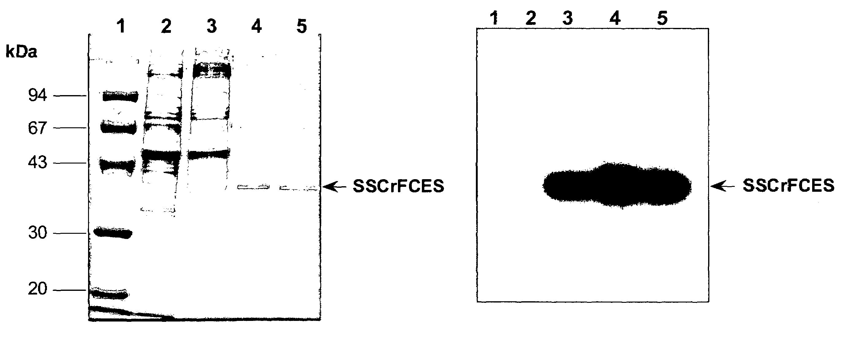
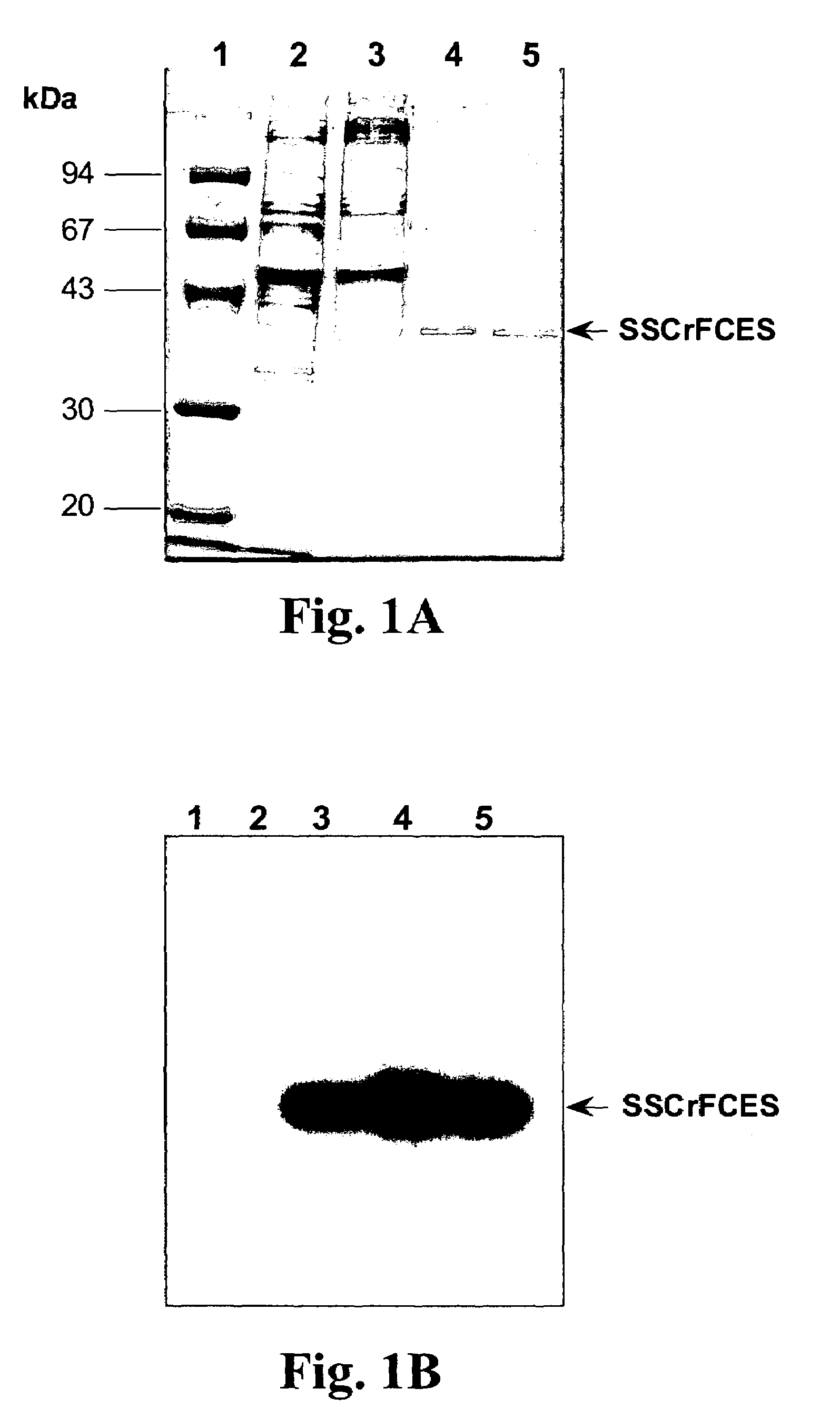
[0089]The medium containing SSCrFCES was initially concentrated and desalted via 3 rounds of ultrafiltration using a 10 kDa cutoff membrane in an Amicon stirred cell (Millipore). Affinity chromatography purification under denaturing conditions yielded a 38 kDa protein of interest, in addition to a 67 kDa protein. Western blot analysis indicated that the 67 kDa protein does not contain the carboxyl poly-His tag. Thus this larger protein is likely due to non-specific adsorption to the resin.

(b) Purification of SSCrFCES by Preparative Isoelectric Membrane Electrophoresis

[0090]Typically, 2 liters of conditioned medium were initially subjected to successive ultrafiltration using a 100 kDa and 10 kDa molecular weight cutof...

example 2

ELISA-Based Lipid A Binding Assay

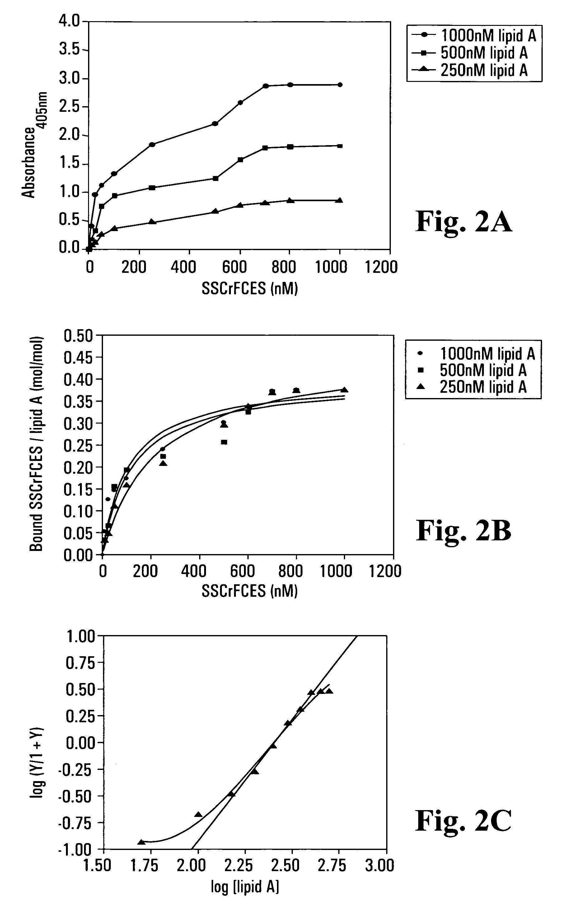
[0094]A Polysorp™ 96-well plate (Nunc) was first coated with 100 μl per well of various concentrations of lipid A diluted in pyrogen-free PBS. The plate was sealed and allowed to incubate overnight at room temperature. The wells were aspirated and washed 6 times with 200 μl per wash solution (PBS containing 0.01% Tween-20 and 0.01% thimerosal). Blocking of unoccupied sites was achieved using wash solution containing 0.2% BSA for 1 hour at room temperature. Subsequently, blocking solution was removed and the wells washed as described above. Varying concentrations of SSCrFCES were allowed to interact with bound lipid A at room temperature for 2 hours.

[0095]Bound SSCrFCES was detected by sequential incubation with rabbit anti-SSCrFCES antibody (1:1000 dilution) and goat anti-rabbit antibody conjugated with HRP (1:2000 dilution) (Dako). Incubation with each antibody was for 1 h at 37° C. with washing between incubations as described above. In the final s...

example 3

Surface Plasmon Resonance (SPR) Studies on Biospecific Binding Kinetics Between Lipid A and: CrFCES; SSCrFCsushi-1,2,3-GFP; SSCrFCsushi-1-GFP; SSCrFCsushi-3-GFP; and Synthetic Peptides

[0099]Recognition of lipid A by the abovenamed secreted recombinant proteins and peptides was, performed with a BIAcore X™ biosensor instrument and an HPA sensor chip. Briefly, lipid A at 0.5 mg / ml in PBS was immobilized to a HPA sensor chip (Pharmacia) according to the manufacturer's specification. In all experiments, pyrogen-free PBS was used as the running buffer at a flow rate of 10 μl / min.

[0100]With purified SSCrFCES, 4 μg / ml was injected into the flow cell at a rate of 10 μl / min, and the binding response was measured as a function of time. Following injection of SSCrFCES, a solution of INDIA™ HisProbe™-HRP antibody, diluted in PBS to 400 μg / ml, was also injected to cause a shift in SPR in order to further confirm that SSCrFCES binds to lipid A. For regeneration, 100 mM of NaOH solution was inject...


PUM

PropertyMeasurementUnit
Fractionaaaaaaaaaa
Molar densityaaaaaaaaaa

Abstract

Recombinant fragments of Factor C are disclosed. These proteins and peptides show great potency in recognizing, binding to, neutralizing and removing endotoxin. These molecules can thus be used for anti-microbial, anti-endotoxin, and anti-sepsis therapy. SSCrFCES is a 38 kDa protein representing the LPS-binding domain of Factor C. The ability of SSCrFCES to bind lipid A was analyzed using an ELISA-based assay as well as surface plasmon resonance. Surface plasmon resonance similarly carried out for SSCrFC-sushi-1,2,3-GFP, SSCrFC-sushi-1GFP, and SSCrFC-sushi-3GFP confirmed their superior affinity for endotoxin. The 50% endotoxin-neutralizing concentration of SSCrFCES against 200 EU of endotoxin is 0.069 μM, suggesting that SSCrFCES is an effective inhibitor of LAL coagulation cascade. Although partially attenuated by human serum, as low as 1 μM of SSCrFCES inhibits the LPS-induced secretion of hTNF-α and hIL-8 by THP-1 and human pheripheral blood mononuclear cells with a potency more superior than polymyxin B. SSCrFCES is non-cytotoxic, with a clearance rate of 4.7 ml / minute. The LD90 of SSCrFCES for LPS lethality in mice is achieved at 2 μM. These results demonstrate the endotoxin-neutralizing capability of SSCrFCES in vitro and in vivo, as well as its potential for use in the treatment of endotoxin-induced septic shock. Also embodied in this application is the use of the sushi peptides and their mutant derivatives as potent antimicrobials. Further embodied in this application is the use of sushi peptides or sushi recombinant proteins to remove endotoxin from liquids.

Description

CROSS-REFERENCES TO RELATED APPLICATIONS[0001]This is a divisional of U.S. patent application Ser. No. 09 / 626,795, filed Jul. 26, 2000, now issued as U.S. Pat. No. 6,719,973, which is a continuation-in-part of U.S. patent application Ser. No. 09 / 219,868, filed Dec. 24, 1998, now abandoned, and which claims the benefit of U.S. Provisional Application No. 60 / 159,569 filed Oct. 15, 1999. The entire disclosure of each of 09 / 219,868 and 60 / 159,569 are hereby expressly incorporated by reference.FIELD OF THE INVENTION[0002]The present invention relates to the use of recombinant polypeptides and synthetic peptides derived from a horseshoe crab Factor C as well as computationally designed peptide analogues, all of which have endotoxin-binding domain(s). The recombinant proteins may be expressed from insect cell clones, either as is or as fusion proteins, e.g. with green fluorescent protein (GFP). The extreme sensitivity of the present recombinant Factor C to LPS, with its unique LPS-binding ...

Claims


Application Information

Patent Timeline
IPC IPC(8): G01N1/00C02F3/34C12Q1/00G01N33/00G01N33/566A61K38/00C12N9/64
CPCA61K38/16C12N9/6424C07K2319/00C07K2319/02Y10T436/25Y10T436/25375Y10T436/25125A61P31/00A61P31/04
Inventor DING, JEAK L.HO, BOWTAN, NGUAN S.
Owner NAT UNIV OF SINGAPORE
Learn More