Recombinant sendai virus vector for introducing exogenous genes to airway epithelia

a technology of exogenous genes and sendai virus, which is applied in the direction of viruses/bacteriophages, biocide, genetic material ingredients, etc., can solve the problems of inability to transfect cells which are not first removed from the body, inability to use vivo approaches, and loss of new introduced gene function as those cells, etc., to achieve mild affect, not affecting sev infection efficiency, and higher expression

Inactive Publication Date: 2008-01-01
DNAVEC RES
View PDF23 Cites 17 Cited by
  • Summary
  • Abstract
  • Description
  • Claims
  • Application Information

AI Technical Summary

Benefits of technology

[0078]Effect of mucus on gene transfer efficiency of each vector was examined using a sheep tracheal strip model, which was prepared by a known method (Kitson, C. et al., Gene Ther. 6: 534-546, 1999). After killing, the epithelial layer of resected sheep trachea was dissected to muscle and adventitia, and was cut into 0.5 cm2 square pieces subsequently confirmed the ciliary beating under the phase-contrast microscope. In some tissue, mucus depletion was followed to the known method (Kitson, C. et al., Gene Ther. 6: 534-546, 1999). These tissues were placed in the air-liquid interface. Ten μl of SeV-luc or GL-67-luc vector solution was applied to the apical surface to perform transfection. After 48 hours, the pieces were subjected to luciferase assay as described in Example 2. The results are shown in FIG. 9.
[0079]As shown in FIG. 9, mucus was not markedly affect SeV-mediated gene transfer compared to GL-67-luc-mediated gene transfer.
[0080]The procedure of 5-1 was repeated except that various concentrations of bovine salivary gland mucin were applied just before gene transfer. The results are shown in FIG. 10.
[0081]As FIG. 10 shows, gene transfer of GL-67-luc was inhibited by addition of mucin. Luciferase activities of the mucin added samples were not significantly different from that of fresh samples, suggesting barrier activity of the mucin, but not mucus viscosity, to cationic lipid-mediated gene transfer. On the other hand, serous mucin components do not affect SeV infection efficiency, while mucus viscosity mildly affect to SeV-mediated gene transfection.
[0082]Sheep tracheal epithelia were transfected with SeV-luc or AdCMV-luc in the same manner as in 5-1. After gene transfer, the edge of the tissue was dissected and cut, and the luciferate activity of edge and mid portion was measured separately. The results are shown in FIG. 11. The same gene transfer procedure as above was repeated using SeV-GFP and high titer adenovirus serotype 5 carrying GFP driven by CMV-IE promoter, AdCMV-GFP (Kramel Biotech International Ltd.), in place of SeV-luc and AdCMV-luc. Two days after gene transfer, green fluorescent protein (GFP) signals were observed under fluorescent phase-contrast microscope. The results are shown in FIG. 12.
[0083]As shown in FIGS. 11 and 12, AdCMV-luc showed higher expression in injured edge, while relatively little expression in uninjured mid-portion of sheep tracheal tissue. In contrast, SeV-luc-treated tissue showed no significant difference in gene expression between edge and mid portions.

Problems solved by technology

For example, if only differentiated, replicating cells are infected, the newly introduced gene function will be lost as those cells mature and die.
Ex vivo approaches also can be used to transfect only a limited number of cells and cannot be used to transfect cells which are not first removed from the body.
However, intraperitoneal administration did not.
It was demonstrated, however, that adenovirus-mediated gene transfer to airway epithelia produced low gene transfer efficiency; low rate of uptake of adenoviral particles to the apical plasma membrane could be a cause of inefficient gene transfer, and lack of both the αβγ3 integrins and the CAR receptors which are the receptors for adenovirus, in apical surface of airway epithelial cells (Goldman, M. et al., Gene Ther.
To date, no report is available for vector systems and gene transfer methods enabling efficient introduction of exogenous genes to airway epithelia.

Method used


Image

  • Recombinant sendai virus vector for introducing exogenous genes to airway epithelia
  • Recombinant sendai virus vector for introducing exogenous genes to airway epithelia
  • Recombinant sendai virus vector for introducing exogenous genes to airway epithelia

Examples


example 1

Construction and Reconstitution of Recombinant Sendai Virus Vector

[0062]A recombinant Sendai virus was constructed by the known method (Kato, A. et al., EMBO J. 16: 578-598, 1997, Hasan, M. K. et al., J. Gen. Verol. 78: 2813-2810, 1997). First, 18 bp of spacer sequence (5′-(G)-CGGCCGCAGATCTTCACG-3′) (SEQ ID NO: 3) with the NotI restriction site was inserted into the proximal locus between the leader sequence and the 5′-end of the sequence encoding N-protein of cloned SeV genomic cDNA, pSeV(+), to obtain plasmid pSeV18+b(+), which also contains a self-cleaving ribozyme site from antigenomic strand of hepatitis delta virus. Whole cDNA of E. coli lacZ containing nuclear localising signal, luciferase, green fluorescent protein (GFP), and E. coli lacZ were amplified by polymerase chain reaction using the primers with the NotI site and new sets of SeVE and S signal sequence-tags for exogenous genes, and inserted into the NotI site of the cloned genome. The whole length of template SeV gen...

example 2

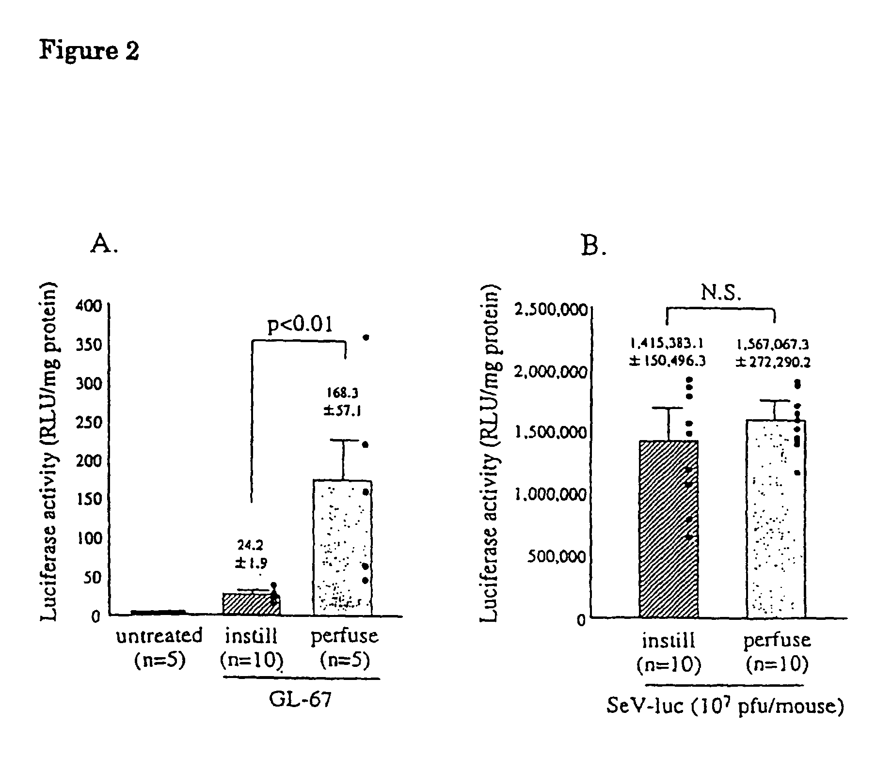
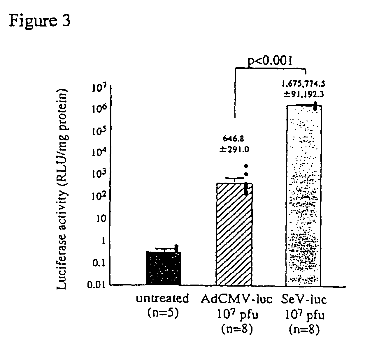
In vivo Gene Transfer to the Mouse Nose and Lung by Nasal Instillation or Nasal Perfusion

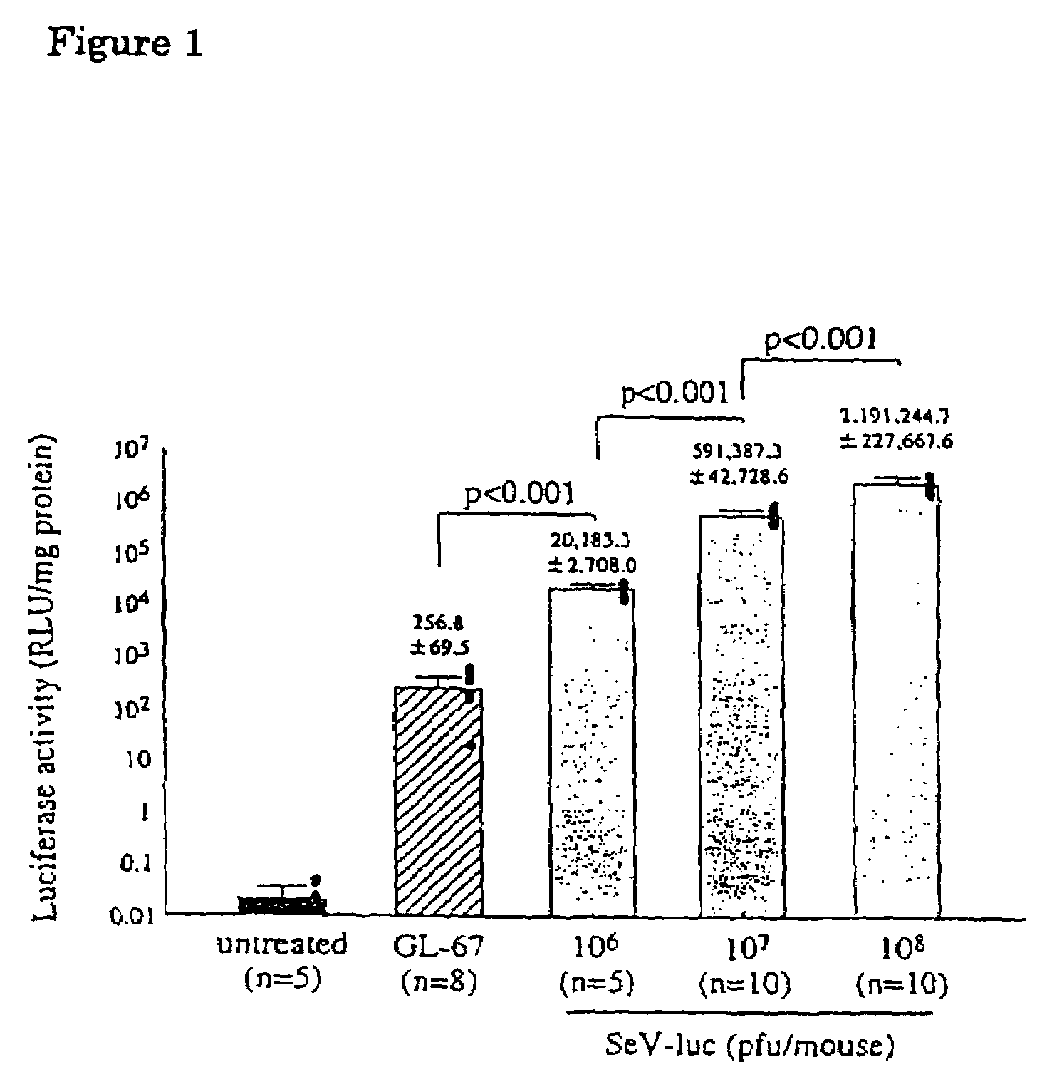
2-1. Comparing Sendai Virus Vector with Cationic Lipid

[0063]pCMV-luciferase was constructed by insertion of HindIII-BamHI fragment of pGL3-control vector (Promega), into the multicloning site of pcDNA3 (Invitrogen) to be driven by human cytomegalovirus immediate early (CMV-IE) promoter. pCMV-luciferase was then complexed with GL-67-DOPE-PEG (Genzyme Co. Ltd.) to obtain GL-67-pCMV-luc.

[0064]To examine gene transfer efficiency of the vectors to the lung and the effect of contact time on gene transfer efficiency, the vectors were administered to nasal cavity by nasal instillation and nasal perfusion. First, male balb / c mice (6-8 weeks) were instilled intranasally with 100 μl of various concentration of the Sendai virus vector containing luciferase (SeV-luc) prepared in Example 1 or GL-67-pCMV-luc (80 μg DNA / mouse) by the known method (Yonemitsu, Y. et al., Gene Ther. 4: 631-638, 1997).

[0065]Nasal p...

example 3

Gene transfer to Lung of Ferret

[0074]Ferrets (500-600 g weight) were anaesthetised and instilled intranasally with 3 ml of purified SeV-LacZ in BSS with either 3×10⁸ or 3×10⁹ pfu / ml (n=3 each group), as in Example 2. Controls (n=2) received 3 ml of SeV-Luc (109 pfu / ml). Forty-eight hours post-infection, ferrets were sacrificed, the trachea cannulated in situ and the lungs inflated with ice cold fixative solution (2% formalin, 0.2% glutaraldehyde, 2 mM MgCl2, 5 mM EGTA in PBS, pH 7.3). The trachea and lungs were excised en bloc and underwent X-Gal staining as described in Example 2. Each lung was dissected into 7 parts: trachea, 4 right lobes (upper (R1), mid (R2, R3), and lower (R4)) and 2 left lobes (upper (L1) and lower (L2)), and β-gal positive cells in the airway epithelia and submucosal glands were quantified microscopically by point counting using a graticulated lens. Ten ×20 magnification fields / airway were assessed to obtain the percentage of blue cells / airway and 3 to 8 air...


PUM

PropertyMeasurementUnit
concentrationaaaaaaaaaa

Abstract

Provided are a recombinant Sendai virus vector for introducing exogenous genes to airway epithelia and a method for introducing exogenous genes using the vector. The recombinant Sendai virus vector enables efficient gene transfer to native mucus-layered airway epithelial cells by briefly contacting the vector with the cells. Furthermore, the vector can introduce genes to not only apical surfaces but also submucosal glands where CFTR primarily expresses. The vector can thus be used for gene therapy of CF, a CFTR-deficient disease.

Description

[0001]This application claims priority from international application serial number PCT / JP00 / 07737, filed on Nov. 2, 2000, which in turn claims priority from Japanese application serial number JP 11-359218, filed on Dec. 17, 1999, and U.S. provisional application Ser. No. 60 / 163,055, filed on Nov. 2, 1999, the disclosures of which are hereby incorporated by reference.TECHNICAL FIELD[0002]The present invention relates to a recombinant Sendai virus vector for introducing exogenous genes to airway epithelia and a method for introducing exogenous genes using the vector.BACKGROUND ART[0003]With the advent of molecular cloning techniques, an expanding array of genes with mutations responsible for important human diseases have been identified and isolated. Absent or mutated genes in human patients can be replaced by ex vivo techniques, which include transformation of cells in vitro with naked DNA, DNA encapsulated in liposomes, appropriate integration vectors followed by introduction into ...

Claims


Application Information

Patent Timeline
Patent Type & Authority Patents(United States)
IPC IPC(8): A01N63/00A61K31/70A01N43/04A01N65/00C12N15/00C12N5/00C12N5/02
CPCA61K48/0075C12N15/86C12N2760/18843C12N2760/18871
Inventor YONEMITSU, YOSHIKAZUHASEGAWA, MAMORUALTON, ERIC W F W
Owner DNAVEC RES
Why Eureka
Try Eureka