Orthopaedic implant fixation using an in-situ formed anchor

a technology of orthopaedic implants and anchors, applied in the field of orthopaedic implants, can solve the problems of fractures under normal physiologic loading conditions, decreased bone mass, and increased risk of osteoporosis, and achieves the effects of reducing the likelihood of detrimental migration, reducing the likelihood of fracture, and reducing the pressur

Inactive Publication Date:
View PDF26 Cites 62 Cited by
  • Summary
  • Abstract
  • Description
  • Claims
  • Application Information

AI Technical Summary

Benefits of technology

[0017]The present invention is also advantageous because relatively low pressures are required to fill a relatively large bone cavity with an in-situ hardenable material. This will result in the effective use of a wide range of in-situ hardenable materials to include materials with higher injection viscosity. Lower pressures result in improved injection over a greater distances, injection through smaller diameter tubes and needles, and the potential for simpler, low-pressure injection systems, such as syringes. In addition, lower pressure results in a decreased likelihood of detrimental migration of in-situ hardenable material to unintended areas.
[0018]The advantages of the present invention include simplicity, as a bone cavity can be used as a mold to form an uncomplicated anchor interlocked to a preformed element. The present invention can be applied to numerous implants or implant system components to include, but not limited to, fasteners, pins, nails, intramedullary nails, and suture anchors. Applications include bone fracture fixation, bone fracture prevention, and soft-tissue repair. These and additional advantages will become evident from a consideration of the following description and drawings.

Problems solved by technology

However, strength can be compromised by osteoporosis, a metabolic disease characterized by a decrease in bone mass.
Osteoporosis leads to skeletal fractures under light to moderate trauma and, in its advanced state, can lead to fractures under normal physiologic loading conditions.
Whether treating a fracture associated with osteoporosis or another disorder of the musculoskeletal system, implant attachment to weakened osteoporotic bone can be problematic.
Inadequate attachment of an orthopaedic implant to osteoporotic bone can result in less effective, or ineffective implant fixation.
Tightening bone screws is generally subjective and the appropriate fixation is especially difficult to judge when securing a bone screw to osteoporotic cancellous bone.
Over-tightening can lead to stripping of bone and inadequate fixation, while under-tightening can also lead to inadequate fixation.
These time consuming repair techniques have experienced some success; however, the necessity for repair emphasizes the potential ineffectiveness of screw thread purchase in osteoporotic bone.
Also, bone screws can occasionally loosen, losing their effectiveness.
Further, loose bone screws can ultimately back-out and migrate to an undesirable position or location.
Foremost, the addition of apertures to a screw thread substantially weakens the bone screw.
Another disadvantage is the potential for uneven distribution of bone cement within cancellous bone, caused in part by bone pore regions not directly adjacent to apertures receiving a disproportionate amount of the injected cement.
In addition, extruding directly into bone can require relatively high pressures depending on the bone characteristics and the viscosity of the injectable material.

Method used


Image

  • Orthopaedic implant fixation using an in-situ formed anchor
  • Orthopaedic implant fixation using an in-situ formed anchor
  • Orthopaedic implant fixation using an in-situ formed anchor

Examples


Embodiment Construction

[0032]Throughout the following description and the drawings, like reference numerals are used to identify like parts of the present invention. The term “proximal”, as is traditional, will refer to the portion of the structure which is closer to the operator while the term “distal” will refer to the portion which is further from the operator.

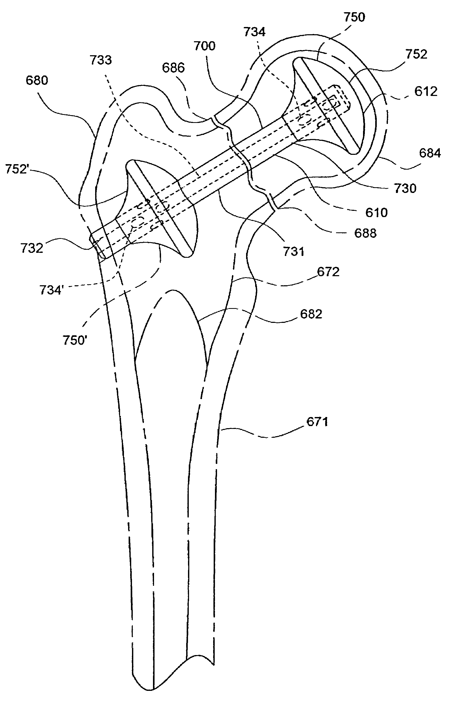
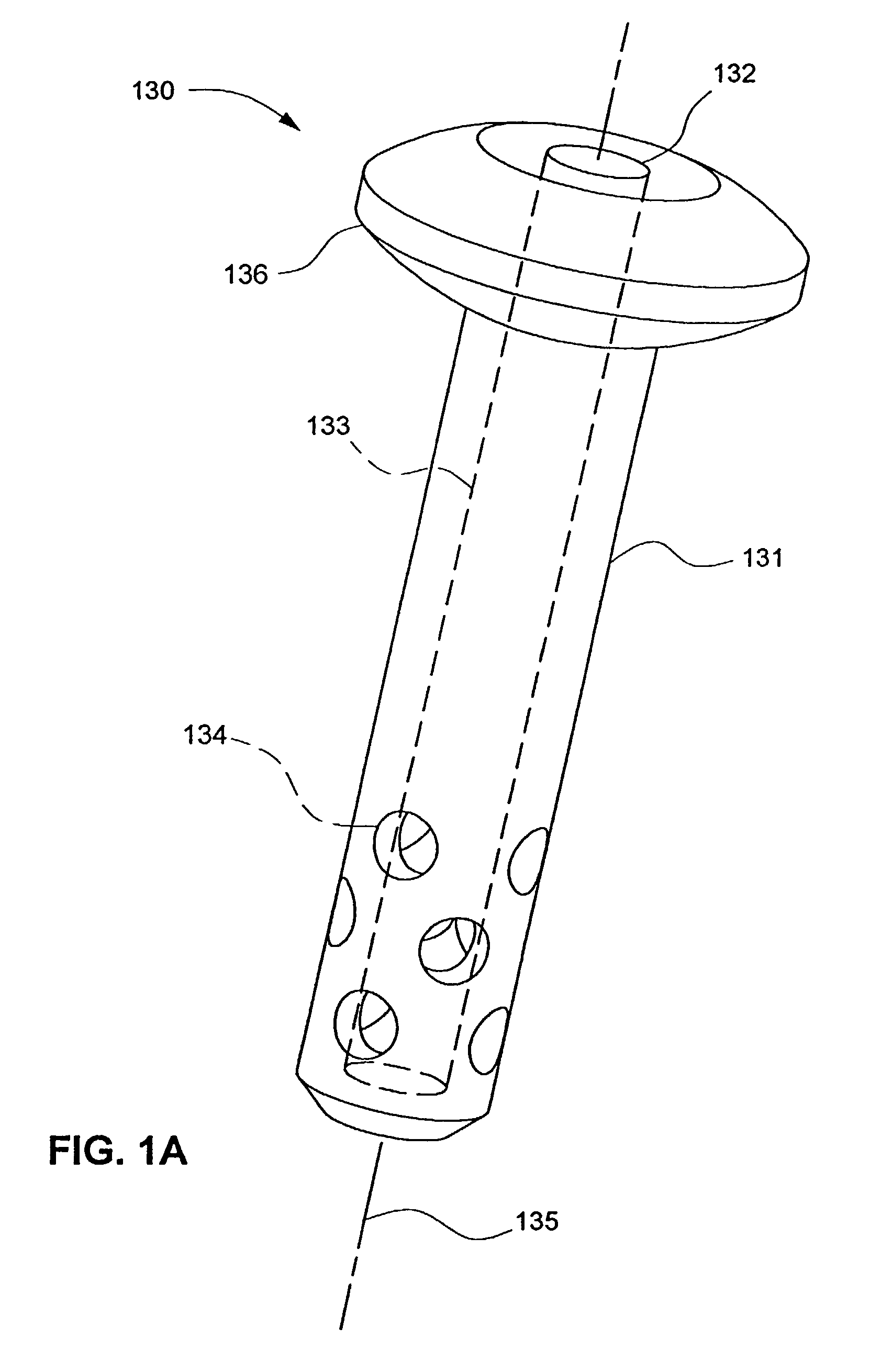
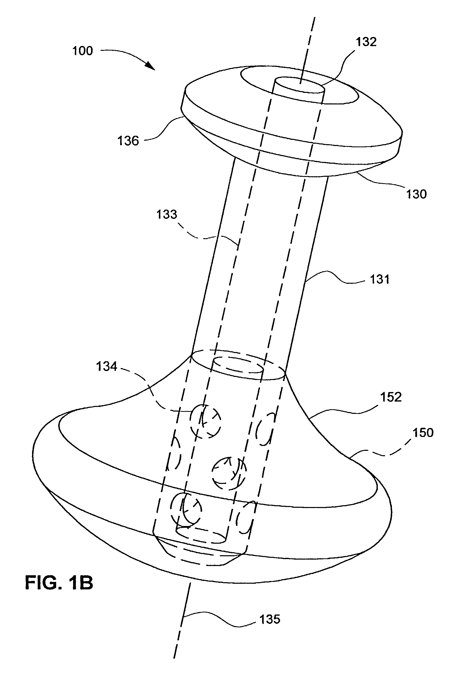
[0033]Numerous orthopaedic implants are broadly categorized as bone fasteners, to include screws, pins, and nails. These implants, pervasive in orthopaedic surgery, can be used as stand-alone devices to attach fractured or fragmented bone, or bone fasteners can be used as a component in an assembly or construct, such as a bone plate and screw construct. In order to focus on the spirit of the invention, the preferred embodiments of the present invention called bone fasteners will generally be shown as a stand-alone device without additional implant components and, further, without a reference to a specific application. Those skilled in the art wil...


PUM


Abstract

An orthopaedic implant fixation using a surgically created bone cavity as a mold for forming an anchor from an in-situ hardenable material. An in-situ formed anchor of the present invention is especially useful for attaching an implant to osteoporotic cancellous bone. The injectable nature of the in-situ formed anchor allows implants to be adapted to minimally invasive surgical techniques. The present invention can be adapted to numerous implants or implant system components to include fasteners, pins, nails, intramedullary nails, and suture anchors. Applications include bone fracture fixation, bone fracture prevention, and soft-tissue repair.

Description

CROSS-REFERENCE TO RELATED APPLICATIONS[0001]This application claims the priority of U.S. Provisional Patent Application Ser. No. 60 / 336,557 filed Nov. 1, 2001.FIELD OF THE INVENTION[0002]The present invention generally relates to orthopaedic implants and, more particularly, to fixation of orthopaedic implants to bone.BACKGROUND OF THE INVENTION[0003]Bone structures are typically comprised of two types of bone, cortical bone and cancellous bone. Cortical bone can be characterized as a rigid and dense material, whereas cancellous bone can be characterized as a structured material with a high degree of visible porosity. Cortical bone and cancellous bone combine to form structures that are strong and lightweight. However, strength can be compromised by osteoporosis, a metabolic disease characterized by a decrease in bone mass. It is estimated that osteoporosis affects approximately 15-20 million people in the United States. Although osteoporosis can affect persons of all ages and both ...

Claims


Application Information

Patent Timeline
Patent Type & Authority Patents(United States)
IPC IPC(8): A61B17/56A61B17/04A61B17/72A61B17/74A61B17/86A61B17/88
CPCA61B17/0401A61B17/7098A61B17/742A61B17/746A61B17/8811A61B17/72A61B17/725A61B17/864A61B17/866A61B2017/0412A61B2017/0414A61B2017/0427A61B2017/0458
Inventor MIDDLETON, LANCE M.
Why Eureka
Try Eureka