Vascular graft sterilization and decellularization

a vascular graft and allograft technology, applied in the field of sterilization and decellularization of allografts or xenografts, can solve the problems of significant economic cost, reduced graft patency, and insufficient autograft material, so as to reduce the number of potential harmful organisms, effective sterilization and decellularization of tissue, and effective removal

Active Publication Date: 2010-02-09
RTI BIOLOGICS INC
View PDF28 Cites 10 Cited by
  • Summary
  • Abstract
  • Description
  • Claims
  • Application Information

AI Technical Summary

Benefits of technology

[0011]The present technology is related to the field of sterilization and decellularization of allografts or xenografts, specifically to processes that achieve effective removal of the cells contained within a vascular tissue matrix and an effective reduction in potential harmful organisms to create grafts suitable for human implantation. As a non-limiting example, human saphenous vein samples are introduced to a sequence of chemicals with optional mechanical stimulation which provides effective sterilization and decellularization of the tissue. Since it is difficult to predict frequencies and concentrations of pathogens and infection windows may exceed the time between contraction and the time of death, inactivation of a highly resistant spore is used to determine sterility assurance. The level of decellularization is demonstrated with an immunohistochemistry stain for antigens located on the cell membranes within the tissue. The processing conditions of the present technology leave a neutralized scaffold with minimal detrimental biochemical effects on the extracellular matrix, no need for blood typing, and optimal biological functionality.
[0012]In one aspect of the present technology, a vascular graft is processed at a temperature effective for decellularization in combination with predetermined concentrations of cleaning agents (e.g. enzymes and detergents) to provide effective decellularization with minimal tissue damage. In an additional embodiment, a chelating agent is also used to enhance cell removal.
[0014]In a further aspect, a process comprises distending vascular tissue, such as by providing a pressure gradient across a wall of the vascular tissue. Distension of venous tissue by pressurizing the lumen can be used to expand the pore volume and allow more efficient penetration of cleaning agents. This distension also reduces the amount of time used to reach effective graft decellularization. For example, in at least one such embodiment, a process is provided for preparing a vascular graft comprising at least one blood vessel, the process comprising providing a pressure gradient across a wall of the blood vessel, and contacting the blood vessel with one or more cleaning agents.

Problems solved by technology

This procedure offers benefits to the patient but incurs significant economic cost on the national level.
Commonly, insufficient autograft material is available and a patient may receive cadaver tissue (allograft).
However, allograft conduits have poor patency rates resulting in numerous revision surgeries, amplifying the economic impact.
A reduction in graft patency is a result of an immunological response to cellular material contained within and on the allograft leading to intimal hyperplasia and eventual graft occlusion.
Allografts can have complications leading to early stenosis and graft failure thought to be related to an immune reaction similar to graft rejection.
Grafts most commonly fail due to the development of fibrous intimal hyperplasia where there is an excess proliferation of smooth muscle cells.
Current processing methods only treat the tissue without removing a substantial amount of endogenous material that may harbor contaminants, such as blood and lipids, as well as antigen containing cells and cellular debris that elicit an immune reaction.
Rejection plays a significant role in failure and leads to allosensitization (Timaran, 2002).
Therefore, a great deal of effort is placed in determining an ABO-compatible donor as well as lymphocyte cross-matching (Prager, 2002).
Stripping this layer of cells from the conduit removes the need for donor matching, but it is believed by some that a lack of surviving cells and endothelial integrity may play a role in graft degeneration and early and late patency (Prager, 2002).
Currently, the demand for vascular allografts far exceeds the supply.
Current processing methods such as using an antimicrobial soak can result in a substantial discard rate due to positive first and final cultures from bacterial and fungal contamination resistant to the cocktail aimed at achieving sterility.
Therefore, vascular transplantation carries some risk of disease transmission to the recipient considering the occurrence of false negative test results, human errors in screening and processing, and virological testing within the window period of contraction and detection of diseases.
These examples include transmission of hepatitis C, bacteria, and fungi, and have resulted in serious morbidity and even mortality (Kainer, 2002).
Traditional methods of sterilization, such as irradiation and ethylene oxide, have significant deleterious effects on tissue integrity that limit or preclude their use on allograft tissue.
However, patients who do not have sufficient or acceptable autografts, or are unable to receive synthetic grafts are must use allografts.
A failure of this type could be catastrophic and lead to morbidity, such as loss of limb, or even mortality.

Method used


Image

  • Vascular graft sterilization and decellularization
  • Vascular graft sterilization and decellularization
  • Vascular graft sterilization and decellularization

Examples


example 1

Processing Parameters
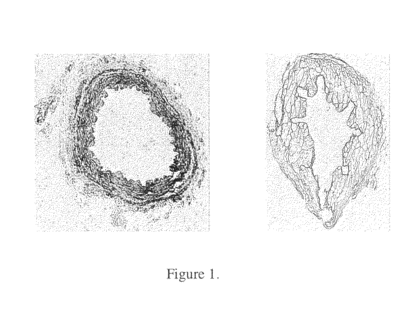
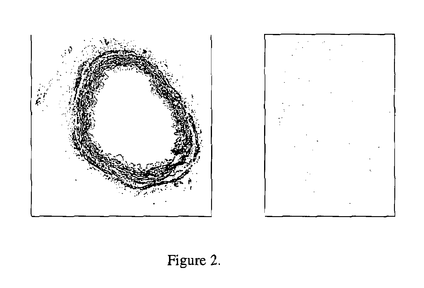
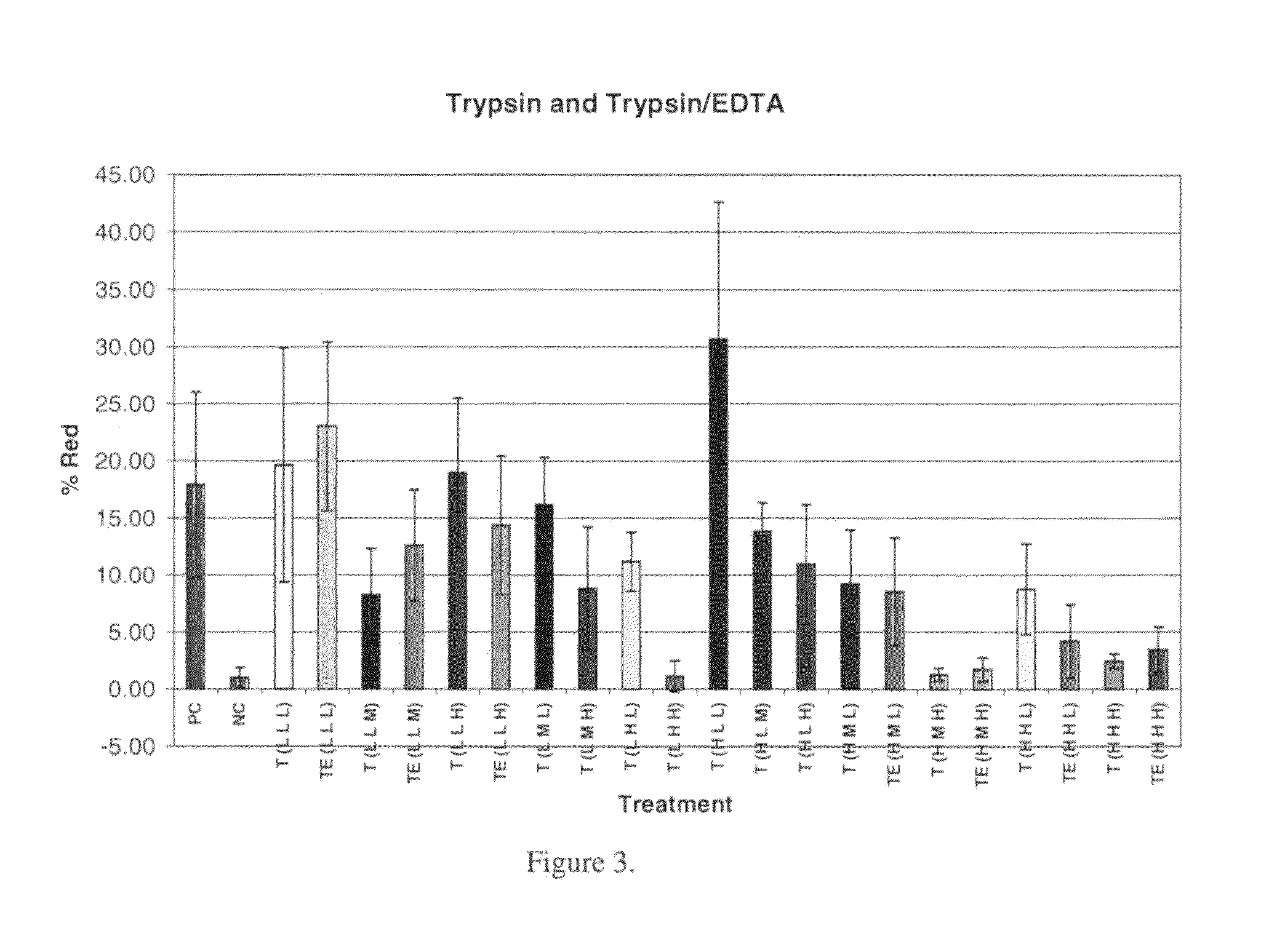
Cell Removal:

[0195]The effectiveness of cell and cellular debris removal was evaluated using two detergent and two enzyme solutions at various concentrations (Table 2). The detergents used in this study were Triton-X 100 at 0.25% (v / v) and 1.0% (v / v) and sodium deoxycholate (Cholate) at 0.25% (w / v) and 1.0% (w / v). Due to viscosity issues with sodium deoxycholate at low temperatures, tests were repeated at 0.5% (w / v). One treatment group combined Triton-X 100 with sodium deoxycholate. Trypsin alone at 0.05% (w / v) or 1.0% (w / v) and in combination with 0.02% (w / v) EDTA were the enzyme solutions used in the decellularization process. EDTA was also combined with Triton for a comparison since the Trypsin samples appeared grossly degraded after treatment.

[0196]Samples of human saphenous veins 10 mm in length were placed in each of the test solution at 37° C. or 48° C. for both 6 and 24 hours while continuously shaking. Samples were then rinsed in PBS at room temperatur...

example 2

Reduction Curves (Survivor Curves) for Spores

[0210]In order to determine the reduction in spores after processing with cleaning agents, an effective and consistent method was developed. Initially, a 107 spores / ml titre of B. stearothermophilus was used to enumerate the remaining population after treatment on both TSA media plates and sheep's blood agar (BA) plates. It was determined that residual hydrogen peroxide inhibited the growth at concentrations of 0.6% or higher for TSA plates and 6% or higher for blood plates (FIG. 12). It is believed that the catalase contained in the blood agar plates inactivated the hydrogen peroxide. A similar investigation was performed on PAA, but no inhibition was observed within the processing concentrations (FIG. 13). Even though the blood agar plates allowed for dilutions as low as 1:10, the titre used was too low to detect more than a 3 log reduction in the process limiting the data collection for the survivor curves. Therefore, the titre used in...

example 3

Derivation of Governing Equations

[0226]The vein wall is divided into five layers: endothelial cell monolayer, intima, internal elastic lamina (IEL), media and adventitia. Since the endothelial layer acts as an active barrier to transport it is likely to offer more resistance to flow than bulk tissue (Kenyon, 1979). The study mentioned above on rabbit aortic tissue found that removal of the endothelial cells increased the macromolecular uptake at both 70 and 160 mm Hg of applied pressure (Meyer, 1996). The resistance due to this layer will not be a factor in this study since the grafts will be deendothelialized. The layer in contact with the luminal fluid will then be the intima. The intima itself provides very little resistance to flow and it has been shown that it provides no support to matrix stress while consolidation occurs at the interior wall at the border between the intima and rigid IEL (Kenyon, 1979). When it is combined with the IEL there is a greater pressure drop (ΔP / L) ...


PUM


Abstract

The present technology is related to the field of sterilization and decellularization of allografts or xenografts, specifically to processes that achieve effective removal of the cells contained within a vascular tissue matrix and an effective reduction in potential harmful organisms to create grafts suitable for human implantation. In some embodiments, vascular grafts, vascular tissue and / or blood vessels are contacted with cleaning solution under conditions suitable conditions to reduce immune reaction in patients. More specifically, the present technology is directed to sterilization and decellularization of vascular grafts.

Description

CROSS-REFERENCE TO RELATED APPLICATION[0001]This application claims priority to U.S. Provisional Application No. 60 / 742,828, filed on Dec. 5, 2005, entitled “Vascular Graft Sterilization and Decellularization”, which is incorporated by reference herein in its entirety.FIELD OF THE INVENTION[0002]The present technology is related to the field of sterilization and decellularization of allografts or xenografts, specifically to processes that achieve effective removal of the cells contained within a vascular tissue and an effective reduction in potentially harmful organisms to create grafts suitable for human implantation. More specifically, the present technology is directed to methods to treat vascular allografts or xenografts to reduce immune reaction. Even more specifically, the present technology is directed to sterilization and decellularization of vascular grafts.BACKGROUND OF THE INVENTION[0003]Vascular disease is one of the most common diseases worldwide and, with the increasin...

Claims


Application Information

Patent Timeline
Patent Type & Authority Patents(United States)
IPC IPC(8): A61F2/04
CPCA61L2/0088A61L2/0094A61L27/3687A61L27/507A61L2430/40
Inventor SQUILLACE, DONNA
Owner RTI BIOLOGICS INC
Why Eureka
Try Eureka