Device and method for in-line blood testing using biochips

a biochip and in-line technology, applied in the field of blood screening, can solve the problem of creating a sampling risk by using such small fractions

Inactive Publication Date: 2010-08-31
NOVARTIS VACCINES & DIAGNOSTICS INC
View PDF81 Cites 5 Cited by
  • Summary
  • Abstract
  • Description
  • Claims
  • Application Information

AI Technical Summary

Benefits of technology

[0006]The present invention provides a new platform technology for screening blood. The technology ensures that a much larger fraction of blood collected, even every drop, is tested. Under previous screening methods, only very small fractions of roughly 450 ml of collected blood was used for screening. For example, 0.5 ml was used for NAT testing, 20 μl each for anti-HIV I / II, anti-HCV, anti-HBC, ALT, and syphilis testing, and 100 μl each for HbsAg and HIV p24 testing. Using such small fractions creates a sampling risk, because the fractions may not be representative of the entire volume of collected blood. That is, disease agents that are relatively scarcely scattered in the collected blood may not be contained within a small fraction or volume. The present invention addresses this problem by ensuring a more thorough and more comprehensive screening of collected blood. The present invention also enables testing for hundreds of agents or molecules to be performed at once, rather than testing for a small number of agents or molecules in separate assays.
[0010]The present invention provides at least the following advantages over currently known procedures for screening blood:
[0013](3) The invention can employ low-density biochips, so adding additional targets, such as components of new emerging diseases, is easier and more efficient.
[0014](4) The assays of the invention are rapid, due to minimized reaction times resulting from the use of micro-volumes of sample in a biochip processor.
[0015](5) The “laboratory on a chip” screening system of the invention provides a confined environment for running the multiple assays. The screening capture unit and biochip processing unit are both self-sealed, consume little space and diminish the opportunity for human error and / or contamination that may be caused by exposing the sample to the surrounding environment or by extra handling of the sample required to conduct multiple individual assays.

Problems solved by technology

Using such small fractions creates a sampling risk, because the fractions may not be representative of the entire volume of collected blood.

Method used


Image

  • Device and method for in-line blood testing using biochips
  • Device and method for in-line blood testing using biochips
  • Device and method for in-line blood testing using biochips

Examples


example 1

[0106]This example demonstrates the use of a biochip coated with antibody to capture corresponding antigen in blood and detection of the target antigen by mass spectroscopy.

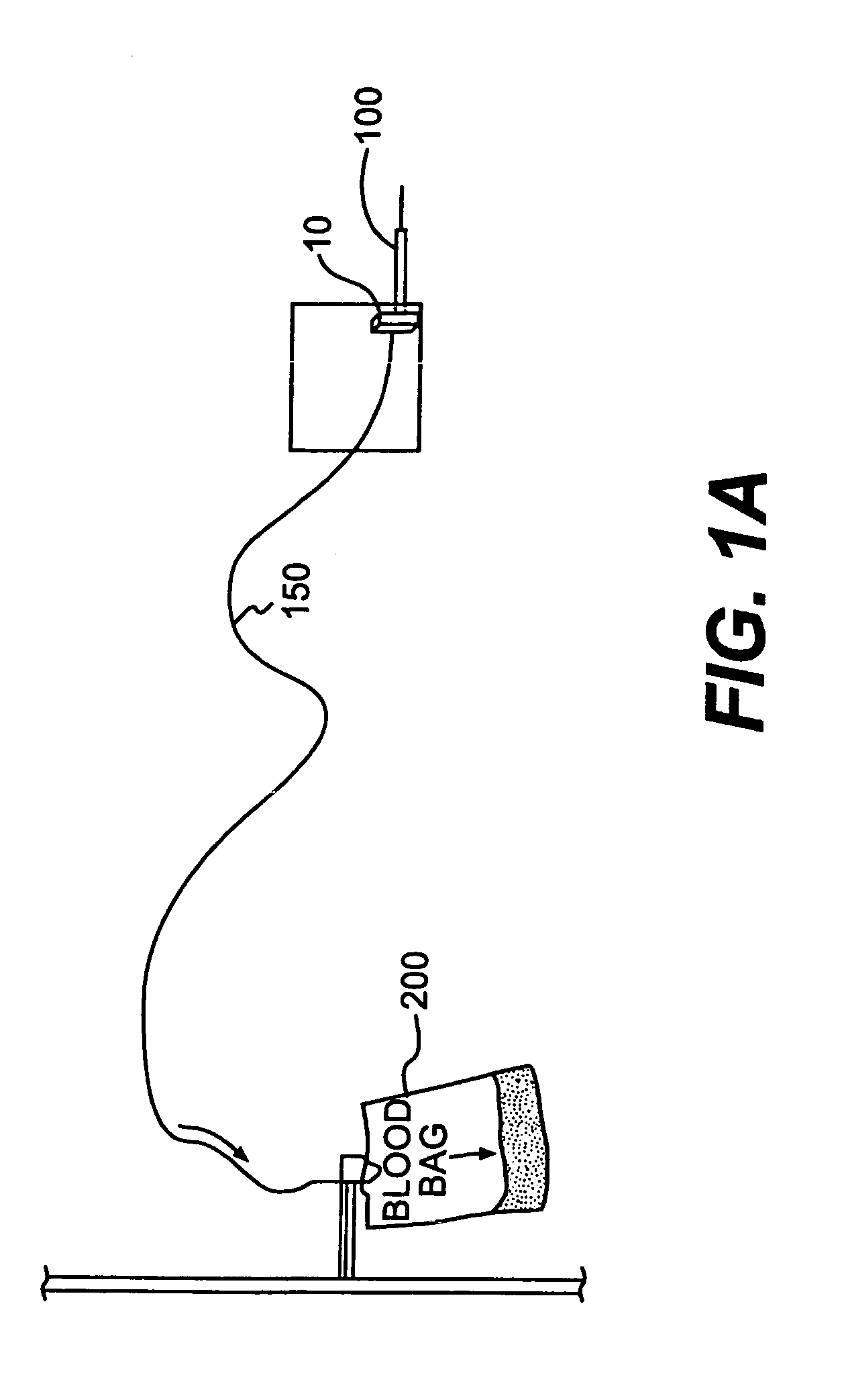
[0107]A biochip (Ciphergen PS20 Protein chip) was covalently bound to a mouse monoclonal antibody against HIV P-24 core protein. The biochip then was placed in a tube where 12 ml of human blood was circulated a flow rate of about 30 ml / min at 37° C., to mimic the flow rate and temperature of a normal blood drawing. See FIG. 13. HIV P-24 antigen was spiked in the blood at a concentration of 0-200 ng / ml. After 10 min of circulation, the chip was removed and the bound antigen was detected by mass spectroscopy (Ciphergen Protein Chip Reader).

[0108]The results showed that antigen can be detected at a concentration of about 8 ng / ml in a 12 ml blood sample. FIG. 14 depicts a typical result.

example 2

[0109]This example demonstrates the use of a biochip coated with antigen to capture corresponding antibody in blood and detection of the target antibody by mass spectroscopy.

[0110]A biochip (Ciphergen PS20 Protein chip) was covalently bound to HIV P-24 core protein. The biochip then was placed in a tube where 12 ml of human blood was circulated at a flow rate of about 30 ml / min at 37° C., to mimic the flow rate and temperature of a normal blood drawing. Mouse monoclonal antibody against HIV P-24 was spiked in blood at a concentration of 0-20 ug / ml. After 10 min. of circulation, the chip was removed and the bound antibody was detected by mass spectroscopy (SELDI-MS from Ciphergen).

[0111]The results showed that antibody can be detected at a concentration of about 1 ug / ml in a 12 ml blood sample. FIG. 15 depicts a typical result.

example 3

[0112]This example demonstrates the use of an antibody against viral surface protein to capture a virus in blood, followed by detection of viral nucleotides using a nucleic acid technology assay.

[0113]West Nile virus was captured by an Elisa assay. An Elisa plate was coated with West Nile Virus Monoclonal Antibody 3A3 (from BioReliance, Rockville, Md.) as a capture antibody. Purified West Nile virus samples then were added. After incubation, a polyclonal antibody (rabbit anti mouse West Nile virus, from BioReliance, Rockville, Md.) was added and incubated. The detection antibody was goat anti rabbit antibody conjugated with HRP. Other anti-West Nile virus antibodies, such as mouse monoclonal antibody 3.91D, also have been shown to capture West Nile virus. See, e.g., Hunt et al., 2002.

[0114]Table 1 (below) summarizes results of the virus-capture procedure.

[0115]

TABLE 1Capture virus using monoclonal antibody.12 Viral3 Vero4 SampleVirusAntigen PreM-3Cell Supernatantdiluent1.1000.2950.2...


PUM

PropertyMeasurementUnit
temperatureaaaaaaaaaa
pore sizeaaaaaaaaaa
pore sizeaaaaaaaaaa

Abstract

A device for in-line blood screening and testing using biochips is disclosed. The screening methods include nucleic acid amplification techniques and antibody / antigen assays to detect target molecules and agents indicative of infectious diseases or metabolic diseases.

Description

BACKGROUND OF THE INVENTION[0001]This invention relates to the field of blood screening and provides improvements that enable more comprehensive, accurate and convenient screening to be performed, for detecting the presence of targeted molecules that indicate the presence of a pathogen, infectious agent or metabolic disease. More specifically, the invention provides a method and system for in-line blood screening, wherein an in-line removable screening capture device with biochips is provided between a blood collection needle and a blood collection bag, such that collected blood flows through the screening capture device and contacts the biochips before being collected in the blood collection bag. Alternatively, the biochips are provided in an inlet of the blood collection bag, inside the blood collection bag or inside a separate diversion blood collection bag.[0002]Currently, blood banks use several individual assays to test for the presence of multiple agents or molecules associat...

Claims


Application Information

Patent Timeline
Patent Type & Authority Patents(United States)
IPC IPC(8): C12Q1/68A61B5/00A61M5/32B01L3/00B01L7/00C07H19/00C07H21/02C07H21/04C12M1/34C12P19/34C12QG01N33/48G01N33/53
CPCB01L3/5027G01N33/5302A61B5/14557B01L7/52B01L2200/027B01L2200/10B01L2300/0636B01L2300/0825B01L2300/0877B01L2300/0887
Inventor CHIEN, DAVID Y.PHELPS, BRUCE H.FONG, YIU-LAN
Owner NOVARTIS VACCINES & DIAGNOSTICS INC
Why Eureka
Try Eureka