Whole blood preparation for cytometric analysis of cell signaling pathways

a cytometric analysis and whole blood technology, applied in the field of sample preparation for cytometry, can solve the problems of inability to address methodologies, undetectable significant information on immune cell population variations that exist in both defined cellular populations and across different cell subtypes, and inability to provide information about the functional responses of those cells to stimuli immediately reflective of intracellular events

Active Publication Date: 2010-09-28
UNIV HEALTH NETWORK +1
View PDF14 Cites 9 Cited by
  • Summary
  • Abstract
  • Description
  • Claims
  • Application Information

AI Technical Summary

Benefits of technology

[0008]This invention is directed to a method for preparation of a biological sample for measurement of protein epitopes that allows for the preservation of intracellular protein epitopes and detection of signal transduction pathways based on the ability to capture transient activation states of the epitopes. The method provided by the invention allows for the rapid fixation of biological samples containing red blood cells, to ensure that epitopes of signal transduction molecules and other intracellular protein epitopes are preserved in the active state. The method of the invention further allows for lysis of red blood cells, thereby making it a useful method for cytometric analysis of biological samples, including, for example, whole blood, bone marrow aspirates, peritoneal fluids, and other red blood cell containing samples. The invention also provides a method to recover or “unmask” epitopes on intracellular antigens that have been made inaccessible by the cross linking fixative necessary to fix the sample. Significantly, the methods of the invention allow preservation and analysis of phospho-epitope levels in biological samples taken directly from patients to determine disease-specific characteristics.

Problems solved by technology

Although surface staining may be an effective means of characterizing cells, it does not provide information about the functional responses of those cells to stimuli that are immediately reflective of intracellular events.
Therefore significant information on immune cell population variations that exist in both defined cellular populations and across different cell subtypes is undetectable and cannot be addressed by methodologies that require cell lysis for protein analysis.
Analysis of signal transduction pathways by flow cytometry presents technical problems that are not currently encountered in routine clinical applications.
The phosphorylation states of individual signaling elements are rapidly modified in response to specific kinases and phosphatases, and therefore subject to artifactual changes during sample storage and preparation.

Method used


Image

  • Whole blood preparation for cytometric analysis of cell signaling pathways
  • Whole blood preparation for cytometric analysis of cell signaling pathways
  • Whole blood preparation for cytometric analysis of cell signaling pathways

Examples


example i

Red Blood Cell Lysis, Fixation and Permeabilization of Whole Blood Samples

[0061]This example describes and compares preparation of whole blood samples by hypotonic lysis followed by fixation and detergent lysis subsequent to fixation.

[0062]Briefly, Phorbol myristate acetate (PMA, Sigma Chemical Corp., St. Louis, Mo.) was prepared as a 40 M working solution in 100% anhydrous ethanol and used in whole blood at a final concentration of 400 nM. Triton® X-100 (polyethylene glycol p-(1,1,3,3-tetramethylbutyl)-phenyl ether) and other detergents used in the examples were purchased as the Surfact-Pak™ Detergent Sampler kit from Pierce Biotechnology (Rockford, Ill.). The Sampler kit contains seven different non-ionic detergents (Tween®-20, Tween®-80, Triton® X-100, Triton® X-114, Nonidet® P-40, Brij-35® and Brij-58®, all supplied as 10% solutions in water), and three powders (nonionic Octyl-B-Glucoside, and Octyl-B-Thioglucopyaranoside, and zwitterionic CHAPS). Powdered detergents were dissol...

example ii

Impact of Fixation Agent and Detergent Concentrations on White Blood Cell Light Scatter

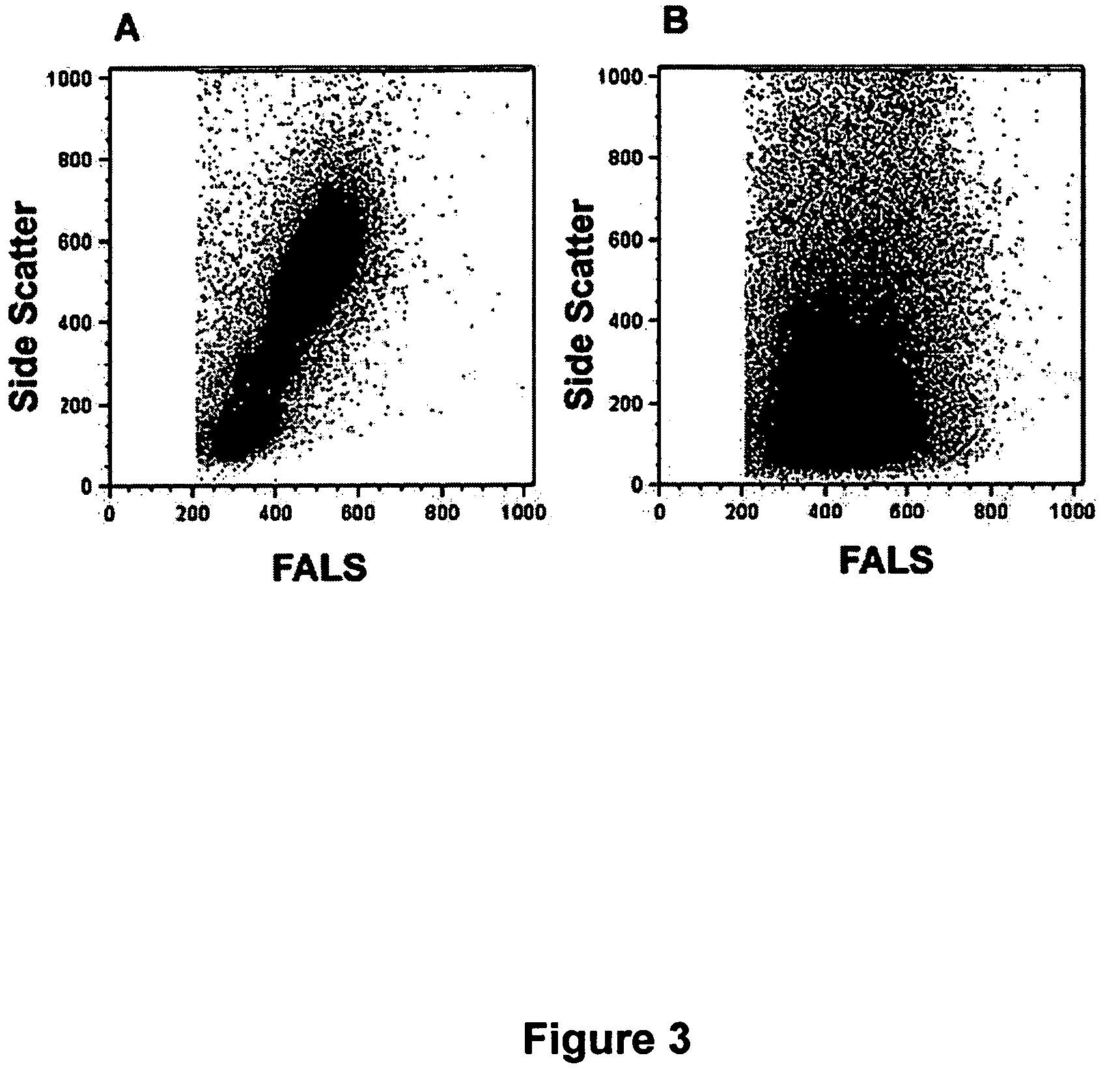
[0082]This example demonstrates the impact of concentration and incubation times for both fixative agent and detergent on the resolution and recovery of white blood cells.

[0083]Based on previous studies that suggested that higher concentrations of cross linking fixative helps retain light scatter profiles and resolution of white blood cell populations, the impact of different fixative concentrations was investigated. Whole blood samples were fixed at room temperature or at 37 deg. C. for 10 minutes in increasing concentrations of formaldehyde (from 1% to 10% final concentration), then immediately incubated with 1 ml 0. 1% TX-100 (at room temperature). As shown in FIG. 6, increasing formaldehyde concentration from 2% to 4% (for room temperature incubation) significantly improved the resolution of white blood cell populations (left panels). Similarly, treatment of whole blood samples with formaldehy...

example iii

Impact of Alcohol Unmasking Agent on P-ERK and Impact on CD3 Expression

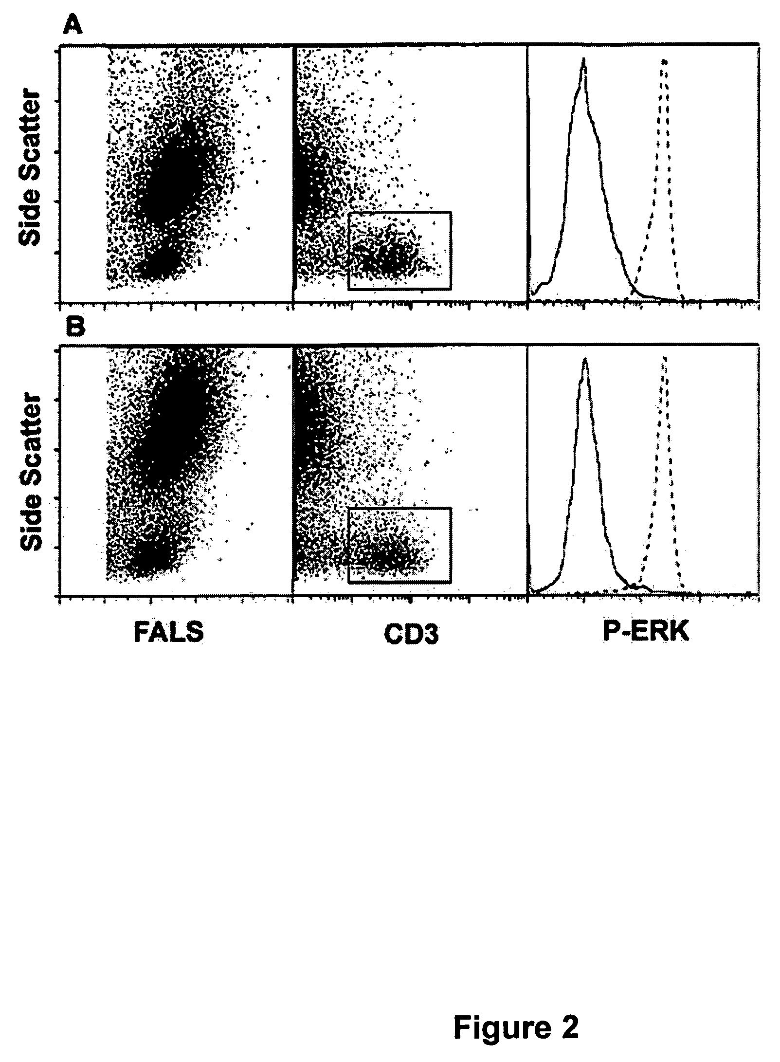
[0088]This example demonstrates the impact of alcohol on lymphocyte recovery and unmasking of surface epitopes.

[0089]In order to “unmask” phospho-protein (and other) epitopes rendered unreactive following fixation with cross linking fixatives, a series of experiments was undertaken to evaluate the impact of alcohol treatment following detergent treatment. Following 30 minute exposure to detergent, fixed whole blood samples were washed with cold (4 deg C.) buffer (PBS w / o Ca++ or Mg++) and resuspended in a series of different concentrations of methanol or ethanol at 4 deg C. One aliquot of each sample was held overnight at 4 deg. C. to investigate the impact of storage in alcohol solutions. As before, samples were stained (following removal of alcohol by centrifugation and washing in PBS) with antibodies to CD3-PE (to evaluate T-lymphocyte recovery and staining) and phospho-ERK (to evaluate epitope unmasking).

[009...


PUM

PropertyMeasurementUnit
temperatureaaaaaaaaaa
time periodaaaaaaaaaa

Abstract

This invention is directed to a method for preparation of a biological sample for measurement of protein epitopes that allows for the preservation of intracellular protein epitopes and detection of signal transduction pathways based on the ability to capture transient activation states of the epitopes. The method provided by the invention allows for the rapid fixation of biological samples containing red blood cells, to ensure that epitopes of signal transduction molecules and other intracellular protein epitopes are preserved in the active state. The method of the invention further allows for lysis of red blood cells, thereby making it a useful method for cytometric analysis of biological samples, including, for example, whole blood, bone marrow aspirates, peritoneal fluids, and other red blood cell containing samples. The invention also provides a method to recover or “unmask” epitopes on intracellular antigens that have been made inaccessible by the cross linking fixative necessary to fix the sample. Significantly, the methods of the invention allow preservation and analysis of phospho-epitope levels in biological samples taken directly from patients to determine disease-specific characteristics.

Description

FIELD OF THE INVENTION[0001]This invention relates generally to the field of sample preparation for cytometry and, more specifically, to methods capable of biological sample preparation for signal transduction measurements by flow cytometry and image cytometry.BACKGROUND OF THE INVENTION[0002]Flow cytometry has become an indispensable tool in clinical and basic immunological research due to its ability to distinguish subsets in heterogeneous populations of cells. Recently, major advances have been made in both flow cytometry instrumentation and applications, expanding the number of possible simultaneous analysis parameters to thirteen or more. With more parameters available, researchers have begun to identify more well-defined and biologically interesting subsets of lymphocytes samples based upon surface epitope staining.[0003]Flow cytometry is routinely used for the identification of cellular populations based on a surface phenotype and also used for cellular based assays such as c...

Claims


Application Information

Patent Timeline
Patent Type & Authority Patents(United States)
IPC IPC(8): A01N1/02G01N1/00
CPCG01N33/80Y10T436/25Y10T436/107497Y10T436/25125Y10T436/2525Y10T436/108331Y10T436/25375
Inventor CHOW, SUEHEDLEY, DAVIDSHANKEY, T. VINCENTGROM, PATRICIA
Owner UNIV HEALTH NETWORK
Why Eureka
Try Eureka