
Embolization device

a technology of embolization device and collagen, which is applied in the field of medical devices, can solve the problems of obliteration of aneurysms, collagenous biomaterial embolization device, and inability to use collagen as embolization device, so as to increase the production of interleukin-10, promote the remodeling of surrounding tissue, and increase the immune respons

Inactive Publication Date: 2010-12-28
COOK BIOTECH
View PDF196 Cites 32 Cited by
  • Summary
  • Abstract
  • Description
  • Claims
  • Application Information

AI Technical Summary

Benefits of technology

[0011]The foregoing problems are solved and a technical advance is achieved in an illustrative embolization device advantageously utilizing a collagen based material. This collagen based material can be a biomaterial called Small Intestine Submucosa (SIS). SIS has been shown to be a remarkable biomaterial that promotes remodeling of the surrounding tissue, such as cellular invasion, host incorporation, and absorption of the SIS material into the local tissue. Furthermore, SIS has been shown to be acellular, strong, and exhibit a sidedness in that it has a differential porosity of its mucosal and serosal sides. SIS also does not trigger any negative immune system response as evidence suggests that it has no viral activity when checking for enveloped, non-enveloped, DNA, and RNA virus. Studies also show that SIS increases the Th-2 immune response by increasing the production of interleukin-10 over interferon-γ, which indicates that the immune response is more accommodation than rejection. Due to these and other properties, SIS makes for an excellent implantable biomaterial for use as an embolization or aneurysm treatment device. Furthermore, collagen is not currently being used as an embolization device or as an aneurysm lumen filling device, and in particular, SIS has not yet been used as such a device.
[0012]In accordance with the present invention, provided is a collagenous implantable biomaterial, that is further advantageous in that it is promotes healing of the occluded area and healing of the aneurysm. Furthermore, as described herein, the collagenous biomaterial embolization device has been shown to obliterate aneurysms. Another ideal characteristic is that the device can cause new endothelial growth across the neck of the aneurysm, thus resulting in obliteration of the aneurysm.

Problems solved by technology

Furthermore, collagen is not currently being used as an embolization device or as an aneurysm lumen filling device, and in particular, SIS has not yet been used as such a device.
Furthermore, as described herein, the collagenous biomaterial embolization device has been shown to obliterate aneurysms.
Another ideal characteristic is that the device can cause new endothelial growth across the neck of the aneurysm, thus resulting in obliteration of the aneurysm.

Method used


Image

  • Embolization device
  • Embolization device
  • Embolization device

Examples


example 1

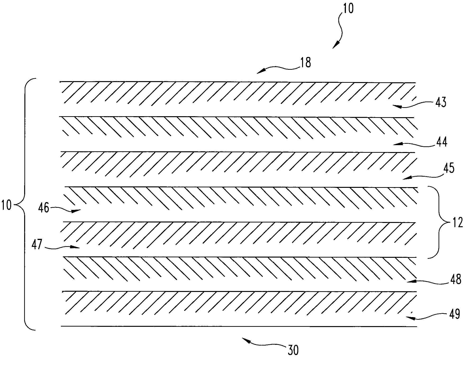
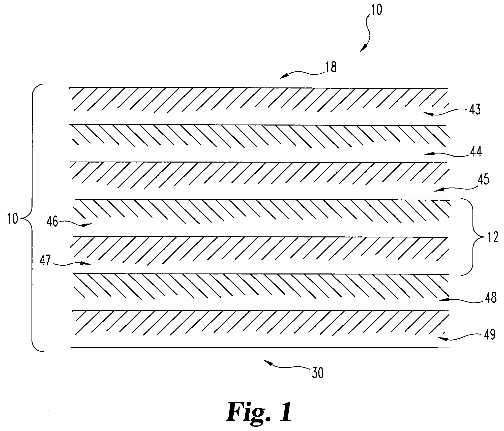
[0074]Thirty feet of whole intestine from a mature adult hog is rinsed with water. This material is then treated in a 0.2% by volume peracetic acid in a 5% by volume aqueous ethanol solution for a period of two hours with agitation. The tela submucosa layer is then delaminated in a disinfected casing machine from the whole intestine. The delaminated tela submucosa 12 is rinsed four (4) times with sterile water and tested for impurities or contaminants such as endotoxins, microbial organisms, and pyrogens. The resultant tissue was found to have essentially zero bioburden level. The tela submucosa layer separated easily and consistently from the whole intestine 10 and was found to have minimal tissue debris on its surface.

example 2

[0075]A ten foot section of porcine whole intestine is washed with water. After rinsing, this section of tela submucosa intestinal collagen source material is treated for about two and a half hours in 0.2% peracetic acid by volume in a 5% by volume aqueous ethanol solution with agitation. Following the treatment with peracetic acid, the tela submucosa layer is delaminated from the whole intestine. The resultant tela submucosa 12 is then rinsed four (4) times with sterile water. The bioburden was found to be essentially zero.

example 3

[0076]A small section of the tela submucosa intestinal collagen material was subcutaneously implanted in a rat. Within 72 hours, significant angiogenesis was observed.


PUM


Abstract

A medical device (40) including a collagenous biomaterial (10) for causing embolus and treating an aneurysm. The collagenous biomaterial (10) includes a small intestine submucosa (12) that is configured into various components (16) for causing a vascular occlusion or a packing aneurysm lumen.

Description

REFERENCE TO RELATED APPLICATIONS[0001]This application is a continuation of U.S. patent application Ser. No. 09 / 448,915 filed Nov. 24, 1999, now abandoned, which claims priority to U.S. Provisional Application Ser. No. 60 / 110,434 filed Dec. 1, 1998, each of which is hereby incorporated by reference in its entirety.TECHNICAL FIELD[0002]The invention relates generally to a medical device and, in particular, to an embolization device.BACKGROUND OF THE INVENTION[0003]During the course of medical diagnosis, treatment, and the follow-up of various medical conditions, it is often necessary for the physician to block a certain blood vessel, deprive a certain area of life-sustaining blood, or fill a cavernous area in the blood vessel. For example, where a certain blood vessel is perforated, blood will flow out of the vessel into the local area causing a hemorrhage. For this condition, the physician will need to, inter alia, plug the perforation or occlude the vessel upstream of the perforat...

Claims


Application Information

Patent Timeline
IPC IPC(8): A61M29/00A61F2/06A61B17/12A61B19/00A61K35/38A61L27/36A61L31/00
CPCA61B17/12022A61B17/12109A61B17/12113A61B17/1214A61B17/12145A61B17/1215A61B17/12163A61B17/12168A61B17/12172A61B17/12181A61B17/1219A61K35/38A61L27/3604A61L27/3629A61L27/3641A61L27/3645A61L31/005A61L31/18A61B19/54A61L2430/36A61B90/39
Inventor MORAN, CHRISTOPHER J.BLEYER, MARK W.KOZMA, THOMAS G.PATEL, UMESH H.
Owner COOK BIOTECH
Learn More